

We use cookies to ensure our website works properly and to personalise your experience. Cookies policy
Department of Pharmaceutical Chemistry, Al Shifa College of Pharmacy, Perinthalmanna, Malappuram, Kerala.
Fused 1,2,4-triazolo[3,4-b] [1,3,4] thiadiazines pair the metabolic stability of the 1,2,4-triazole ring with the conformational flexibility of a fused 1,3,4-thiadiazine, and over the last decade the scaffold has moved from a broadly bioactive template into an anticancer chemotype. This review surveys the molecular design rationale, synthetic routes (classical cyclocondensation, click-chemistry appendage, multicomponent and green protocols) and structure-activity trends reported for this core, focusing on substitution at the 3- and 6-positions and on its engagement of oncogenic kinases, chiefly EGFR and CDK2, with VEGFR-2 as a target for which data are still lacking. Individual series show cytotoxicity comparable to reference cytotoxic agents in hepatocellular, lung, colorectal and glial cancer models, alongside documented activity against tubulin polymerization, COX-2, STAT3 signalling and topoisomerase II?. Direct kinase engagement by the fused bicyclic core was long an open question, extrapolated mainly from monocyclic triazole chemistry; a small but growing body of target-annotated evidence now links the fused core itself to EGFR and CDK2 inhibition, with several 2023-2025 series reporting low- to sub-micromolar, and in some cases low-nanomolar, biochemical kinase IC50 values competitive with erlotinib and roscovitine. Integrated in silico-in vitro workflows – molecular docking, molecular dynamics, ADMET prediction, apoptosis and cell-cycle analysis – increasingly accompany these synthesis papers. Even so, the evidence base is still dominated by in vitro and enzymatic assay data, with few in vivo efficacy studies and no VEGFR-2- or FAK-specific data identified for the fused core; these gaps are identified as the main obstacles separating this chemotype from clinical translation.
Cancer remains among the leading causes of death worldwide, and rising global incidence and mortality keep the search for chemotypes with better potency, selectivity and drug-likeness at the centre of medicinal chemistry [1]. Conventional cytotoxic chemotherapy is limited by narrow therapeutic indices and acquired resistance. Targeted kinase inhibitors have changed how several tumour types are treated, but the number of oncogenic kinases that can be drugged with existing chemical matter is still small next to the scale of the problem: a 2024 audit put the number of FDA-approved small-molecule kinase inhibitors at roughly eighty, most of them for oncology [2]. This gap between clinical need and available chemotypes keeps a steady stream of heterocyclic scaffold papers coming, each looking for new ways to engage kinase active sites, allosteric pockets or downstream signalling nodes.
Heterocyclic rings are central to modern oncology drug discovery. Most small-molecule kinase inhibitors in clinical use carry at least one nitrogen- or sulfur-containing heterocycle that hydrogen-bonds directly in the hinge region or sits in a hydrophobic back pocket. Within that landscape, the 1,2,4-triazole ring is one of the most heavily used five-membered heterocycles in anticancer design, prized for metabolic stability, a useful balance of hydrogen-bond donor/acceptor character, and easy synthesis via cyclocondensation, while its 1,2,3-triazole isomer is readily installed as a linker by copper-catalysed azide-alkyne cycloaddition (CuAAC, "click") chemistry [3-4]. The 1,3,4-thiadiazole and its six-membered thiadiazine relative add a further layer of utility: the exocyclic and ring sulfur atoms bring extra polarizability and hydrogen-bond-accepting character, and thiadiazole/thiadiazine agents have turned up across a wide pharmacological range, from carbonic anhydrase, cholinesterase and phosphodiesterase-4 inhibition through to direct cytotoxicity [5-8].
Fusing these two rings into the bicyclic 1,2,4-triazolo[3,4-b] [1,3,4] thiadiazine system was originally driven by antimicrobial, antitubercular, analgesic/anti-inflammatory, antioxidant and enzyme-inhibitory activity [9-11]; only more recently has the chemotype been deliberately steered toward oncology targets, a shift traced in recent synthetic and pharmacological surveys of the scaffold [12]. Several design features make that redirection attractive. First, the bicyclic core is conformationally constrained enough (the thiadiazine ring is only modestly puckered) to support well-defined binding geometries, while the 3- and 6-positions (and, in the 7H-tautomer, the 7-position) give orthogonal vectors for substituent decoration (Figure 1) – allowing chemists to tune a "left-hand" and "right-hand" pharmacophore independently, a strategy already used in furan/thiophene-, coumarin-, benzimidazole-, indole- and naphthoquinone-hybridised series [13-16]. Second, the core is reachable in two or three synthetic steps from cheap carboxylic acid or hydrazide starting materials, keeping analogue libraries manageable even for academic groups without dedicated medicinal chemistry infrastructure. Third, several independent series have reported low-micromolar to sub-micromolar cytotoxicity against solid-tumour cell lines together with mechanistic evidence – tubulin destabilisation, Akt/ASK-1 modulation, COX-2 inhibition, STAT3 pathway suppression, topoisomerase IIβ inhibition – that ties the chemotype to druggable, cancer-relevant pathways rather than nonspecific cytotoxicity [17-23].
EGFR and VEGFR-2 are the oncogenic kinases most often proposed as rational targets for triazole- and thiadiazine-based hybrids, both well validated in solid tumours and both amenable to structure-based design around ATP-competitive or type-II allosteric pockets; CDK2 and FAK appear somewhat less often but are increasingly explored in triazole-hybrid series aimed at cell-cycle arrest and anti-migratory activity [24-26]. For several years, direct kinase-panel data for the fused triazolothiadiazine core specifically stayed sparse, with most of the mechanistic depth for this exact scaffold instead coming from tubulin-binding, COX-2/aromatase and stress-kinase (Akt/ASK-1, STAT3) studies. That picture has changed materially since 2023: two independent groups have now reported biochemical EGFR and dual EGFR/CDK2 enzyme-inhibition data – not just cytotoxicity and docking – for triazolothiadiazine and closely fused triazolothiadiazine-triazole/quinazoline hybrids, several with potency matching or exceeding the erlotinib and roscovitine reference standards [27-29]. VEGFR-2- and FAK-specific biochemical data for the bicyclic core, by contrast, are still absent from the literature surveyed here. This distinction matters for how the evidence should be read, and Sections 4.6, 4.7 and 7 return to it explicitly.
This review aims to (i) summarise the molecular design rationale and ring-fusion chemistry behind the triazolothiadiazine scaffold; (ii) compare the synthetic strategies – classical cyclocondensation, click chemistry, multicomponent and green/process-intensified methods – used to build this and closely related triazole-thiadiazole/thiadiazine systems; (iii) organise the available structure-activity relationship (SAR) evidence by mechanism and, where reported, by molecular target; (iv) describe the integrated in silico-in vitro workflows increasingly used alongside synthesis papers in this space; and (v) identify the main evidence gaps – in vivo validation, kinase-selectivity profiling, and normal-cell counter-screening – that currently separate this chemotype from clinical translation.
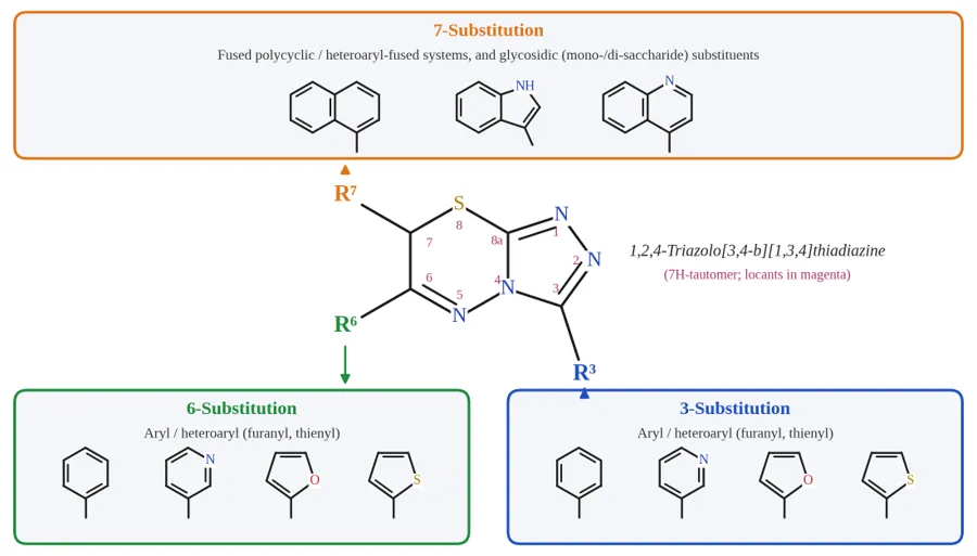
Figure 1: General scaffold of 1,2,4-triazolo[3,4-b][1,3,4]thiadiazine, numbered at the 3-, 6- and 7-positions, annotated with the representative classes of R-group substitution (aryl, heteroaryl/furanyl-thienyl, fused polycyclic and glycosidic) discussed throughout the text.
MATERIALS AND METHODS
This review draws on a structured survey of peer-reviewed literature on 1,2,4-triazolo[3,4-b] [1,3,4] thiadiazines and closely related fused triazole-thiadiazine hybrids, published up to 2026. Literature was retrieved from PubMed, Scopus, Web of Science and publisher databases (ACS, RSC, Elsevier, Springer, Wiley, MDPI) using the search terms triazolothiadiazine, triazolo[3,4-b] thiadiazine, anticancer, kinase inhibitor, EGFR, VEGFR-2, CDK2, tubulin inhibitor and molecular docking, used singly and in combination. Only original research articles and reviews reporting verifiable synthetic, in vitro, in silico or in vivo data on this scaffold or closely related fused hybrids were included; sources without a traceable peer-reviewed origin were excluded. For each included study, the structural class, cell lines or enzymatic targets, potency values (IC50/Ki, reported exactly as stated in the primary source), mechanistic findings, and docking/molecular dynamics/ADMET data were extracted and tabulated. Numeric values are reported only where explicitly stated in the source; where a study reported only a qualitative trend, that is stated as such rather than estimated.
RESULTS AND DISCUSSION
2. Chemical space and molecular design of triazolothiadiazines
2.1 Core structure and tautomerism
The 1,2,4-triazolo[3,4-b] [1,3,4] thiadiazine core is built by fusing a 1,2,4-triazole ring, through its 4-amino and 5-thiol/thione positions, to a six-membered 1,3,4-thiadiazine ring closed across the exocyclic sulfur and the adjacent ring nitrogen. The bicyclic system is most often encountered as the 7H-tautomer, in which the C7 methylene lies between the ring sulfur and C6, leaving C3 (on the triazole ring) and C6 (on the thiadiazine ring) as the two main positions for aryl or heteroaryl substitution (Figure 1) [9,30-31]. This 3,6-disubstitution pattern is the dominant design motif across essentially all series discussed here, and it is what makes the "two-pharmacophore" hybridisation strategy described below possible.
2.2 Ring-fusion patterns and hybridisation strategy
Because the triazole-derived carboxylic acid or acid hydrazide fixes the substituent at C3, while the α-haloketone or α-bromoacetophenone used in the second cyclisation step fixes the substituent at C6, the two positions can be varied independently and combinatorially. This has made triazolothiadiazines a convenient hybridisation platform (Figure 2): furanyl and thienyl heterocycles have been appended to improve π-stacking with hydrophobic kinase or tubulin sub-pockets [13,16]; coumarin has been fused in to combine the chromenone ring's intrinsic bioactivity with the triazolothiadiazine core, giving hybrids evaluated for alkaline phosphatase inhibition and anticancer/antileishmanial activity [14]; benzimidazole has been appended for aromatase-inhibitory hybrids [15]; and indole, naphthoquinone and spirooxindole fusions have each been explored specifically for anticancer evaluation [22,32-33]. Benzofuran- and benzothiazole-linked triazolothiadiazine hybrids extend this same hybridisation logic further, though largely outside the anticancer-specific literature reviewed here [34]. Bis-triazolothiadiazines – two triazolothiadiazine units joined through a common linker – represent a further hybridisation strategy aimed at boosting target engagement through avidity or dual-site binding, and have been evaluated specifically against hepatocellular carcinoma cell lines [32,35].
2.3 Pharmacophore hybridisation and kinase-binding hypotheses
The rationale for pairing triazolothiadiazines with kinase targets follows the broader logic used across the triazole-hybrid anticancer literature: the ring nitrogens, and the thiadiazine sulfur where present, can act as hydrogen-bond acceptors mimicking the adenine-mimetic hinge contacts made by ATP, while an appended aryl or heteroaryl group at C6 can be positioned to occupy the hydrophobic back pocket characteristically opened in type-II kinase inhibitor binding modes [24-26,36]. This hypothesis is well supported for monocyclic 1,2,3- and 1,2,4-triazole-EGFR/VEGFR-2 hybrids, where docking and, in several cases, co-crystallography or kinase-panel data directly confirm hinge-region engagement [24-26]. For the fused triazolothiadiazine core specifically, the corresponding evidence is still mostly indirect: molecular docking studies routinely place triazolothiadiazine derivatives into ATP-competitive pockets of validated cancer targets, but experimental confirmation is more often available for cytoskeletal (tubulin) or stress-signalling (Akt/ASK-1, STAT3) targets than for receptor tyrosine kinases proper [19-21,23]. We treat this as a testable hypothesis awaiting confirmation, not an established mechanism, through the SAR discussion in Section 4.
2.4 Substituent effects at a glance
A few recurring substituent trends emerge across the literature, discussed in more mechanistic depth in Section 4: electron-withdrawing and halogen substituents on the C6-aryl ring tend to track with improved cytotoxic potency in hepatocellular and lung/colorectal cell-line panels [17,37]; furan and thiophene at C3 or C6 outperform simple phenyl in more than one independent series, plausibly reflecting the smaller ring size and different electronics of these five-membered heteroaromatics [13,16]; and fused bicyclic or hybridised right-hand groups (coumarin, naphthoquinone, spirooxindole) tend to push potency further but at some cost to aqueous solubility and synthetic step count [14,22,32-33].
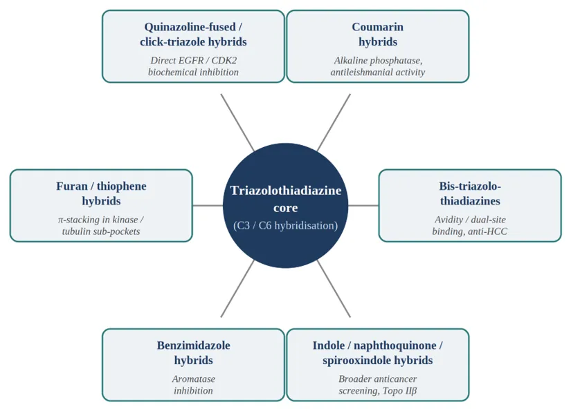
Figure 2: Principal hybridisation strategies applied at the triazolothiadiazine C3/C6 positions (schematic).
3. Synthetic strategies and fusion methodologies
3.1 Classical cyclocondensation
Most triazolothiadiazine anticancer series in the literature are built through a two- or three-step cyclocondensation sequence (Figure 3). In the first step, a substituted carboxylic acid is fused with thiocarbohydrazide under solvent-free thermal conditions, typically at 150-180 °C (or, in an alternative route, a hydrazide is converted with carbon disulfide/potassium hydroxide to the potassium dithiocarbazate, which is then cyclised with hydrazine hydrate), to give a 4-amino-5-substituted-2,4-dihydro-3H-1,2,4-triazole-3-thione intermediate [9,30]. In the second step, this triazole-thione is cyclised onto a phenacyl bromide (α-bromoacetophenone) or, in variant routes, onto chloroacetonitrile in the presence of sodium acetate, closing the thiadiazine ring and installing the C6-aryl substituent in a single operation [9,32]. Where a 6-amino variant is needed, the choice between an α-haloketone and a haloacetonitrile decides whether the resulting 7H-thiadiazine ring carries a C6-aryl group directly or a C6-amino group suited to further Schiff-base elaboration.
Several methodological refinements of this core sequence have been reported: heteropoly acid catalysis has been used to speed up cyclisation and improve yields under milder conditions [30]; visible-light-mediated regioselective routes have been developed to reach 7-aroyl-6-methyl variants that are hard to obtain by thermal cyclisation alone [38]; and alternative cyclising agents, including hydroxy(tosyloxy)iodobenzene (Koser's reagent)-mediated routes, have been used to build symmetrical and unsymmetrical analogues under fairly mild, metal-free conditions [9,31]. Bis(α-bromoketone) precursors let the same chemistry extend to bis-triazolothiadiazines linked through a central aromatic or heteroaromatic spacer, a strategy applied specifically to generate hepatocellular-carcinoma-active compounds [32,35].
3.2 Click chemistry (CuAAC) approaches
Although the core triazolothiadiazine ring itself is built by cyclocondensation rather than 1,3-dipolar cycloaddition, CuAAC "click" chemistry is widely used in the broader triazole-hybrid anticancer literature to append a monocyclic 1,2,3-triazole unit as a conformationally rigid, metabolically stable linker between two pharmacophores – for instance, joining an EGFR-directed quinazolinone or benzimidazole fragment to a second bioactive heterocycle [24-26]. Several triazolothiadiazine-1,2,3-triazole hybrids combine both ring systems in one molecule, using CuAAC to install the triazole linker after the thiadiazine ring has already been assembled by cyclocondensation (Figure 3) – a good illustration of how the two chemistries complement rather than compete with each other.
3.3 Multicomponent reactions
Multicomponent reactions (MCRs) offer a route to rapid analogue diversification by combining three or more reagents – typically a hydrazide, an aldehyde and a bromoketone, or equivalent building blocks – in a single pot, avoiding the intermediate isolation that stepwise cyclocondensation requires (Figure 3). MCR approaches have been used to generate pyrazolyl- and chromenone-substituted triazolothiadiazines and related fused heterocycles with better atom economy than the stepwise route, though the anticancer evaluation of MCR-derived triazolothiadiazine libraries specifically remains less extensively documented than that of the classical cyclocondensation series discussed in Section 4 [9].
3.4 Green and process-intensified methods
Microwave irradiation has been used to cut both the triazole-thione formation and thiadiazine cyclisation steps down from several hours of reflux to minutes, generally with comparable or improved yields; ultrasound-assisted protocols give similar rate acceleration under milder thermal conditions. Solvent-free fusion – already the default for the first (triazole-thione-forming) step of the classical route – and, more recently, ionic-liquid- or heteropoly acid-catalysed cyclisations have been explored specifically to cut solvent waste and catalyst loading in thiadiazine-forming chemistry [30]. Taken together, these green variants generally trade a modest increase in specialised equipment (microwave or sonication reactors) for shorter reaction times, lower solvent volumes and, in several reports, better isolated yield than conventional oil-bath reflux.
TABLE 1: REPRESENTATIVE SYNTHETIC ROUTES TO THE TRIAZOLOTHIADIAZINE CORE AND RELATED FUSED SCAFFOLDS
|
Sr. No. |
Synthetic route |
Key reagents and conditions |
Product ring system |
Reported yield |
Principal advantage / limitation |
|
1 |
Classical two-step cyclocondensation |
(i) Carboxylic acid + thiocarbohydrazide, solvent-free fusion, 150-180 °C; (ii) α-bromoacetophenone, NaOAc/EtOH, reflux |
3,6-Disubstituted 7H-triazolo[3,4-b] [1,3,4] thiadiazine |
50-85% |
Cheapest and most widely used; long reflux times and intermediate isolation required |
|
2 |
Heteropoly acid-catalysed cyclisation |
Keggin-type heteropoly acid catalyst, mild thermal conditions |
Triazolo- and triazino-fused thiadiazines |
Good to high |
Faster ring closure at lower temperature; catalyst recovery adds a workup step |
|
3 |
Visible-light-mediated regioselective route |
Photocatalysis, ambient to mild temperature |
7-Aroyl-6-methyl regioisomer |
Moderate |
Accesses regiochemistry unavailable thermally; requires a photoreactor |
|
4 |
Koser's-reagent oxidative cyclisation |
Hydroxy(tosyloxy) iodobenzene, metal-free, mild |
Mono- and bis-triazolothiadiazines |
Moderate to good |
Metal-free and step-economical; hypervalent iodine reagent cost |
|
5 |
Bis(α-bromoketone) linking |
Bis-triazole-thione + bis(α-bromoketone), base |
Bis-triazolothiadiazine (dimeric) |
Moderate |
Enables avidity/dual-site designs; lower yields and poorer solubility |
|
6 |
CuAAC click appendage |
Cu(I) catalyst, terminal alkyne + organic azide, mild heat |
1,2,3-Triazole linker appended to the preformed core |
Good to high (>80%) |
Excellent regioselectivity and modularity; post-functionalisation only, does not build the fused ring |
|
7 |
One-pot multicomponent reaction (MCR) |
Hydrazide + aldehyde + α-bromoketone, single pot |
Pyrazolyl- and chromenone-substituted analogues |
Variable |
Superior atom economy, avoids intermediate isolation; anticancer evaluation of MCR libraries still limited |
|
8 |
Microwave / ultrasound-assisted |
Microwave irradiation or sonication, largely solvent-free |
3,6-Disubstituted triazolothiadiazine |
Equal to or above conventional reflux |
Reduces reaction time from hours to minutes with less solvent; needs specialised equipment |
Compiled from references [9,24-26,28,30-32,35,38].
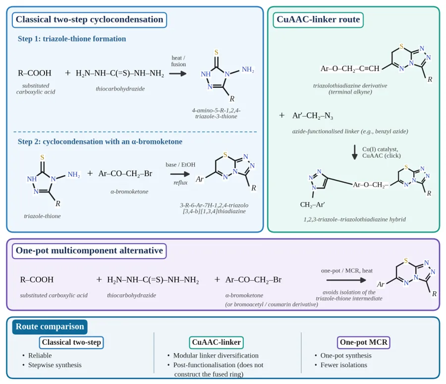
Figure 3: Schematic reaction scheme comparing the classical two-step cyclocondensation route (carboxylic acid + thiocarbohydrazide → triazole-thione; triazole-thione + α-bromoketone → triazolothiadiazine) with CuAAC-linker and multicomponent alternatives.
4. Structure-activity relationships and kinase targeting
This section organises the available SAR evidence, starting with the mechanistic/target categories for which experimental data exist specifically for the triazolothiadiazine core (tubulin, COX-2/stress-kinase, STAT3, topoisomerase IIβ; Figure 4), and then moving to the broader EGFR/VEGFR-2/CDK2-directed triazole-hybrid literature, which is more developed for monocyclic triazole chemotypes than for the fused thiadiazine system itself.
4.1 Hepatocellular and liver-targeted series
Liver cancer has been a particularly active application area for triazolothiadiazines derived from non-steroidal anti-inflammatory drug (NSAID) scaffolds. An early study of NSAID-motif triazolothiadiazines screened the series against epithelial cancer cells; compounds with IC50 values below roughly 5 µM induced apoptosis and SubG1 cell-cycle arrest in liver cancer cells, and the two compounds studied mechanistically (1g and 1h) triggered oxidative-stress-induced apoptosis through ASK-1 activation and Akt inhibition [17]. The same group and collaborators later took an initial panel of 32 triazolothiadiazine NSAID derivatives and selected three compounds, on the basis of their IC50 values, for molecular assays across nine HCC cell lines (Huh7, HepG2, Hep3B, PLC, SK-Hep1, Mahlavu, FOCUS, SNU182 and SNU475); the lead compound (7b; Figure 4) was the most potent, inducing G2/M-phase cell-cycle arrest and apoptosis (Figure 5), with oxidative-stress-induced JNK activation involving the dynamic interplay of ASK1, MKK7 and c-Jun [39]. Compound 7b showed IC50 values in the sub-micromolar to low-micromolar range (roughly 0.2-1 µM in the most sensitive lines), with cytotoxicity comparable to the reference chemotherapeutic camptothecin, and inhibited stemness markers alongside cell death through oxidative-stress-dependent JNK pathway activation, again pointing to the ASK1-MKK7-c-Jun axis as the operative mechanism; in nude mice, 7b treatment also significantly reduced tumour volume and prolonged disease-free survival [39]. Two structurally related leads from the same NSAID-derived series (7a, 7c) showed a wider, less favourable IC50 range (roughly 7-23 µM and 12-50 µM across the same panel), which shows that even within one closely related chemotype, potency can vary by more than an order of magnitude depending on the specific C3/C6 substitution pattern [39]. Separately, bis-triazolothiadiazines built through a bis(α-bromoketone) linking strategy have been evaluated specifically for anti-hepatocellular-carcinoma activity with accompanying molecular docking, though – consistent with the broader pattern noted in Section 2.3 – the docking target used here was not itself a validated oncogenic kinase [32]. Taken together, these liver-focused series are the most mechanistically developed anticancer thread in the triazolothiadiazine literature, and their emphasis on stress-kinase and redox pathways (Akt/ASK-1/JNK) rather than receptor tyrosine kinases is a useful calibration point for readers coming to this scaffold with an EGFR/VEGFR-2-centric expectation.
4.2 Lung and colorectal cancer
A COX-2-directed rationale has also been used to design triazolo[3,4-b]-1,3,4-thiadiazines specifically for non-small cell lung cancer (NSCLC) and colorectal cancer (CRC), reflecting COX-2's established role in tumour initiation, progression and invasion in these tissues. A representative series built from an indole-acetic-acid-derived triazole-thione and evaluated by MTT assay against A549 (lung adenocarcinoma) and Caco-2 (colorectal adenocarcinoma) cell lines, alongside a normal lung fibroblast counter-screen (CCD-19Lu), identified several compounds with favourable selectivity for the cancer lines over the normal control, with mechanistic data pointing toward COX-2 inhibition as a contributing factor [37]. A structurally related indomethacin-based triazolothiadiazine series was separately evaluated against the T98 glioma cell line, again with docking support for a COX-2-mediated mechanism [18]. These NSAID-derived series share a coherent design logic – repurposing a validated anti-inflammatory pharmacophore as the C3 or C6 substituent on the triazolothiadiazine core – that turns up repeatedly across the anticancer papers discussed here.
4.3 Tubulin-targeted series
A separate, mechanistically well-supported thread of triazolothiadiazine anticancer chemistry targets tubulin polymerization rather than any kinase. A systematic structure-activity study of 3,6-diaryl-7H-triazolothiadiazines, designed as rigid vinylogous combretastatin A-4 (CA-4) analogues in which the bicyclic core locks CA-4's otherwise easily isomerised (Z,E)-butadiene linker into a fixed geometry, found this substitution pattern active against tubulin assembly; the most potent compound in the series (Figure 4) showed nanomolar antiproliferative IC50 values (roughly 0.011-0.015 µM) against SGC-7901, A549 and HT-1080 cells, essentially matching CA-4 itself (IC50 roughly 0.009-0.013 µM) [19]. Follow-up work on a related analogue confirmed G2/M cell-cycle arrest and apoptosis induction in SGC-7901 gastric cancer and HeLa cervical cancer cells [20]. A subsequent, larger structure-activity campaign optimised this tubulin-inhibitor series further across fifty-two new analogues, reporting both in vitro potency gains and, notably, in vivo efficacy data in a tumour model – one of the few examples in the triazolothiadiazine literature to move beyond cell-based assays [21]. Most recently, a trimethoxybenzyl-substituted analogue from the same research group (compound B5) extended this series further, combining nanomolar-to-sub-micromolar antiproliferative potency (IC50 = 0.046, 0.57 and 0.96 µM against HeLa, HT-29 and A549, comparable to or better than CA-4), confirmed tubulin polymerization inhibition and G2/M arrest, antivascular activity in wound-healing and tube-formation assays (Figure 5), and – again – in vivo antitumour efficacy without overt toxicity in an A549 xenograft model [40]. Because tubulin inhibitors work through a fundamentally different mechanism from kinase inhibitors (disrupting mitotic spindle assembly rather than ATP-competitive or allosteric enzyme inhibition), this series is best read as a distinct pharmacological class within the broader triazolothiadiazine chemotype rather than as kinase-targeted evidence, scaffold overlap notwithstanding. It nonetheless remains the single most mechanistically and translationally mature thread in the entire triazolothiadiazine anticancer literature, now backed by two in vivo efficacy studies from the same research group [21,40].
4.4 STAT3 pathway and topoisomerase IIβ inhibition
A pyrazole-containing 1,2,4-triazolo[3,4-b] thiadiazine series was specifically optimised as STAT3 pathway inhibitors (Figure 4), using a cell-based reporter assay to guide structure-activity refinement; this is one of the more direct links in the literature between the triazolothiadiazine core and a bona fide oncogenic signalling pathway, since constitutive STAT3 activation drives proliferation and survival across many solid tumours [23]. Separately, naphthoquinone-hydrazino-triazolothiadiazine hybrids were designed with molecular docking support against topoisomerase IIβ (Figure 4) and evaluated for anticancer activity, combining the naphthoquinone fragment's intrinsic redox-cycling bioactivity with the triazolothiadiazine core [22]. Both series apply the hybridisation logic described in Section 2.2 toward a specific, target-annotated anticancer hypothesis rather than generic cytotoxicity screening.
4.5 Broader anticancer screening series
Beyond the target-annotated series discussed above, a substantial body of literature reports triazolothiadiazine and closely related triazolothiadiazole libraries screened for general anticancer or antiproliferative activity without a specific molecular target being pursued mechanistically. These include libraries evaluated for cytotoxicity alongside cholinesterase inhibition [41-42]; anticancer-focused triazole/triazolothiadiazine series from several independent groups reporting cytotoxicity data without extensive mechanistic follow-up [43-46]; coumarin-triazolothiadiazine hybrids evaluated for combined alkaline phosphatase inhibition and anticancer/antileishmanial activity [14]; and spirooxindole-triazolothiadiazine hybrids reported specifically for anticancer evaluation [33]. This category of evidence is useful for establishing that cytotoxic activity is a recurring, reproducible feature of the scaffold across multiple independent groups, but – consistent with the counter-screening caveats raised in Section 6 – it generally does not on its own establish a specific druggable mechanism, and should be weighted accordingly when building a target hypothesis for new analogue design.
4.6 EGFR, VEGFR-2 and the wider triazole-hybrid kinase literature
Direct, biochemically confirmed EGFR engagement by the fused triazolothiadiazine core was, until recently, an open question, extrapolated only from the much larger body of evidence connecting monocyclic 1,2,3-triazole chemistry to EGFR. The 2023-2025 literature shows this gap has begun to close. Three studies from two independent research groups have now reported purified-enzyme EGFR inhibition data – not only cytotoxicity and docking – for triazolothiadiazine and closely fused triazolothiadiazine-hybrid chemotypes.
First, a 1,2,3-triazole-triazolothiadiazine hybrid series built by CuAAC click chemistry onto the triazolothiadiazine core itself was screened against MCF-7, MDA-MB-231 and MDA-MB-415 breast cancer cell lines and counter-screened against the non-cancerous MCF-10A line; the two most active hybrids (bearing 4-fluorophenyl and 3,5-dichlorophenyl substituents) showed EGFR enzymatic IC50 values of 0.419 ± 0.05 µM and 0.312 ± 0.02 µM, essentially matching erlotinib (IC50 = 0.421 ± 0.03 µM) tested under the same conditions, with docking and ADMET analysis supporting drug-likeness [28]. Second, a series of quinazoline-triazolothiadiazine hybrids – combining the quinazoline EGFR pharmacophore directly with the fused thiadiazine ring rather than through a monocyclic triazole linker – was evaluated against three breast cancer lines (MCF-7, MDA-MB-231, MDA-MB-468); the lead compound reached an EGFR IC50 of 0.37 ± 0.03 µM, modestly better than erlotinib (IC50 = 0.42 ± 0.01 µM) in the same assay, with a second compound at 0.51 ± 0.04 µM, and molecular docking (Discovery Studio 2021) used to rationalise the binding mode of the six most potent analogues [29]. Third, and most striking, a mono- and bis-(pyrazolyl-triazolothiadiazine) series evaluated for dual EGFR/CDK2 inhibition (discussed further in Section 4.7) included compounds with EGFR IC50 values in the low-nanomolar range, again outperforming erlotinib under matched assay conditions [27]. Together, these three 2023-2025 papers (from two research groups) provide direct, purified-enzyme confirmation that the fused triazolothiadiazine core – not just its monocyclic triazole relatives – can reach erlotinib-competitive EGFR inhibition, and they substantially strengthen the mechanistic case that was first made on docking grounds alone.
The much larger body of evidence connecting triazole chemistry to EGFR and VEGFR-2 more broadly still comes from monocyclic 1,2,3-triazole hybrids, most often installed via CuAAC as a linker between an EGFR-pharmacophore fragment (quinazoline, quinazolinone or benzimidazole) and a second bioactive heterocycle. A comprehensive 2025 literature review specifically catalogued the growing family of 1,2,3-triazole-EGFR hybrids, noting the field's shift toward addressing acquired resistance in both wild-type and mutant EGFR through hinge-binding triazole-linked chemotypes [24]. Representative recent examples include 1,2,3-triazole/quinazolin-4-one hybrids explicitly designed as dual EGFR/BRAF-V600E inhibitors, several showing low-micromolar antiproliferative activity with cell-cycle arrest in the G1 phase [25], and benzimidazole/1,2,3-triazole hybrids reported as apoptotic, EGFR-directed antiproliferative agents with GI50 values in the low-nanomolar range against breast cancer cell lines [26]. Other recent series in this space have been profiled as multi-target EGFR/VEGFR-2/topoisomerase-II inhibitors built on a quinazoline-1,2,3-triazole hybrid scaffold, with reported low-micromolar EGFR and VEGFR-2 inhibitory activity approaching that of erlotinib and sorafenib, respectively, together with topoisomerase II inhibition.
Even with the EGFR gap substantially narrowed, VEGFR-2 remains an open question for the fused triazolothiadiazine core: at the time of writing we could not find a published biochemical VEGFR-2 IC50 value for the bicyclic triazolothiadiazine scaffold itself, unlike the several monocyclic-triazole and other heterocyclic chemotypes for which such data now exist, including recent triazole-tethered indolinone and triazole-coumarin-glycoside VEGFR-2 inhibitors reporting low-nanomolar potency against the purified enzyme. Given the structural and physicochemical similarity between the EGFR-active triazolothiadiazine hybrids just described and validated VEGFR-2-directed triazole chemotypes, running the existing triazolothiadiazine compound library – much of it already synthesised and characterised – against a VEGFR-2 biochemical assay is a comparatively low-cost, high-value next experiment for this field.
4.7 CDK2, GSK-3β and other kinase targets
Unlike VEGFR-2 and FAK, CDK2-directed evidence for the fused triazolothiadiazine core is no longer absent from the literature. A mono- and bis-(6-pyrazolyltriazolo-thiadiazine) series was purpose-built around a pyrazole appendage previously validated in EGFR/CDK2-directed chemotypes, and screened for cytotoxicity against MCF-7 breast cancer cells with MCF-10A normal-cell counter-screening; three compounds (a simple pyrazolyl-triazolothiadiazine with a free thiol, its N-phenyl analogue, and a bis-triazolothiadiazine linked through a short one-carbon alkylene spacer) showed potent, selective cytotoxicity (MCF-7 IC50 = 3.16, 2.74 and 0.39 µM, versus ≥50, 46.7 and ≥50 µM against MCF-10A) and were carried forward for dual enzymatic EGFR and CDK2 inhibition assays [27]. The most active compound (Figure 4) reached EGFR and CDK2 IC50 values of 19.6 ± 0.64 nM and 87.9 ± 2.8 nM – outperforming both erlotinib (IC50 = 67.3 ± 2.54 nM) and the CDK2 reference roscovitine (IC50 = 140 ± 3.6 nM) at the enzymatic level, and inhibiting EGFR and CDK2 activity by 97.2% and 94.1% at 10 µM – making this one of the most potent biochemical kinase-inhibition values reported in the triazolothiadiazine anticancer literature surveyed here [27]. The structure-activity pattern within this series is instructive: activity rose in the order unsubstituted triazole < N-phenyl triazole < free-thiol triazole among the simple pyrazolyl-hybrids, consistent with the thiol group acting as an extra hydrogen-bond donor and the phenyl group contributing hydrophobic contacts; among the bis-hybrids, the shortest alkylene spacer (a single methylene-to-methylene tether) gave markedly higher potency than the longer-spacer analogues, suggesting that avidity or dual-site engagement is sensitive to spacer length rather than simply favoured by more linker flexibility [27].
A second, independent line of CDK2-adjacent evidence comes from kinase-panel profiling of a hydrazinotriazolothiadiazine series originally developed against the full NCI-60 cancer cell line panel: the most active compound (Figure 4) was subsequently screened against sixteen kinases and found to inhibit glycogen synthase kinase-3β (GSK-3β) with an IC50 of 0.883 µM and 14-fold selectivity over CDK2, together with increased active caspase-3, G2/M cell-cycle arrest and increased Annexin V-positive apoptotic cells in PC-3 prostate cancer cells, with molecular docking and dynamics used to rationalise the binding mode in the GSK-3β ATP-binding site [47]. GSK-3β is not itself part of the EGFR/VEGFR-2/CDK2 axis emphasised elsewhere in this review, but it is a bona fide oncology-relevant serine/threonine kinase, and this sixteen-kinase panel is, as far as we know, the broadest kinase-selectivity screen reported anywhere in the triazolothiadiazine anticancer literature – a useful precedent for the kind of counter-screening recommended more broadly in Sections 6 and 7.
No FAK-specific data for the fused triazolothiadiazine core were found, nor were VEGFR-2 data (Section 4.6); the ring-closure chemistry already established for triazolothiadiazine synthesis (Section 3) could in principle be applied directly to FAK- or VEGFR-2-pharmacophore-bearing precursors already validated in the monocyclic-triazole literature, generating a further wave of target-annotated triazolothiadiazine hybrids for biochemical evaluation, following the template that the EGFR and CDK2 work described above has now successfully set for this scaffold.
TABLE 2: SELECTED TRIAZOLOTHIADIAZINE AND CLOSELY RELATED HYBRIDS WITH REPORTED ANTICANCER OR KINASE ACTIVITY
|
Sr. No. |
Compound / series (ref.) |
Structural feature |
Model system |
Reported potency |
Mechanistic readout |
|
1 |
NSAID-derived lead 7b [39] |
NSAID pharmacophore at C3/C6 |
Nine-line HCC panel (Huh7, HepG2, Hep3B, PLC, SK-Hep1, Mahlavu, FOCUS, SNU182, SNU475) |
IC50 ≈ 0.2-1 µM in the most sensitive lines; comparable to camptothecin |
Oxidative-stress-dependent JNK activation via ASK1-MKK7-c-Jun; G2/M arrest; loss of stemness markers |
|
2 |
Congeners 7a and 7c [39] |
Related NSAID substitution |
Same nine-line HCC panel |
IC50 ≈ 7-23 µM (7a) and 12-50 µM (7c) |
Same pathway, an order of magnitude less potent |
|
3 |
3,6-Diaryl CA-4 mimic [19] |
Diaryl on a conformationally locked core |
SGC-7901, A549, HT-1080 |
IC50 0.011-0.015 µM (CA-4: 0.009-0.013 µM) |
Inhibition of tubulin assembly |
|
4 |
Trimethoxy benzyl analogue B5 [40] |
3,4,5-Trimethoxybenzyl at C3 |
HeLa, HT-29, A549; A549 xenograft |
IC50 0.046, 0.57 and 0.96 µM |
Tubulin polymerisation inhibition; G2/M arrest; antivascular and in vivo antitumour activity |
|
5 |
Furan / thiophene hybrids [13,16] |
Five-membered heteroaryl at C3 or C6 |
PC-3, HepG2, A549, MCF-7 |
Lead IC50 3.7-12.7 µM |
G2/M arrest; docking- and MD-supported binding |
|
6 |
Hydrazino hybrid 5l [47] |
Hydrazino linker to 4-methoxyphenyl |
NCI-60 panel; PC-3 |
GSK-3β IC50 0.883 µM; 14-fold selectivity over CDK2 |
GSK-3β inhibition; caspase-3 activation; G2/M arrest; Annexin V positivity |
|
7 |
Pyrazolyl STAT3 series [23] |
Pyrazole at C3/C6 |
Cell-based STAT3 reporter assay |
Potency gain reported qualitatively |
Suppression of STAT3 pathway signalling |
|
8 |
Naphthoquinone-hydrazino hybrids [22] |
Naphthoquinone appendage |
Cancer cell-line screen |
Activity docking-supported; no enzymatic IC50 reported |
Proposed topoisomerase IIβ inhibition |
|
9 |
Click 1,2,3-triazole hybrids [28] |
4-Fluorophenyl and 3,5-dichlorophenyl |
MCF-7, MDA-MB-231, MDA-MB-415; MCF-10A counter-screen |
EGFR IC50 0.419 ± 0.05 and 0.312 ± 0.02 µM (erlotinib 0.421 ± 0.03 µM) |
Biochemical EGFR inhibition; favourable ADMET profile |
|
10 |
Quinazoline-fused hybrids [29] |
Quinazoline joined directly to the thiadiazine ring |
MCF-7, MDA-MB-231, MDA-MB-468 |
EGFR IC50 0.37 ± 0.03 and 0.51 ± 0.04 µM (erlotinib 0.42 ± 0.01 µM) |
Biochemical EGFR inhibition; hinge-region binding by docking |
|
11 |
Pyrazolyl dual-target series [27] |
Free thiol / N-phenyl triazole; short methylene-linked bis-analogue |
MCF-7 (IC50 0.39-3.16 µM); MCF-10A (46.7 to ≥50 µM) |
EGFR IC50 19.6 ± 0.64 nM; CDK2 IC50 87.9 ± 2.8 nM (erlotinib 67.3 ± 2.54 nM; roscovitine 140 ± 3.6 nM) |
Dual EGFR/CDK2 inhibition; 97.2% and 94.1% enzyme inhibition at 10 µM |
|
12 |
Bis-triazolothiadiazines [32,35] |
Two cores joined by an aromatic spacer |
Hepatocellular carcinoma cell lines |
Cytotoxic; no kinase IC50 reported |
Anti-HCC activity with docking support at a non-kinase target |
Values are reproduced exactly as published, retaining the units and error terms given in the primary source.
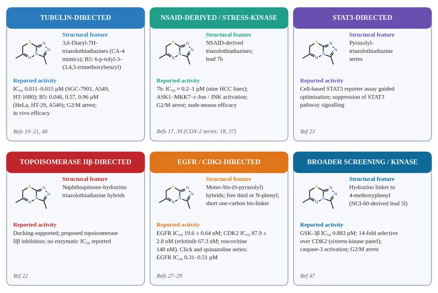
Figure 4: Representative triazolothiadiazine anticancer series grouped by mechanistic category, showing the structural feature and reported activity of each series; compound designations follow the cited sources.
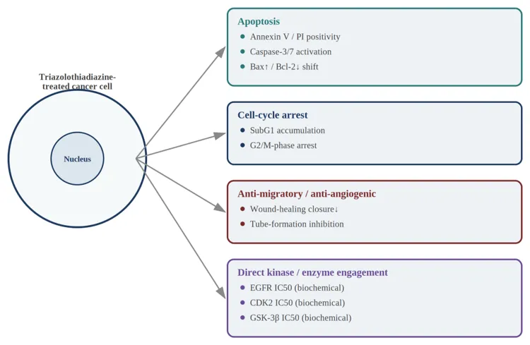
Figure 5: Major in vitro and biochemical effects reported for triazolothiadiazine and related hybrids (Table 2), schematic.
5. Integrated in silico and in vitro evaluation workflows
5.1 Molecular docking practice
Across the triazolothiadiazine and wider triazole-hybrid anticancer literature, molecular docking is used almost universally as a rationalisation and hypothesis-generation tool, typically run with AutoDock Vina or an equivalent scoring engine against a crystal structure retrieved from the Protein Data Bank [48-49], consistent with the broader, well-established role of virtual screening as a hit-prioritisation tool in early-stage drug discovery [50]. Good practice – followed inconsistently across the papers surveyed – includes re-docking the native co-crystallised ligand to validate the docking protocol against the experimental binding pose (usually assessed by root-mean-square deviation, RMSD), and comparing the docking score and interaction pattern of new compounds against a co-crystallised reference inhibitor rather than in isolation. Where this validation step is reported, docking results for triazolothiadiazine and triazole-hybrid series generally reproduce the expected hinge-region hydrogen bonds and hydrophobic back-pocket contacts seen with reference kinase inhibitors (Figure 6) [16,25]; where re-docking validation is missing, docking scores should be read as indicative rather than confirmatory.
5.2 Molecular dynamics
A smaller part of the literature extends static docking with explicit-solvent molecular dynamics (MD) simulations, typically over tens to a few hundred nanoseconds, to check the stability of the predicted binding pose and quantify persistent hydrogen bonds or hydrophobic contacts over the simulation trajectory [16]. MD is particularly useful for triazolothiadiazine ligands because the bicyclic core's moderate flexibility – specifically, rotation about the exocyclic aryl-to-ring bonds at C3 and C6 – means a single static docking pose may not capture the full range of accessible binding conformations; MD trajectories showing a stable, low-RMSD binding pose over the simulated timescale give meaningfully stronger support for a proposed binding hypothesis than docking alone.
5.3 ADMET and drug-likeness prediction
In silico ADMET and drug-likeness prediction, commonly done with tools such as SwissADME, is used across the literature to flag likely solubility, permeability, cytochrome P450 interaction and Lipinski rule-of-five compliance issues before compounds are prioritised for synthesis or further biological testing [51-52]. For the triazolothiadiazine series specifically, the fused bicyclic core with pendant aryl/heteroaryl groups at C3 and C6 tends to sit close to the upper boundary of Lipinski-compliant molecular weight and lipophilicity once larger hybridised right-hand groups (coumarin, naphthoquinone, benzimidazole) are appended, a recurring practical constraint on further hybridisation-based potency optimisation.
TABLE 3: REPRESENTATIVE IN SILICO STUDIES ON TRIAZOLOTHIADIAZINE AND CLOSELY RELATED HYBRIDS
|
Sr. No. |
Modelled target |
Computational method |
Key predicted interactions |
Corresponding experimental potency |
Supporting analysis |
|
1 |
EGFR (PDB 4HJO) [29] |
Molecular docking, Discovery Studio 2021 |
Hinge-region hydrogen bond to the Met769 backbone; hydrophobic back-pocket contacts |
EGFR IC50 0.37-0.51 µM |
Drug-likeness within acceptable limits |
|
2 |
EGFR [28] |
Molecular docking |
Hinge hydrogen bonding plus hydrophobic occupancy |
EGFR IC50 0.31-0.42 µM |
SwissADME pharmacokinetic screen |
|
3 |
EGFR and CDK2 [27] |
Docking guided by enzymatic assay data |
ATP-pocket occupancy at both kinases |
EGFR 19.6 nM; CDK2 87.9 nM |
MCF-10A normal-cell counter-screen |
|
4 |
GSK-3β [47] |
Docking with molecular dynamics refinement |
Stable ATP-binding-site pose over the simulated trajectory |
GSK-3β IC50 0.883 µM; 14-fold over CDK2 |
Sixteen-kinase selectivity panel |
|
5 |
Aromatase and hMAO-B [15] |
Docking against three crystal structures |
Complementary active-site fit |
IC50 0.020 µM (aromatase) and 0.045 µM (hMAO-B) |
NIH3T3 normal-cell counter-screen |
|
6 |
Furan / thiophene series target [16] |
Docking with molecular dynamics |
Hydrogen bonding and π-stacking within the hydrophobic sub-pocket |
Cytotoxicity reported; no enzymatic IC50 |
MD performed; trajectory length not specified |
|
7 |
Topoisomerase IIβ [22] |
Molecular docking |
Binding adjacent to the intercalation site |
No enzymatic IC50 reported |
Binding mode not further validated |
|
8 |
COX-2 [18,37] |
Molecular docking |
Active-site fit consistent with NSAID-derived design |
IC50 0.20-1.20 µM |
Selectivity index >62-250 relative to COX-1 |
Where a study reported only a qualitative docking description, this is stated explicitly rather than converted to a numeric value.
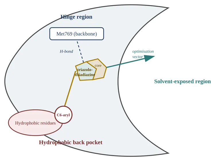
Figure 6: Proposed EGFR ATP-pocket binding mode of triazolothiadiazine-hybrid ligands, based on docking studies reported in Table 3 (refs 27-29). Schematic composite, not an extracted docking pose.
5.4 In vitro assays and mechanistic follow-up
Cell viability is almost always assessed by MTT or the closely related sulforhodamine B (SRB) assay [53], generally across a panel of three to six cancer cell lines with, less consistently, a matched normal-cell counter-screen to establish a selectivity index. Where mechanistic follow-up is pursued – as in the Akt/ASK-1/JNK-directed HCC series (Section 4.1), the STAT3-directed series (Section 4.4) and the tubulin-directed series (Section 4.3) – this typically combines flow-cytometric apoptosis analysis (Annexin V/propidium iodide staining) [54] with cell-cycle distribution analysis and, in a minority of cases, direct enzymatic or reporter-based confirmation of the proposed target engagement (Figure 5). Across the literature reviewed here, mechanistic depth – an apoptosis assay, cell-cycle analysis, and a specific molecular target confirmed biochemically rather than only by docking – is present for a real but still limited subset of published triazolothiadiazine series, with most papers reporting cytotoxicity data supported by docking alone.
5.5 Concordance between in silico and in vitro findings
Where both docking/MD and biochemical or cell-based target-engagement data are reported for the same compound series, concordance is generally good: docking-predicted high-affinity binders tend to be the same compounds showing the strongest cytotoxicity and the clearest mechanistic signal in follow-up assays [16,19-21,23]. This is reassuring, but should be read with some caution, because docking scores and cytotoxicity are both, to a first approximation, correlated with simple physicochemical properties (lipophilicity, molecular size); concordance between the two does not by itself rule out that both are tracking a shared confounding variable rather than genuine, target-specific engagement. Direct biochemical kinase or enzyme inhibition assays, run independently of the cell-based cytotoxicity readout, remain the more reliable form of validation, and are exactly the missing piece – per the gap identified in Section 4.6 – for a confident EGFR/VEGFR-2 mechanistic claim specific to the triazolothiadiazine core.
6. Safety, selectivity, and drug-likeness considerations
Counter-screening against a matched normal cell line (most often a lung, kidney or dermal fibroblast line) is reported for a meaningful subset of the triazolothiadiazine anticancer literature, and where it is reported, favourable selectivity indices are common – several series show markedly lower cytotoxicity toward normal fibroblasts than toward the paired cancer cell line [37]. Normal-cell counter-screening is not universal across the literature surveyed here, however, and its absence in a given paper should be read as a data gap rather than assumed favourable selectivity.
Potential off-target liabilities specific to the triazolothiadiazine and related thiadiazole chemotypes include the general metabolic lability associated with certain sulfur-heterocycle motifs, which can undergo bioactivation to reactive metabolites in some structural contexts, and the possibility of off-target enzyme inhibition given the demonstrated cross-reactivity of closely related triazolothiadiazole/triazolothiadiazine chemotypes with carbonic anhydrase, cholinesterase and monoamine oxidase [5-6,41-42]. This cross-reactivity cuts both ways: it shows the scaffold is a "druggable" chemotype capable of high-affinity enzyme engagement, but it also means anticancer-directed analogues should ideally be counter-screened against these off-target enzyme classes to rule out confounding poly pharmacology, particularly where a specific kinase mechanism is being claimed.
ADMET trends observed in silico across the series reviewed here (Section 5.3) suggest that smaller, less hybridised triazolothiadiazine analogues (simple aryl at C3 and C6, without a fused coumarin, naphthoquinone or benzimidazole extension) are more likely to stay within favourable oral bioavailability space, while the larger hybridised structures associated with the strongest reported potency (Section 4) tend to trade some of that favourable drug-likeness for target engagement – a familiar potency-versus-developability trade-off in medicinal chemistry generally. In vivo efficacy data specific to the triazolothiadiazine core remain limited to the two tubulin-inhibitor optimisation series from the same research group noted in Section 4.3, both of which reported in vivo antitumour efficacy (a tumour-growth model and, more recently, an A549 xenograft model) alongside their in vitro structure-activity data [21,40] and to the NSAID-derived HCC lead 7b, which reduced tumour volume and prolonged disease-free survival in a nude-mouse model [39]; beyond these examples, in vivo validation for this scaffold – including for the newly reported EGFR/CDK2-directed series in Sections 4.6-4.7 – is, at the time of writing, essentially absent from the literature surveyed for this review.
7. Challenges, gaps, and future directions
TABLE 4: COMPARATIVE OVERVIEW OF THE PRINCIPAL MECHANISTIC THREADS DISCUSSED IN SECTION 4
|
Sr. No. |
Mechanistic thread |
Favourable substitution trend |
Selectivity and developability |
Reported target(s) |
Level of evidence |
|
1 |
NSAID-derived stress-kinase series |
NSAID pharmacophore retained at C3/C6 |
Favourable in the subset counter-screened |
Akt / ASK-1 / JNK pathway signalling |
Cell-based with pathway confirmation; in vivo (nude mouse) for 7b |
|
2 |
Tubulin-directed (CA-4 mimics) |
Diaryl and 3,4,5-trimethoxybenzyl |
Tolerated in two in vivo models (same research group) |
Tubulin |
In vitro plus in vivo efficacy |
|
3 |
Furan / thiophene hybrids |
Five-membered heteroaryl in place of phenyl |
Limited ADMET characterisation |
Not specifically assigned |
Cytotoxicity with docking support |
|
4 |
EGFR-directed (click and quinazoline) |
Halogenated phenyl; fused quinazoline |
Selective over MCF-10A (click series) |
EGFR |
Purified-enzyme inhibition |
|
5 |
Dual EGFR/CDK2 series |
Free thiol or N-phenyl triazole; short bis-linker |
Strong selectivity over MCF-10A |
EGFR and CDK2 |
Purified-enzyme inhibition |
|
6 |
GSK-3β-directed |
Hydrazino linkage, NCI-60-derived lead |
14-fold selective over CDK2 |
GSK-3β |
Sixteen-kinase panel |
|
7 |
STAT3 pathway inhibitors |
Pyrazolyl at C3/C6 |
Not characterised in detail |
STAT3 pathway |
Cell-based reporter assay |
|
8 |
Topoisomerase IIβ-directed |
Naphthoquinone appendage |
Aqueous solubility a concern |
Topoisomerase IIβ (predicted) |
Docking only |
|
9 |
Broader anticancer screening |
Coumarin, spirooxindole, benzimidazole |
Variable across series |
None assigned |
Cytotoxicity screening only |
Compiled from references [13-14,16-17,19-23,27-29,33,37,39-40,43-47]
Three linked limitations recur across the triazolothiadiazine anticancer literature and, together, define the scaffold's main path toward more advanced drug-discovery validation.
Limited in vivo data. Apart from the exceptions noted in Section 6 (two from the same tubulin-inhibitor optimisation lineage and one NSAID-derived HCC lead, 7b), essentially all of the anticancer evidence assembled for this scaffold is cell-based. Moving even a small number of the other mechanistically well-supported series – the remaining NSAID-derived HCC leads, the STAT3-directed pyrazolo-triazolothiadiazines, and, most notably now, the newly reported EGFR/CDK2-directed hybrids – into xenograft or other in vivo efficacy models would substantially strengthen the translational case for the chemotype as a whole.
Incomplete kinase-selectivity panel data for the bicyclic core. As discussed in Sections 4.6 and 4.7, direct biochemical EGFR and dual EGFR/CDK2 inhibition data for the fused triazolothiadiazine core are no longer absent from the literature – three 2023-2025 studies from two research groups now report purified-enzyme IC50 values competitive with erlotinib and roscovitine [27-29], and a sixteen-kinase panel on a related hydrazinotriazolothiadiazine lead identified GSK-3β as an additional, selectively engaged target [47]. What is still missing is breadth rather than existence: VEGFR-2 and FAK biochemical data for the bicyclic core were not identified in the literature surveyed, no single study has yet run the same triazolothiadiazine lead across a broad (>20-kinase) selectivity panel the way the GSK-3β study did for a narrower set, and the existing EGFR/CDK2 data sit mostly in breast cancer cell lines rather than the lung, hepatocellular and colorectal contexts that dominate the cytotoxicity literature (Sections 4.1-4.2). Running the existing, already-synthesised triazolothiadiazine leads – particularly the EGFR/CDK2-active pyrazolyl-hybrids [27] – against a broader oncology kinase panel, adding VEGFR-2 and FAK and extending the cell-line panel beyond breast cancer, is a practical next step given how many compounds already exist in the published literature.
Under-explored substitution space. Most published series concentrate substituent variation at the C6-aryl position, with comparatively little systematic exploration of C3 substitution or of the 7-position once the 7H-tautomer's methylene bridge is functionalised. Given the clear substituent-dependent trends already established (furan/thiophene improving activity relative to simple phenyl; halogen/electron-withdrawing groups improving potency in several series), a more systematic, matched-pair SAR study varying C3 and C6 independently across a shared cell-line panel would help disentangle which substituent effects are position-specific versus general.
Looking ahead, a few concrete directions could speed up this scaffold's progress. Covalent or allosteric inhibitor design around the triazolothiadiazine core – exploiting the ring sulfur or an appended Michael-acceptor extension for targeted covalent engagement of a non-catalytic cysteine, following the precedent set by covalent EGFR inhibitors in the wider kinase-inhibitor field – has not, to our knowledge, been explored for this chemotype and represents a design opportunity. Combination-therapy and nano-formulation strategies, already applied successfully to other triazole-hybrid anticancer chemotypes to improve solubility and tumour-selective delivery, could similarly be applied to the more lipophilic, hybridised triazolothiadiazine analogues discussed in Section 6. Finally, AI/machine-learning-guided design, paired with the experimental docking/MD/ADMET pipeline already in routine use across this literature (Section 5), could help prioritise the C3/C6 substituent combinations most likely to satisfy both the potency and drug-likeness constraints identified above before committing synthetic effort – a particularly practical recommendation given how tractable the underlying cyclocondensation chemistry (Section 3) already is for rapid analogue generation.
CONCLUSION
The 1,2,4-triazolo[3,4-b] [1,3,4] thiadiazine scaffold has built up a real, if still uneven, body of anticancer evidence over roughly the past decade. Its strengths are clear: synthetic tractability through well-established cyclocondensation chemistry, a flexible two-position substitution pattern well suited to pharmacophore hybridisation, and, for a meaningful subset of published series, mechanistic depth connecting the chemotype to validated cancer-relevant pathways – most convincingly for tubulin polymerization, Akt/ASK-1/JNK-mediated oxidative stress signalling in hepatocellular carcinoma, STAT3 pathway inhibition, and topoisomerase IIβ inhibition, and now also direct, purified-enzyme confirmation of EGFR and dual EGFR/CDK2 inhibition, with several 2023-2025 compounds matching or exceeding the erlotinib and roscovitine reference standards [27-29], plus a sixteen-kinase panel identifying GSK-3β as an additional selectively engaged target [47]. The scaffold's remaining limitations are narrower than they looked even two or three years ago: VEGFR-2 and FAK biochemical data for the fused bicyclic core were not identified in the literature surveyed, the existing kinase-panel work is still concentrated on a small number of targets and a single cancer type (breast), and, with the exception of two tubulin-inhibitor in vivo efficacy studies from the same research group [21,40] and one nude-mouse study of the HCC lead 7b [39], in vivo validation across the rest of the chemotype remains essentially absent. These narrower, more addressable gaps – rather than any fundamental weakness in the chemotype – are what currently separate triazolothiadiazines from a more clinically credible anticancer lead series. Closing them through broader kinase-panel profiling (adding VEGFR-2 and FAK to the EGFR/CDK2 assays already validated for this core), expanded in vivo validation beyond the tubulin-inhibitor series and HCC lead 7b, and a more systematic, matched-pair approach to C3/C6 substitution would be a natural and achievable next phase for this chemistry, and would let the considerable synthetic and in silico infrastructure already built for this scaffold be brought to bear on a more rigorously target-validated next generation of triazolothiadiazine-based anticancer agents.
ACKNOWLEDGEMENT
The authors thank the Department of Pharmaceutical Chemistry, Al Shifa College of Pharmacy, Perinthalmanna, for providing literature access and infrastructural support for this review.
CONFLICT OF INTEREST
The authors declare no conflict of interest.
REFERENCES



Megha Santhosh M, Padmaraj C P, Hiba Abdul Razak, Fathima C O, Amrutha P Anil, Alagha K, Artha Rajagopal K, Triazolothiadiazines as Anticancer Agents and Emerging Kinase Inhibitors: Synthetic Strategies, Structure-Activity Relationships and In Silico Insights – A Review, Int. J. of Pharm. Sci., 2026, Vol 4, Issue 10, 599-623. https://doi.org/10.5281/zenodo.23168968
 10.5281/zenodo.23168968