

We use cookies to ensure our website works properly and to personalise your experience. Cookies policy
Global College of Pharmaceutical Technology, Nadia, West Bengal 741102
Cytotoxic chemotherapy remains central to cancer treatment, but its clinical value is limited by poor cancer selectivity and systemic toxicity. Converting an active drug into a prodrug — an inert derivative that is later transformed into the active agent inside the body — is one of the more effective ways to raise the selectivity and overall efficacy of a chemotherapeutic. Beyond the traditional goal of adjusting basic pharmaceutical properties, prodrugs can now be engineered so that cancer-directed ligands, transporter substrates, or polymers are attached to the drug to boost its accumulation at the cancer site. A separate strategy relies on enzymes that are unusually abundant in cancer tissue: these enzymes, delivered either as antibody conjugates or expressed from a transferred gene, convert the inactive prodrug into its cytotoxic form specifically inside or near the cancer cells.
Standard chemotherapy agents work by halting cell division or by disrupting a specific stage of the cell cycle rather than by selectively recognizing malignant cells [1]. Because of this mechanism, they also damage healthy tissues that divide quickly — bone marrow, hair follicles, the lining of the gut, red blood cell precursors, and lymphoid tissue — which restricts how long a patient can safely stay on treatment. A further complication is that most solid human cancers actually grow slowly, so conventional chemotherapy is often poorly suited to eliminating them [2]. Clinicians frequently compensate by escalating the dose, but higher doses raise the risk of serious harm to normal proliferating tissue, sometimes forcing treatment to stop before the cancer is fully eradicated.
Turning a chemotherapeutic into a targeted prodrug is one of the more promising ways to address this selectivity problem. A prodrug is a pharmacologically inactive, chemically altered version of a drug that is converted back into the active compound once inside the body [3]. In its classical form, this chemical modification is used simply to improve traits such as aqueous or lipid solubility, chemical stability, absorption across the gut wall or into the brain, or to reduce unwanted properties like bad taste, local irritation, pain on injection, premature metabolism, or general toxicity [4,5]. The present discussion moves beyond these classical goals to focus on prodrug designs aimed specifically at improving cancer selectivity.
To achieve cancer selectivity, a prodrug can be built to recognize antigens, peptide transporters, or enzymes that cancer cells express at unusually high levels relative to normal tissue. This is typically done by attaching a cancer-directed ligand or a polymer to the cytotoxic drug through a linker that can later be broken [6]. A fully engineered targeted prodrug can therefore be thought of as having up to four parts: the active drug or a close derivative of it; a chemically fragile linker that connects a reactive group on the drug (hydroxyl, carboxylic acid, amine, carbonyl, or phosphate) to the rest of the molecule (the "promoiety"); an optional spacer — either a polymer or an enzyme-sensitive segment — that frees the drug once the right enzyme is present; and a targeting group that directs the whole assembly to cancer cells (Fig. 1).
2. Chemical Linkers Used in Prodrug Design
Any prodrug needs a reactive site on the parent drug where the promoiety can be chemically attached. The connecting linker is usually chosen so that it breaks down either spontaneously or under a defined trigger — an enzyme, or a shift in pH — releasing the free drug at the right time and place. The promoiety itself is what gives the conjugate its improved drug-like behavior or its ability to reach the intended tissue [3].
Ester bonds are the linkage used most often because the necessary hydroxyl or carboxylic acid groups are common on both drugs and promoieties, and because esterase enzymes are present throughout the body and readily hydrolyze the bond [7]. Depending on the exact chemical environment, an ester linkage can persist from minutes to hours [8,9]. The paper illustrates this variability with two PEGylated camptothecin derivatives that share a similar parent drug and the same type of ester bond: one, EZN-2208, has a plasma half-life of roughly twelve minutes [10], while the other, IT-101, lasts closer to 1.7 hours [11,12].
The likely explanation is structural — EZN-2208 uses a 40 kDa, four-arm branched PEG core coupled directly to SN-38 through a short spacer, which leaves the ester linkage relatively exposed to circulating esterases, whereas IT-101 self-assembles into a micelle-like structure (via an inclusion complex between cyclodextrin and camptothecin) that physically shields the ester bond from the same enzymes. Not all ester linkages behave the same way either; carbamate esters, for instance, tend to be considerably more stable than carboxyl, phosphate, or carbonate esters [3].
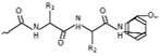
Amide bonds, formed between an amine and a carboxylic acid, are chemically sturdier than esters and can survive in plasma for many hours or even days without a dedicated enzyme present. Because of this stability, amide-linked prodrugs are usually engineered to be broken specifically by one particular enzyme, which enhances selectivity while limiting premature toxicity. Peptide linkers such as GFLG (cleaved by lysosomal enzymes concentrated in cancers) and SSKYQ (a substrate recognized only by prostate-specific antigen) exemplify this strategy. GFLG appears in a number of drug conjugates that have reached clinical testing, including PK1, PK2, PNU166945, and DX-8951 [13], while SSKYQ-based prodrugs such as L-377202 have advanced into Phase I/II trials [14].
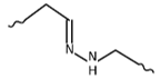
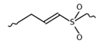
Other, less common linkage chemistries include oxime and imine bonds [15], disulfide bonds [16], and non-cleavable thioether bonds [17]. Whatever chemistry is used, the connecting bond generally needs to survive circulation in the blood and only break once the prodrug has reached its target. Linker choice is therefore an important optimization step. Hydrazone linkages, for example, are stable near neutral pH but degrade once the pH drops to around 5, which happens naturally once the prodrug is taken up into acidic endosomes and lysosomes [18]. Peptide-based linkers behave similarly in the sense that they resist degradation in serum but are readily cut by cancer-associated proteases once inside the cell [19]. Disulfide linkers add another layer of selectivity because they are opened specifically by intracellular thiols such as glutathione, which is present at much higher concentration inside cancer cells than in normal tissue [20]. Interestingly, a linker that resists breaking down entirely can still be useful if the targeting component itself carries some therapeutic activity of its own [17].
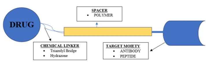
Fig. 1 — General design of a prodrug [components: chemical linker (e.g., triazolyl bridge, hydrazone), spacer (polymer), and targeting moiety (antibody, peptide)].
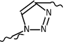
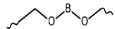
Table 1 — Newer linkers used in prodrug and bioconjugate design
|
Name |
Linker class |
Chemistry |
|
Triazolyl bridge |
1,2,3-Triazole linker |
|
|
Stimuli-responsive (pH) |
Hydrazone |
|
|
Stimuli-responsive (pH) |
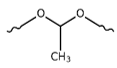
Acetal / ketal |
|
|
Stimuli-responsive (redox/thiol) |
Vinyl sulfone |
|
|
Photocleavable |
o-Nitrobenzyl |

|
|
Stimuli-responsive (ROS) |
Boronate ester |
|
|
Enzyme-cleavable (glycosidase) |
β-Glucuronide |

|
|
Enzyme-cleavable / self-immolative (ADC) |
Val–Cit–PAB dipeptide |
|
3. Targeting-Ligand Conjugated Prodrugs
Because cancer cells frequently over-express specific surface antigens, receptors, or transporters, a variety of targeting groups — antibodies, antibody fragments, peptides, aptamers, and small-molecule ligands — can be attached to a cytotoxic drug to steer it toward the cancer.
3.1 Antibody–Drug Conjugates
Monoclonal antibodies were the earliest targeting ligands adopted for prodrug design because of their strong binding affinity. An antibody–drug conjugate (ADC) is generally biologically silent until receptor-mediated endocytosis carries it into the cell and the active drug is released [21]. The ratio of drug molecules per antibody has to be carefully tuned to balance potency against selectivity [22]. Despite the appeal of using cancer-specific antibodies to spare healthy tissue, several practical hurdles remain, including immunogenicity, imperfect selectivity, and difficulty penetrating the dense interior of solid cancers. Humanized antibodies have largely solved the immunogenicity problem associated with earlier murine antibodies, and ongoing discovery of new cancer-restricted antigens continues to improve selectivity. Poor tissue penetration remains harder to fix, since therapeutic agents must cross multiple biological barriers and overcome elevated pressure inside the cancer interstitium [23]; using smaller antibody fragments such as scFv or Fab can help these conjugates penetrate more effectively than an intact antibody [24].
Antigens exploited for antibody–drug conjugates include CD19, CD22, CD33, HER2, TAG-72, MUC16, PSMA, EGFR, and αvβ integrin, among others, each with different degrees of cancer specificity [25]. MUC16, for instance, is largely restricted to ovarian and related reproductive cancers [26], and PSMA is preferentially found in prostate cancer tissue [27], while TAG-72 shows up across many adenocarcinomas — colorectal, pancreatic, gastric, ovarian, endometrial, breast, and non-small-cell lung — regardless of the organ of origin [25]. Drug classes that have been linked to antibodies in this way include antifolates [22], vinca alkaloids [28], anthracyclines [18], taxanes [29], monomethyl auristatin E [18], calicheamicin [30,31], and the maytansine derivative DM1 [32].
Roughly twenty antibody–drug conjugates had entered clinical trials as of the mid-2000s, and gemtuzumab ozogamicin (Mylotarg) was, at that time, the only one to receive FDA approval.
It links calicheamicin to a humanized anti-CD33 antibody via a hydrolyzable linker [33]. CD33 is present on the leukemic cells of over 90% of AML patients but is absent from normal tissue and from pluripotent hematopoietic stem cells, giving Mylotarg reasonably good target selectivity and allowing it to be internalized after binding [34]. Dosing studies showed that CD33 binding sites became saturated around 7.5 mg/m², and at a 9 mg/m² dose the drug displayed a mean maximum plasma concentration near 5.9 mg/mL, a half-life of about 72 hours, and broadly similar pharmacokinetics in children and adults [35]. In one Phase II trial, roughly a quarter of 277 patients responded to treatment [33].
Mylotarg received accelerated FDA approval in 2000, but was voluntarily withdrawn from the market in 2010 after a post-approval confirmatory trial (SWOG-S0106) failed to show a clinical benefit and showed a higher rate of fatal toxicity relative to standard chemotherapy. Following further trials that identified a lower, safer dosing regimen, the FDA re-approved Mylotarg in September 2017 for newly diagnosed and relapsed/refractory CD33-positive AML — the approval date reflected in Table 2 below.
Table 2 — FDA-Approved Antibody-Drug Conjugates (ADCs) for Cancer Treatment
|
Prodrug |
Antigen |
Antibody (Payload) |
Tumor Target |
Approval Status |
|
Mylotarg* |
CD33 |
Gemtuzumab ozogamicin |
Acute myeloid leukemia (AML) |
Approved 2017 |
|
Adcetris |
CD30 |
Brentuximab vedotin |
Hodgkin lymphoma, systemic ALCL |
Approved 2011 |
|
Kadcyla |
HER2 |
Trastuzumab emtansine (T-DM1) |
HER2-positive breast cancer |
Approved 2013 |
|
Besponsa |
CD22 |
Inotuzumab ozogamicin |
B-cell precursor ALL |
Approved 2017 |
|
Polivy |
CD79b |
Polatuzumab vedotin |
Diffuse large B-cell lymphoma |
Approved 2019 |
|
Padcev |
Nectin-4 |
Enfortumab vedotin |
Metastatic urothelial cancer |
Approved 2019 |
|
Enhertu |
HER2 |
Trastuzumab deruxtecan |
HER2-positive breast cancer |
Approved 2019 |
|
Trodelvy |
TROP2 |
Sacituzumab govitecan |
Triple-negative breast cancer |
Approved 2020 |
|
Zynlonta |
CD19 |
Loncastuximab tesirine |
Large B-cell lymphoma |
Approved 2021 |
|
Tivdak |
Tissue factor |
Tisotumab vedotin |
Cervical cancer |
Approved 2021 |
|
Elahere |
Folate receptor alpha |
Mirvetuximab soravtansine |
Platinum-resistant ovarian cancer |
Approved 2022 |
|
Datroway |
TROP2 |
Datopotamab deruxtecan |
HR+/HER2− breast cancer |
Approved 2025 |
|
Emrelis |
c-Met |
Telisotuzumab vedotin |
Non-small cell lung cancer (NSCLC) |
Approved 2025 |
* Originally approved 2000 under accelerated approval; voluntarily withdrawn 2010 after a confirmatory trial showed no clinical benefit and excess fatal toxicity; re-approved 2017 at a revised, lower dose.
Several other antibody-conjugated drugs remain in later-stage trials, most notably trastuzumab-DM1 (T-DM1) and inotuzumab ozogamicin (CMC-544) [37,38]. HER2, the target of trastuzumab, is over-expressed in roughly a fifth to a quarter of breast cancers and is linked to more aggressive disease, making it an attractive therapeutic target; trastuzumab itself was approved by the FDA in 1998 for HER2-positive metastatic breast cancer [17,39]. Because a substantial share of patients still fails to respond to trastuzumab alone, combining it with a cytotoxic payload has been an appealing next step. T-DM1 links trastuzumab to DM1, a potent antimitotic maytansine derivative, and researchers found that a non-reducible thioether linker outperformed cleavable disulfide alternatives in this particular conjugate [17]. In a Phase I trial, the maximum tolerated dose was 3.6 mg/kg, giving a half-life of 3.5 days and markedly slower clearance than at lower doses [40]; in a subsequent Phase II trial, roughly a third of 110 patients showed meaningful improvement with tolerable side effects [37].
CMC-544 pairs a humanized anti-CD22 antibody with N-acetyl gamma calicheamicin dimethylhydrazide via an acid-sensitive linker — the same drug and linker chemistry as Mylotarg, but directed against CD22, a marker restricted to mature and malignant B lymphocytes. Binding to CD22 on malignant B cells triggers rapid internalization and payload release [38]. Early studies put its maximum tolerated dose at 1.8 mg/m² with an initial half-life near 24 hours that lengthened to about 4 days after repeated dosing, alongside markedly slower clearance [41]; it has since advanced to Phase III testing for B-lymphoid malignancies.
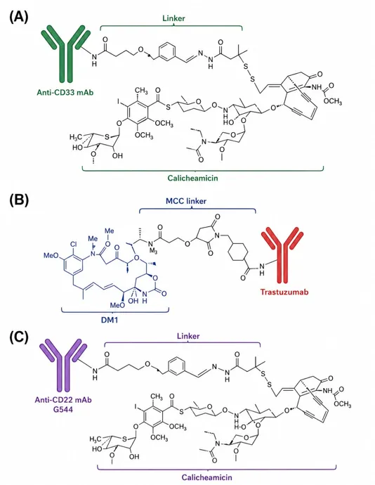
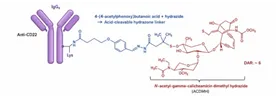
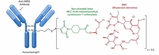
Fig. 2 — Structure of Mylotarg, Trastuzumab-DM1, CMC-544, Besponsa, and Kadcyla
3.2 Peptide–Drug Conjugates
Peptides offer several practical advantages over antibodies as targeting groups: they are much smaller, penetrate cells more readily, are easier to synthesize, and are simpler to chemically modify [39]. They can be designed to recognize either a cancer-associated antigen or an over-expressed transporter.
Minko and colleagues linked an LHRH (luteinizing hormone-releasing hormone) peptide to camptothecin through a PEG spacer, exploiting the fact that LHRH receptors, while present at low levels in healthy prostate, breast, and ovarian tissue, are considerably more abundant on breast, prostate, ovarian, and endometrial cancer cells [42,43]. This construct showed markedly better efficacy with reduced harm to healthy organs, and its small size gave it an advantage in reaching both solid cancer masses and individual metastatic cells.
Separately, a cyclic RGD peptide, E[c(RGDyK)]₂, was conjugated to paclitaxel to exploit αvβ3 integrin, a receptor that is elevated on metastatic cancer cells and activated endothelium but largely absent from resting, healthy vasculature; in vivo testing showed selective cancer accumulation within four hours of dosing [44].
A third example targets GRP78 (glucose-regulated protein 78), which is over-expressed on colon, skin, prostate, and breast cancer cells but only weakly expressed in normal tissue [45,46]. Pep42, a 13-residue cyclic peptide, binds GRP78 and is internalized into lysosomes containing cathepsin B, so a Val-Cit linker — stable in plasma but cleavable by cathepsin B — was used to attach Pep42 to paclitaxel and doxorubicin [47]. Both conjugates showed increased cytotoxicity relative to the free drugs in osteosarcoma cell testing [45].
3.3 Aptamer–Drug Conjugates
Aptamers are short single-stranded DNA or RNA sequences that fold into three-dimensional shapes capable of binding a target with very high affinity, sometimes in the nanomolar-to-picomolar range [48]. Compared with antibodies, they are smaller, easier to manufacture with tight quality control, less likely to provoke an immune reaction, chemically stable, and straightforward to modify [49,51]. Aptamers are typically discovered through SELEX, an iterative screening process applied to large nucleic-acid libraries [50].
One well-studied example targets PSMA, which is abundant on prostate cancer cells and on the cancer vasculature of some non-prostate cancers such as breast and lung [52–54]. An RNA aptamer against PSMA (the A10 aptamer) was attached to cisplatin-loaded polymeric nanoparticles; the resulting conjugate was roughly four times more potent than unmodified nanoparticles and about tenfold more potent than free cisplatin in PSMA-positive prostate cancer cells [55,56].
In another example, doxorubicin was linked to the sgc8c DNA aptamer, which recognizes protein tyrosine kinase 7 (PTK7), a receptor highly expressed on T-cell acute lymphoblastic leukemia cells, for treating that disease [57,58]. A control aptamer with a different binding target produced far less cellular uptake, confirming that the sgc8c conjugate's activity depended on specific target recognition [57].
3.4 Folic Acid–Drug Conjugates
Folic acid binds its receptor with extremely high affinity and is one of the most widely used small-molecule targeting groups. The folate receptor is over-expressed on many malignant cell types — breast, ovarian, lung, kidney, and endometrial cancers among them — while remaining largely confined to a small subset of normal epithelial tissue elsewhere in the body. Folic acid also has low immunogenicity and simpler chemistry than antibody-, peptide-, or aptamer-based targeting [20,59–61].
Researchers have linked folic acid to camptothecin through a disulfide-containing peptide spacer, giving a conjugate that is both more soluble and more selective than the free drug; it showed strong activity (IC₅₀ near 10 nM) against a folate-receptor-rich cervical cancer cell line, and pre-saturating the receptor with excess free folic acid abolished this activity, confirming receptor-mediated uptake [59]. In a separate design, doxorubicin was modified with both folic acid and D-α-tocopheryl polyethylene glycol succinate (TPGS) to improve solubility and membrane permeability alongside cancer targeting; the resulting conjugate extended circulation time, was roughly 45 times more potent than unmodified doxorubicin, and produced less accumulation in cardiac tissue, the organ most vulnerable to doxorubicin toxicity [61].
4. Membrane-Transporter-Associated Prodrugs
Membrane transporters move nutrients such as sugars, nucleosides, amino acids, and small peptides across the cell membrane, and historically they have been exploited to improve oral absorption of poorly permeable drugs. More recently, several transporters that are unusually active in cancer cells have become targets in their own right [4].
Peptide transporters — particularly PEPT1 and PEPT2, which move di- and tripeptides in a sequence-independent but stereoselective way, alongside the related PHT1/PHT2 histidine transporters — are the most extensively used of these [62]. PEPT1 is notably over-expressed in several cancer cell lines, including pancreatic lines AsPC-1 [63] and Capan-2 [64] and the fibrosarcoma line HT-1080 [65], while remaining largely absent from normal cells.
Bestatin, a peptide-mimetic anticancer agent and known PEPT1 substrate, accumulated selectively in PEPT1-expressing HeLa cells and suppressed cancer growth in a naturally PEPT1-over-expressing line, confirming that its uptake depends specifically on this transporter [66]. Separately, a PEPT1-recognized substrate was attached to floxuridine via an ester bond, giving a prodrug that accumulated preferentially in PEPT1-positive cancer cells and selectively slowed their growth while sparing PEPT1-negative cells [64].
Gemcitabine, used clinically against breast, lung, ovarian, pancreatic, and bladder cancers, has also been modified with amino-acid ester groups; radiolabeled uptake studies showed that these derivatives are recognized by PEPT1, PEPT2, or both [67].
The sodium-dependent multivitamin transporter (SMVT), which normally carries biotin, lipoate, pantothenate, and certain peptides across the gut lining [68,69], has likewise been exploited: a biotinylated, PEG-conjugated camptothecin increased cytotoxicity in both drug-sensitive and multidrug-resistant human ovarian cancer cell lines that express high SMVT levels [70], and biotinylated PAMAM dendrimer and HPMA polymer conjugates showed similarly enhanced uptake in HeLa and murine ID8 ovarian cancer cells, respectively, via the same transporter [71,72].
Table 3 — Membrane transporter-conjugated prodrugs
|
Organ |
Disease condition |
Transporter |
Prodrug |
|
HeLa-PEPT1 tumor cells |
— |
PEPT1 |
Bestatin |
|
Colon, liver |
Colon cancer metastasis; hepatic metastasis |
PEPT1 |
Floxuridine |
|
Breast, lung, ovary, pancreas, bladder |
Solid tumor |
PEPT1 and PEPT2 |
Gemcitabine |
|
Ovary |
Ovarian cancer cell |
SMVT |
Biotinylated camptothecin |
|
ID8 murine cell line |
— |
SMVT |
Biotinylated HPMA |
|
HeLa cell line |
— |
SMVT |
Biotinylated PAMAM |
|
Liver |
Hepatocellular carcinoma |
PEPT1 |
Doxorubicin-Gly-Gly-Gly |
|
Intestine (oral delivery) |
General/systemic |
PEPT1 |
Thiodipeptide-drug conjugates (ibuprofen, gabapentin, etc.) |
|
Brain / CNS |
Glioma, various cancers |
LAT1 |
Phenylalanine-drug conjugates |
|
Liver (HepG2) |
Hepatocarcinoma |
GLUT1 |
PAMAM-Camptothecin (GPCC) |
|
Lung (A549) |
Lung cancer |
GLUT1 |
Glucose-PEG-Azo-IR808-S-S-Paclitaxel |
|
Pancreas |
Metastatic pancreatic cancer |
GLUT |
Glufosfamide |
|
Colon, breast, leukemia |
Solid tumors, leukemia |
GLUT1 |
Pt(II) glucose/ mannose/ galactose glycoconjugates |
|
Various (MCF-7 and others) |
Solid tumors |
GLUT1 and OCT2 |
Pt(IV) glycoconjugates |
5. Polymeric Prodrugs
5.1 PEG–Drug Conjugates
Polyethylene glycol (PEG) is an FDA-approved, water-soluble, biologically inert polymer that clears through the kidneys below roughly 50 kDa and through the liver above that threshold [73]. While PEGylation was originally developed for large biologics such as antibodies and growth factors, it has increasingly been applied to small-molecule anticancer drugs as well, generally following one of two strategies distinguished by PEG molecular weight.
High-molecular-weight PEG conjugates (roughly 30–60 kDa or larger) work mainly by enlarging the overall size of the prodrug, which extends circulation time and reshapes biodistribution so that more drug accumulates passively in cancers through the enhanced permeation and retention (EPR) effect. NKTR-102, a PEGylated irinotecan built on a cleavable linker, is a leading example. In mouse xenograft models, it extended the half-life of the active irinotecan metabolite SN-38 roughly ninetyfold in colorectal cancers and tenfold in lung cancers, and increased SN-38 exposure within the cancer by several hundredfold and nearly sixtyfold in those two models, respectively [74]; it also outperformed unmodified irinotecan in a colorectal cancer model [75]. In Phase I human testing, NKTR-102 extended plasma half-life to roughly 50 days compared with under two days for free irinotecan, and produced a partial or significant response in about 38% of patients across several cancer types, including breast, ovarian, and cervical cancer [76]. Related large-PEG conjugates include docetaxel (NKTR-105), SN-38 (EZN-2208), and camptothecin (pegamotecan) [10].
The architecture of the PEG chain — linear, branched, or dendritic — affects how much drug it can carry. A linear methoxy-PEG of 40 kDa, for example, achieves only about 1.25% drug loading by weight, a linear diol PEG of the same size reaches about 1.7% (as in pegamotecan), and a four-arm branched PEG can reach 4.3–5.0% (as in EZN-2208) [77,78].
Low-molecular-weight PEG conjugates, using PEG chains under about 5 kDa, are instead used to improve water solubility, oral bioavailability, or to limit penetration across barriers such as the blood–brain barrier. NKTR-118, a PEG-naloxol conjugate developed for opioid-induced constipation, illustrates this approach: Phase I data showed roughly a tenfold improvement in oral bioavailability relative to naloxone, along with markedly reduced penetration of the blood–brain barrier, which helps preserve the analgesic effect of concurrent opioid therapy [79,80].
5.2 Other Polymer–Drug Conjugates
PEG's relatively limited number of attachment sites caps how much drug it can carry, so polymers with more functional groups — such as N-(2-hydroxypropyl)methacrylamide (HPMA) and polyglutamic acid (PGA) — have been explored as alternatives, some reaching drug loadings as high as 37% by weight [81].
FCE28068 (PK1), an HPMA-doxorubicin conjugate built on a GFLG peptide linker with about 8% drug loading, allowed doxorubicin doses as high as 1680 mg/m² without the cardiotoxicity typically associated with the free drug, and displayed a distribution half-life of 1.8 hours and an elimination half-life of 93 hours [82]. Anticancer activity appeared across colorectal, non-small-cell lung, and breast cancer in Phase I testing, though only the lung and breast cancer groups showed partial responses in Phase II [83].
PGA, unlike PEG or HPMA, is biodegradable, which allows chains longer than 60 kDa to be used safely even though they exceed the renal clearance threshold, and it typically achieves very high drug loading. A PGA–paclitaxel conjugate (CT-2103, Xyotax) reaches about 37% drug loading, and a related PGA–camptothecin conjugate (CT-2106) reaches 33–35%; both have entered Phase I/II clinical testing [81,84–86].
5.3 Polymeric Prodrug Nanoparticles
Some polymer–drug conjugates self-assemble into nanoscale structures such as micelles, typically built from a block copolymer with a hydrophilic outer shell — often PEG — surrounding a hydrophobic drug-containing core. These particles, generally 20–100 nm across, are large enough to benefit from the EPR effect and stable enough to circulate for extended periods, unlike free polymer chains that are cleared quickly by the kidneys. Several such micelles — a paclitaxel micelle (NK105), a cisplatin micelle (NC-6004), and an SN-38 micelle (NK012) — are in clinical testing [87].
A related design uses a linear, cyclodextrin-containing polymer. IT-101, built from a roughly 70 kDa chain, self-assembles into 30–40 nm micelles held together by an inclusion complex between cyclodextrin and camptothecin rather than by conventional hydrophobic packing; as a result, organic solvents such as DMSO do not disrupt the structure, though soluble adamantane can displace camptothecin and collapse the micelle [12]. In Phase I testing, IT-101's half-life reached about 40 hours, and three patients (with pancreatic, renal, and non-small-cell lung cancer) who completed a six-cycle regimen showed progression-free survival of 18, 14, and 11 months, respectively [88]; a Phase II ovarian cancer trial began in 2008.
Prodrugs can also be physically encapsulated rather than covalently built into a nanocarrier. Because paclitaxel is poorly lipid-soluble, it was first converted into a cholesterol conjugate (Tax-chol) to increase its lipophilicity, then encapsulated into lipid nanoparticles with over 90% efficiency and good colloidal stability; adding folic acid to the particle surface substantially increased both cellular uptake and cytotoxicity in folate-receptor-positive cells [89].
5.4 Pharmacokinetics of Polymeric Prodrugs
Because polymeric prodrugs are either intrinsically large (10–100 kDa) or self-assemble into nanoscale particles, they tend to circulate much longer than free drug and benefit passively from the EPR effect — NKTR-102, for instance, extends SN-38 exposure in colorectal cancer xenografts via its markedly prolonged plasma half-life [74].
The tradeoff is that clearance becomes a design concern: because large, non-biodegradable molecules are difficult to eliminate from tissue, most PEG-based prodrugs are kept under the roughly 60 kDa renal filtration cutoff.
6. Enzyme-Cleavable Prodrugs
Using an enzyme that is concentrated specifically in cancer tissue to release the active drug is one of the more advanced targeting strategies, because it confines drug activation almost entirely to the cancer and minimizes exposure elsewhere. In practice, though, relatively few enzymes are both cancer-restricted and catalytically efficient enough to be useful.
Prostate-specific antigen (PSA) and PSMA are the two most extensively exploited. PSA is a serine protease that normally breaks down seminal fluid proteins, and its over-expression is an established marker of prostate cancer [91]. Importantly, PSA's enzymatic activity is essentially confined to prostate cancer tissue, because circulating PSA in the blood is neutralized by natural inhibitors (α1-antichymotrypsin and α2-macroglobulin); prodrugs designed around PSA-cleavable peptide sequences therefore accumulate mainly at the cancer. Two peptide sequences, HSSKLQ and SSKYQ, are the most widely used PSA substrates, with SSKYQ being roughly tenfold more catalytically efficient than HSSKLQ [92]. Drugs conjugated through these sequences include 5-fluorodeoxyuridine [93], paclitaxel [94], cyclopamine [92], doxorubicin [95], and thapsigargin [96]. A related class of PSA substrates incorporates non-natural amino acids to resist unwanted cleavage by other plasma proteases while preserving PSA recognition [97].
One well-characterized example, L-377202, links doxorubicin to a modified PSA-substrate peptide capped with a glutaryl group to block exopeptidase degradation and improve solubility, while non-natural amino acid substitutions minimize an unwanted histamine-release side effect and improve both stability and PSA specificity [14,97]. In preclinical testing it matched doxorubicin's potency in PSA-positive prostate cancer cells while sparing normal epithelial cells and PSA-negative cancer cells, and it produced roughly fifteenfold greater anticancer activity than doxorubicin in a mouse model, alongside five- to tenfold higher drug concentration in cancer tissue and tenfold lower accumulation in the heart [14].
PSMA, a large type-II membrane glycoprotein, is expressed at levels roughly a thousandfold higher in prostate cancer — especially advanced, androgen-resistant disease — than in normal tissue, and its elevated expression predicts recurrence after primary treatment, making it an attractive prodrug target [98,99]. As an exopeptidase, PSMA naturally hydrolyzes N-acetylaspartylglutamate, but only a single PSMA-cleavable substrate peptide has so far been identified, which limits how widely this enzyme can currently be exploited in prodrug design [100,101].
7. Enzyme-Activated Prodrugs
Some anticancer drugs might only reach their full therapeutic potential at doses well above what is currently tolerated in the clinic. Prodrugs activated by cancer-restricted enzymes can, in principle, deliver more than fifty times the standard maximum dose directly to cancer cells [1]. This depends on enzymes that are either unique to the cancer or present there at much higher levels than elsewhere [6], but naturally cancer-restricted enzymes at sufficiently high levels are uncommon [102], so two engineered strategies have been developed instead.
7.1 Antibody-Directed Enzyme Prodrug Therapy (ADEPT)
In ADEPT, the activating enzyme itself — rather than the drug — is first delivered to the cancer by linking it to a cancer-specific antibody, either through direct chemical conjugation or as a recombinant fusion protein. Once the enzyme–antibody conjugate has localized to the cancer, the inert prodrug is administered separately and is converted to active drug specifically in that microenvironment. A major advantage of this approach is catalytic amplification: a single enzyme molecule can generate hundreds of active drug molecules per second, and because the released drug can diffuse to neighboring cells, not every cancer cell needs to take up the enzyme–antibody conjugate directly [1]. A range of enzyme, antibody, and prodrug combinations has been tested against cancer types that otherwise resist conventional chemotherapy [103].
The main drawbacks are immunogenicity of the enzyme–antibody conjugate, the risk that unbound conjugate circulating in the blood could activate the prodrug prematurely, and inconsistency in how the conjugate is manufactured [104,105]. Humanized antibodies have addressed much of the immunogenicity concern associated with earlier murine antibodies and bacterial enzymes [106]. For example, a murine anti-TAG-72 antibody used in an ovarian cancer trial provoked a human anti-mouse antibody response [107], whereas a humanized version of the same anti-TAG-72 antibody produced no detectable immune response while retaining high specificity and affinity [108]. That humanized antibody, when conjugated to β-galactosidase, selectively activated a geldanamycin prodrug; the released active drug bound Hsp90, reduced AKT levels by up to 70%, and was more than twenty-five times as potent as the unactivated prodrug [109].
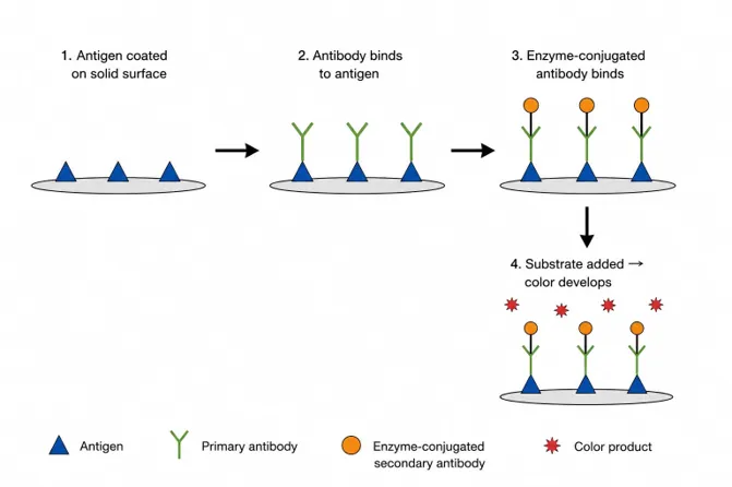
Fig. 3 — Schematic diagram of ADEPT
7.2 Gene-Directed Enzyme Prodrug Therapy (GDEPT)
GDEPT — also called VDEPT or GPAT — instead delivers the gene encoding the activating enzyme directly into cancer cells, after which the inactive prodrug is given systemically and is converted to active drug by the enzyme the cancer cells now express. The key difference from ADEPT is where activation occurs: extracellularly in ADEPT, but intracellularly in GDEPT, meaning that enzyme/prodrug pairs suited to one approach are not necessarily suited to the other [1,6].
Bacterial or viral enzymes are generally preferred for GDEPT because intracellular expression of a non-human enzyme provokes only a mild immune response, because these enzymes typically have no human counterpart to interfere with, and because bacterial proteins tend to fold correctly without the more demanding post-translational processing that a complex eukaryotic enzyme might require [1,111].
A useful side effect of GDEPT is the so-called bystander effect, in which active drug released from a transduced cell also kills neighboring cells that never received the enzyme-encoding gene themselves. Proposed explanations include active drug trapped in dying cells being taken up by neighboring cells through phagocytosis [112], diffusion of active drug through gap junctions connecting adjacent cells [113], and simple passive diffusion of a small, soluble active metabolite such as 5-fluorouracil across cell membranes [114].
A Phase I breast cancer trial demonstrated that GDEPT could be given safely without local or systemic complications: a gene vector used the erbB-2 promoter to express cytosine deaminase selectively in erbB-2-positive cancer cells, converting the systemically administered prodrug 5-fluorocytosine into toxic 5-fluorouracil specifically at the cancer; cancer regression was observed without complications such as insertional mutagenesis, anti-DNA antibody formation, local infection, or cancer ulceration [115]. A related approach combined the erbB-2 promoter with a MUC1 enhancer to drive expression of the suicide gene thymidine kinase, selectively killing MUC1-positive pancreatic and breast cancer cells with the prodrug ganciclovir [116,117].
Cancer selectivity in GDEPT can be further improved by incorporating a cancer-specific regulatory element into the gene vector [118,119]; for example, PSA- or PSMA-specific promoters have been used to restrict enzyme expression to prostate cancer cells [120]. Cytochrome P450 (CYP) is another enzyme exploited this way, activating drugs such as ifosfamide [121], AQ4N [122], and the anthracycline MMDX [123]; intracanceral P450 expression substantially increased MMDX's anticancer activity in a mouse xenograft model [123].
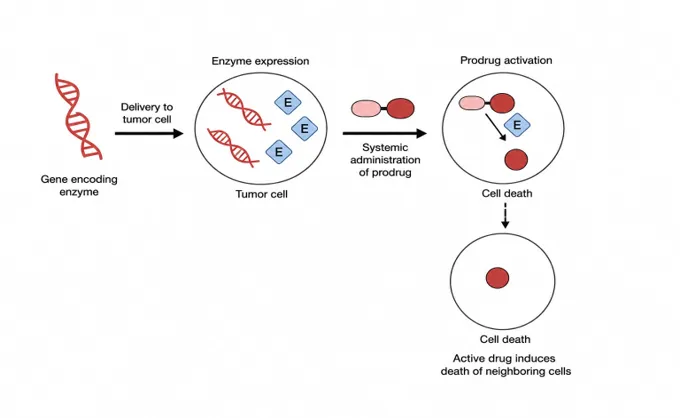
Fig. — Schematic diagram of GDEPT
CONCLUSION
Prodrug chemistry offers a versatile way to improve a drug's pharmaceutical properties through chemical modification or conjugation to a promoiety, but targeted prodrug design specifically remains a relatively young field with much still to establish. The withdrawal of Mylotarg, the first FDA-approved antibody–drug conjugate, from the market in 2010 due to disappointing efficacy and safety in post-approval trials — and its later re-approval in 2017 at a revised dose — is a reminder both of how much work remains and of how targeted-prodrug programs can recover from early setbacks. Even so, many other targeted prodrugs have advanced into clinical testing once preclinical proof-of-concept was established. Immunogenicity, cell permeability, and target specificity remain the three central obstacles to building an effective targeted prodrug.
Humanized antibodies, along with alternative ligands such as peptides and aptamers, have gone a long way toward solving the immunogenicity problem, and smaller antibody fragments or ligands generally improve tissue penetration as well. At the core of any targeted prodrug strategy is the need for a cancer-specific antigen that a natural or engineered ligand can recognize, so identifying and validating such antigens — and confirming their specificity in animal models rather than relying solely on in vitro data — remains critical, since many candidate antigens turn out to also be expressed, at some level, in normal tissue. Likewise, discovering high-affinity peptide or aptamer ligands is a slow and difficult screening process. Continued progress toward identifying cancer-specific ligands that combine high affinity, high specificity, strong stability, low immunogenicity, and good tissue permeability will be essential for targeted prodrugs to reach their full clinical potential.
REFERENCES


Himadri Shekhar Banerjee, Arnab Sarkar, Development of Novel Prodrug Systems to Improve Bioavailability and Therapeutic Efficacy in Cancer Chemotherapy, Int. J. of Pharm. Sci., 2026, Vol 4, Issue 10, 677-695. https://doi.org/10.5281/zenodo.23169716
 10.5281/zenodo.23169716