

We use cookies to ensure our website works properly and to personalise your experience. Cookies policy
Department of Pharmaceutics, Karnataka college of Pharmacy, Bengaluru 560064
Diabetes mellitus, a chronic metabolic disease with long-term health consequences that is becoming more and more prevalent, is characterized by prolonged hyperglycemia. It is estimated that 25% of individuals with diabetes mellitus have poor healing of diabetic wounds, which often results in lower limb amputation and the significant financial and psychological costs that go along with it. The hyperglycemic environment that promotes the formation of biofilms makes treating diabetic wounds difficult. In this review, we provide updates on recent developments in our knowledge of the pathophysiology of diabetic wounds, with particular attention to impaired angiogenesis, neuropathy, suboptimal chronic inflammatory response, barrier disruption, and subsequent polymicrobial infection. Next, we discuss current and upcoming treatment approaches intended to address the different pathologies linked to diabetic wounds. Future therapeutic approaches must address several factors that contribute to poor healing in diabetic wounds given the startling rise in the prevalence of diabetes and, consequently, diabetic wounds(1).
Diabetes mellitus, a chronic metabolic disease with long-term health consequences that is becoming more and more prevalent, is characterized by prolonged hyperglycemia. It is estimated that 25% of individuals with diabetes mellitus have poor healing of diabetic wounds, which often results in lower limb amputation and the significant financial and psychological costs that go along with it(2).
Diabetes mellitus is a group of metabolic diseases characterized by impaired insulin synthesis or activity, leading to hyperglycemia. Diabetes mellitus has a substantial effect on patient’s expectations for quality of life and survival. And it is a complex metabolic disease that affects more than 340 million individuals worldwide(3).
Wound infections are common in diabetic patients. The development of drug-resistant strains and the continuous spread of illnesses show how important it is to comprehend bacterial activity in the host in order to develop new therapeutic strategies(4).
The World Health Organization (WHO) reports that the number of people with diabetes is increasing worldwide, especially in middle-income countries(5).
Patients with diabetes have wounds that are more difficult to heal than normal wounds, particularly diabetic foot ulcers, which can quickly worsen and result in amputation. Diabetic wounds cannot be healed by standard therapies, nor can their many consequences be managed(6).
The restoration of anatomic integrity with comparable function is the outcome of the complex and dynamic process of wound healing. Rapid and thorough healing without the spread of infection and sepsis is the most important prerequisite for wound care. In most cases, acute wounds heal without any problems. Age-related changes in normal physiological processes, such as blood circulation deprivation, obesity, illnesses like diabetes, and stressful environmental conditions, are the main cause for worry. Wounds are classified as either acute or chronic based on their capability for healing. Tissue injuries that do not heal in a systematic sequence and take more than 12 weeks to recover are considered chronic wounds(7).
Chlorhexidine is a cationic compound consisting of two symmetric 4-chlorophenyl rings and two bisguanide groups connected by a core hexamethylene chain. Chlorhexidine has a pH of 5.5 to 7. The most popular formulation is digluconate salt because of its superior stability and water solubility. Chlorhexidine is a broad-spectrum antibacterial agent that combats both Gram-positive and Gram-negative bacteria(8).
Many civilizations have employed Aloe vera (L.) Burm. f. (Liliaceae) in traditional medicine for a variety of therapeutic uses. It's has been applied to heal burns and wounds, among other conditions (Grindlay and Reynolds, 1980). Fresh gel, juice, or formulated products have been utilized for general health, medicinal, and cosmetic objectives. Aloe vera has been demonstrated to have hypoglycaemia and antidiabetic qualities in addition to its ability to heal wounds (Ajabnoor, 1990; Beppu et al., 1993). Despite its lengthy history of usage as a folk treatment, the biochemical intricacies of how it affects physiological and pathophysiological processes are still unknown(9).
HISTORY OF DIABETIC WOUND:
One well-known effect of diabetes mellitus is delayed and persistent wound healing, which is a major cause of morbidity in diabetic patients. Reiber and colleagues described how peripheral neuropathy, vascular insufficiency, decreased immune response, and increased susceptibility to infection all contribute to poor wound healing in diabetic patients. These pathogenic factors increase the risk of wound chronicity, prolong inflammation, and delay tissue regeneration. The authors also emphasized the connection between diabetic wounds and increased hospital stays, higher medical costs, and a lower standard of living. Their findings highlighted the need for effective wound care strategies targeted at infection prevention and improved healing results in diabetic patients(10).
Diabetes mellitus frequently results in chronic sores due to long-term metabolic issues caused by persistent hyperglycemia. Diabetic wounds heal more slowly and are more prone to infection due to a number of factors, including impaired angiogenesis, decreased immune cell activity, and insufficient collagen synthesis. These elements obstruct the proliferative, remodeling, and inflammatory stages of the wound healing cascade. These pathophysiological anomalies provide a therapeutic challenge and highlight the need for advanced wound care techniques that address both infection control and microenvironment regulation(11).
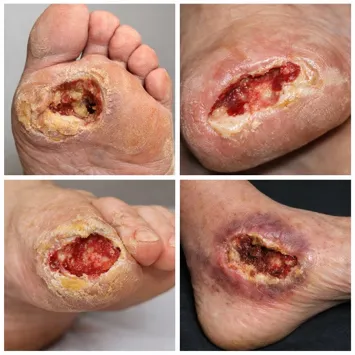
Fig no 1: Diabetic wound
PATHOGENESIS:
Hemostasis, inflammation, proliferation, and remodeling are the successive phases of wound healing, a complex biological process involving coordinated interactions between skin cells, extracellular matrix components, and systemic stimuli. Platelet activation and the initiation of the coagulation cascade occur immediately following tissue damage in order to halt blood loss and produce a transient matrix for cell migration(12).
Hyperglycemia in DM patients can lead to peripheral neuropathy, atherosclerosis, and decreased skin cell activity, all of which can impede wound healing and the formation of DFUs. The majority of the literature, and therefore this section, concentrates on the detrimental consequences of hyperglycemia as it pertains to the development and progression of DFUs, even though hypoglycemia has also been linked to the vascular complications of diabetes. Hyperglycemia hinders the healing process by causing atherosclerosis, which stops circulating nutrients from getting to wounds. Furthermore, it has been discovered that hyperglycemia may contribute to endothelial cell dysfunction in DM patients. Endothelial cells are essential for the healing of DFUs through pressure-induced vasodilation, a reaction that is typically protective for the skin(2).
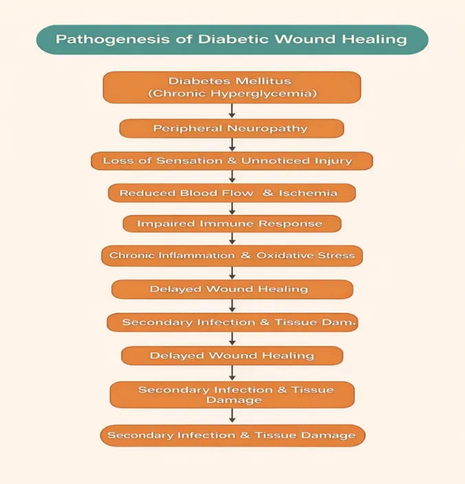
Diabetic wound healing is a complicated pathological process brought on by immunological, vascular, and metabolic problems brought on by long-term hyperglycemia. Leukocyte function is compromised, oxidative stress is increased, and the inflammatory stage of wound healing is prolonged when blood glucose levels are persistently elevated. The proliferative and remodeling stages are severely disrupted in diabetes circumstances due to decreased angiogenesis, poor fibroblast proliferation, and delayed collagen deposition. Wound closure is further delayed by peripheral neuropathy and inadequate microcirculation, which provide a hypoxic environment that encourages bacteria colonization and biofilm development. As a result, diabetic foot ulcers are a typical manifestation of chronic, non-healing diabetic wounds(13).
Table no 1. Pathogenesis of diabetic wound healing.(7)(14)
CHOICE OF WOUND CARE IN DIABETIC WOUND HEALING:
The choice of wound care is crucial for diabetic wound healing because of the condition's chronic inflammation, high susceptibility to infection, and delayed tissue regeneration. Recent studies have shown that effective diabetic wound care requires maintaining a moist wound environment, controlling the microbial load, and promoting angiogenesis and cell proliferation. Hydrogels and nanogel-based dressings are examples of advanced wound care systems that have attracted attention since 2020 because of their ability to provide longer drug release, improved biocompatibility, and greater contact with the wound milieu. Because these solutions can both promote tissue regeneration and provide antibacterial medications, they are more effective than conventional dressings for chronic diabetic wounds(15).
Effective wound care decisions are essential for diabetic wound healing because diabetes-related metabolic issues cause slow tissue repair, reduced new blood vessel development, prolonged inflammation, and a high risk of infection. Recent research indicates that effective treatment of diabetic wounds requires methods that preserve wound moisture, prevent bacterial growth, and encourage cell proliferation and tissue healing. In this context, contemporary wound dressings such as hydrogels and nanogel compositions have attracted a lot of attention. These innovative technologies enable the continuous and targeted release of antimicrobial and therapeutic drugs, enhance formulation stability, and lessen cellular toxicity. Consequently, multifunctional dressings outperform traditional wound care materials in the healing of chronic diabetic wounds(7).
DIFFICULTY IN DIAGNOSING INFECTION IN DIABETIC WOUND INFECTION:
Diagnosing diabetic wound healing is challenging due to the complexity and diversity of diabetes-related tissue damage. Clinical evaluation is often complicated by peripheral neuropathy, which reduces pain perception and delays wound discovery. Impaired blood circulation and microvascular dysfunction make it difficult to accurately assess tissue perfusion and healing capacity. Moreover, persistent inflammation and recurrent infections mask early signs of improvement or deterioration. Conventional diagnostic techniques might not be able to distinguish between acute and chronic wound states, even though biochemical markers of healing are not commonly used in clinical practice. These limitations result in delayed diagnosis, poor healing outcomes for diabetic wounds, and incorrect treatment decisions(16).
Identifying and evaluating diabetic wound healing is often difficult because diabetes alters normal inflammatory and sensory mechanisms. Loss of sensation caused by peripheral neuropathy can delay wound recognition and mask the true extent of tissue damage. Reduced blood supply and ischemic conditions further complicate evaluation by limiting visible signs of tissue repair. Moreover, impaired immune responses diminish classic signs of infection, making clinical diagnosis less reliable. Diagnostic tools such as imaging techniques have limited effectiveness during early stages, and microbiological findings may reflect bacterial presence rather than active infection. Consequently, these challenges frequently lead to delayed therapeutic intervention and extended healing duration in diabetic wounds(17).
ROLE OF CHLORHEXIDINE GLUCONATE IN DIABETIC WOUND HEALING:
Chlorhexidine gluconate is a widely used antiseptic with broad-spectrum activity against gram-positive and gram-negative bacteria, fungi, and some viruses. In diabetic wounds, infection control is crucial because microbial burden delays healing. Chlorhexidine reduces bacterial colonization at the wound site and provides prolonged antimicrobial action due to its substantivity, which is beneficial for chronic wounds. Maintaining a clean wound environment supports granulation and epithelialization. However, its concentration must be carefully controlled to avoid effects on cell viability. Incorporation into gels or nanogels enables sustained antimicrobial delivery while minimizing local irritation(18).
Chlorhexidine gluconate is a broad-spectrum antiseptic commonly used in wound care. In diabetic wounds, high microbial load delays healing, and chlorhexidine helps reduce infection by disrupting microbial cell membranes. Its prolonged antimicrobial action maintains wound cleanliness, supporting tissue repair. However, the concentration must be carefully controlled to avoid cytotoxicity. Incorporation into gels or nanogels enables controlled antimicrobial delivery, making chlorhexidine suitable for diabetic wound management(19).
ROLE OF ALOE VERA IN DIABETIC WOUND HEALING:
Aloe vera supports diabetic wound healing through its anti-inflammatory, antioxidant, and antimicrobial properties. It contains bioactive compounds that enhance fibroblast activity, collagen synthesis, and angiogenesis while maintaining a moist wound environment. These actions reduce inflammation, lower infection risk, and support improved healing in chronic diabetic wounds(20).
Aloe vera is commonly used in diabetic wound treatment because of its anti-inflammatory, antimicrobial, and antioxidant properties. Diabetic wounds often remain inflamed and are highly prone to infection. The bioactive constituents of Aloe vera, including polysaccharides and vitamins, help control inflammation and reduce microbial burden at the wound site. It also supports moisture retention, which protects the tissue from dryness. Studies have shown that Aloe vera is well tolerated, making it a useful supportive option in diabetic wound care(21).
Diabetic wounds often show prolonged inflammation and a high risk of infection. Aloe vera contains polysaccharides and phenolic compounds that help reduce inflammatory reactions and inhibit microbial growth at the wound site. It also maintains wound moisture, preventing tissue dryness. Due to its good tolerability and safety profile, Aloe vera is commonly applied as a supportive agent in diabetic wound management(20).
NANOGELS:
Structure
Nanogels are three-dimensional, nanoscale, cross-linked polymer networks with high water absorption capacity. They consist of hydrophilic or amphiphilic polymer chains that form a porous matrix that could contain drugs or other bioactive materials. Because of their flexibility, which allows for swelling, controlled drug release, and close contact with biological tissues, they are suitable for wound healing applications(22).
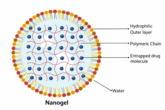
Fig no 2: Structure of nanogel
CLASSIFICATION OF NANOGELS:
|
Sr. No. |
Basis of Classification |
Type of Nanogel |
Description / Examples |
Reference |
|
Structure-based nanogels |
1. Simple nanogels |
Basic polymeric nanogels with uniform structure, such as cholesterol-bearing pullulan nanogels. |
(23) |
|
|
2. Hollow nanogels |
Nanogels with an empty core, useful for high drug loading. |
(24) |
||
|
3. Core–shell nanogels |
Nanogels having a distinct core and outer shell, often used for controlled drug release. |
(25) |
||
|
4. Hairy nanogels |
Nanogels with polymer chains extending from the surface, improving stability and interaction. |
(26) |
||
|
5. Multilayer nanogels |
Nanogels composed of multiple polymer layers for enhanced functionality. |
(27) |
||
|
6. Functionalized nanogels |
Surface-modified nanogels such as PEG-based nanogels for improved targeting. |
(28) |
||
|
Response-based nanogels |
1. Stimuli-responsive nanogels |
Nanogels that respond to pH, temperature, enzymes, or redox conditions |
(29)(30) |
|
|
2. Non-responsive nanogels |
Nanogels that release drugs without responding to external stimuli |
|||
|
Cross-linking based nanogels |
Physically cross-linked nanogels |
Formed through hydrogen bonding, electrostatic forces, or hydrophobic interactions |
(30)(31) |
|
|
Chemically cross-linked nanogels |
Formed by covalent bonds such as disulfide, amide, or photo-induced cross-linking |
DRUG RELEASE MECHANISMS FROM NANOGELS:(32)(33)(34)(35)(36).
When an aqueous medium comes into contact with a nanogel, it enters the porous structure of the gel. The nanogel absorbs the liquid and swells, which helps in the gradual release of the drug trapped inside. The rate and extent of drug release mainly depend on the swelling behaviour of the nanogel and its interaction with the surrounding environment. Based on the release behaviour, drug release from nanogels can be broadly classified into three main mechanisms: diffusion-controlled, swelling-controlled, and chemically controlled release.
1. Diffusion-Controlled Release
In this mechanism, the aqueous medium penetrates the porous network of the nanogel, allowing the drug to slowly diffuse out. As the nanogel swells, the drug moves through the polymer mesh and is released gradually. The release rate depends on the pore size and the swelling capacity of the nanogel.
2. Swelling-Controlled Release
Here, drug release is mainly governed by the extent of nanogel swelling. Water absorption causes the nanogel to expand, increasing porosity and facilitating drug diffusion. Highly cross-linked nanogels swell less and release the drug slowly, whereas loosely cross-linked nanogels swell more and release the drug faster.
3. Chemically Controlled Release
In chemically controlled systems, the drug is either covalently or non-covalently attached to the nanogel matrix. External stimuli such as pH, temperature, enzymes, or ionic strength trigger bond cleavage within the nanogel, resulting in drug release. For example, pH-responsive nanogels release drugs in acidic environments, enzyme-responsive systems degrade in the presence of specific enzymes, and thermoresponsive nanogels release drugs in response to temperature changes.
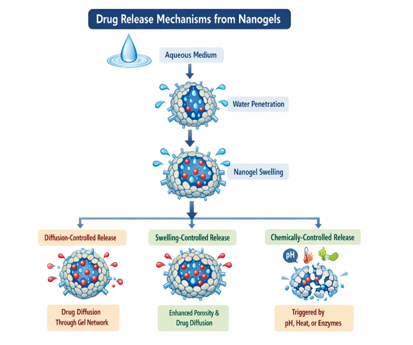
Fig no 3: Drug release mechanism of nanogel
NANOGELS FOR THE TRETMENT OF DIABETIC WOUND HEALING:
Recent studies have shown that diabetic wounds can be effectively treated with nanogel-based wound dressings. In the Luo et al. study, a nanogel dressing was developed to respond to changes in pH and enzyme levels in the diabetic wound environment. The nanogel was able to release therapeutic chemicals in a controlled manner at the wound site. This helped control inflammation and reduce bacterial infection. The study also discovered increased tissue regeneration and wound closure in diabetic lesions treated with the nanogel dressing. These findings suggest that by combining antibacterial activity with tissue repair support, nanogel systems can improve healing in the treatment of diabetic wounds(37).
Inadequate blood flow, infection risk, and delayed healing are the main objectives of effective diabetic wound care. Proper wound care includes maintaining a moist wound environment, preventing infections, and performing routine debridement. Advanced wound dressings have attracted attention because they facilitate faster tissue healing and reduce issues. Nanogel-based dressings are especially beneficial due to their high-water content, good biocompatibility, and controlled delivery of therapeutic drugs. These techniques allow for the direct delivery of antimicrobial, anti-inflammatory, and pro-healing substances to the wound site. By improving drug stability and local availability, nanogels decrease bacterial growth while promoting angiogenesis and tissue regeneration. Patients with diabetes benefit more from these innovative treatments than from conventional wound dressings.(38)
METHODS OF PREPARATION OF NANOGEL:
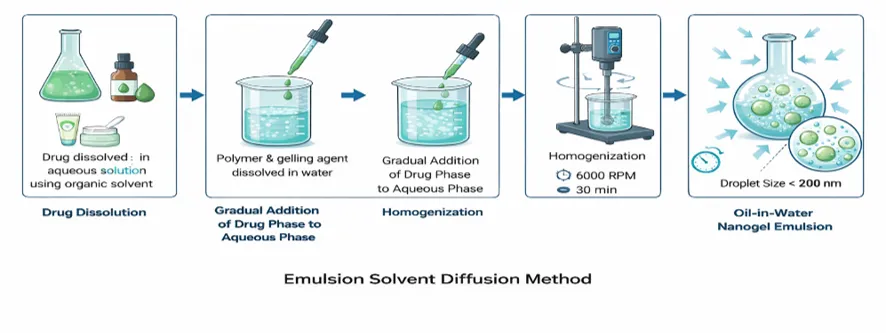
I. Emulsion Solvent Diffusion Method
The drug is dissolved in the aqueous solution using an organic solvent. A polymer and gelling agent are dissolved in water to create the drug phase. After that, it is dropwise added to the aqueous phase. This mixture is homogenized at 6000 rpm for 30 minutes to improve stability and solubility and create an oil-in-water emulsion with droplet sizes typically less than 200 nm(39). The drug is dissolved in the aqueous solution using an organic solvent. A polymer and gelling agent are dissolved in water to create the drug phase. After that, it is gradually added to the aqueous phase. To increase stability and solubility and produce an oil-in-water emulsion with droplet sizes usually less than 200 nm, this mixture is homogenized at 6000 rpm for 30 minutes(40).
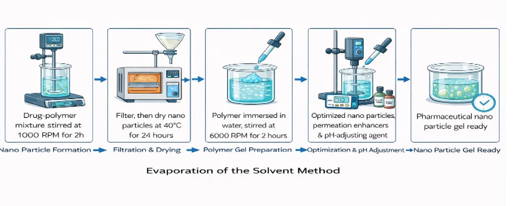
II. Evaporation of the Solvent Method
Initially, an aqueous phase is filled with a drug-polymer mixture, which is then continuously stirred at 1000 rpm for two hours(41). After filtering, the resultant nanogel particles are dried in a hot air oven at 40°C for a full day. For optimal dispersion, the polymer should be immersed in water for two hours before gel formation and then agitated at 6000 rpm(42). The optimized nanogel particle suspension and permeation enhancers are added to the aqueous dispersion after the mixture's pH has been adjusted using a pH-modifying agent(43).
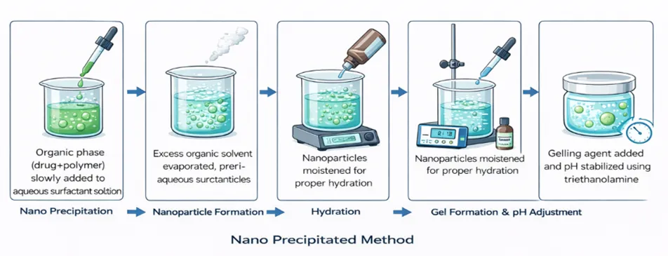
III. Nano Precipitated Method
In this method, the organic phase containing the drug and polymer is slowly added to an aqueous solution containing a surfactant. As the organic solvent diffuses into the aqueous phase, the polymer begins to precipitate, leading to the formation of nanoparticles. After complete removal of the organic solvent, the formed polymeric nanoparticles are collected for further use(44). After allowing the nanoparticles to hydrate properly, the gelling agent is added along with the required amount of nanoparticle dispersion. The pH of the formulation is then adjusted and maintained at the desired level using triethanolamine(45).
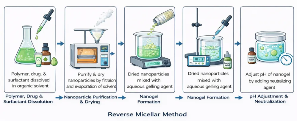
IV. Reverse Micellar Method
In the first step, the polymer, drug, and surfactant are dissolved together in a suitable organic solvent. A cross-linking agent is then added slowly, and the mixture is allowed to react over an extended period, typically overnight, to ensure proper cross-linking(46). The prepared nanoparticles are first purified and then subjected to solvent evaporation to obtain them in a solid state. These dried nanoparticles are subsequently dispersed in an aqueous gelling agent to form a nanogel. Finally, a neutralizing agent is added to adjust and maintain the pH of the formulation(47).
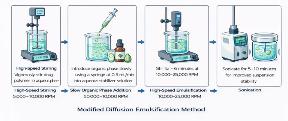
V. Modified Diffusion Emulsification method
The drug–polymer mixture is vigorously stirred in the aqueous phase at a high rotational speed ranging from 5,000 to 10,000 rpm, which helps in the formation and uniform dispersion of the organic phase(48). The organic phase is introduced slowly into the aqueous stabilizer solution using a syringe fitted with a needle at a controlled rate of 0.5 mL per minute. The mixture is then stirred for about six minutes at high speeds ranging from 10,000 to 25,000 rpm. To further improve the stability of the suspension, sonication is carried out for 5 to 10 minutes.
ADVANTAGES OF NANOGELS:
Nanogels are a great way to deliver medications because of their many advantages.
1. These include methods for preparing nanogels that are highly biocompatible,
biodegradable, and recyclable(49).
2. Nanogels can also provide more precise sustained drug release because they are created by putting together a polymer system that controls the preparation's particle size(50).
3. Both parenteral and mucosal administration of nanogels are simple(51).
4. Nanogels can carry both water-soluble and fat-soluble drugs(52).
5. Nanogels remain stable in the bloodstream and the body’s internal fluids and do not trigger unwanted immune reactions(53).
6. Nanogels can reach more areas in the body than hydrogels when given intravenously(54)
LIMITATIONS OF NANOGEL:
1. Nanogels have limitations because their preparation is complex, and complete removal of solvents or surfactants is difficult, which may cause safety concerns(55).
2. The use of nanogels is limited by high preparation costs and difficulties in large-scale production(56).
3. The application of nanogels in diabetic wound healing is limited by formulation complexity and concerns related to long-term stability and safety(57).
FUTURE SCOPE:
Future studies should aim to design multifunctional nanogels that can effectively manage infection, inflammation, and oxidative stress in diabetic wounds. Enhancing the stability, safety, and targeted delivery of nanogels will be important for their successful clinical use. In addition, well-planned clinical trials and standardized production methods are required to ensure consistent quality and cost-effectiveness. The development of smart and responsive nanogels may also allow personalized treatment approaches and improve therapeutic outcomes in chronic diabetic wound management(58).
Ongoing research on polymeric nanomedicines for diabetic wound healing should prioritize improved safety, formulation stability, and production scalability. Designing smart, stimuli-responsive nanogels that allow targeted and controlled drug release may further enhance treatment outcomes. In addition, well-designed clinical trials are necessary to confirm long-term efficacy, biocompatibility, and practical applicability in routine diabetic wound care(57).
Future research on nanomaterial-based wound dressings is expected to focus on developing safer, cost-effective, and multifunctional systems for diabetic wound care. Advanced nanogels and nanocomposites with improved biocompatibility, controlled drug release, and enhanced antimicrobial activity may significantly improve healing outcomes. Combining natural polymers, bioactive agents, and smart stimuli-responsive materials could help overcome current limitations and enable personalized wound management strategies for chronic diabetic wounds(59).
CONCLUSION:
Diabetic wound healing remains a major clinical challenge due to delayed tissue repair, chronic inflammation, and increased risk of infection. Conventional wound dressings often fail to provide effective healing in diabetic conditions. Nanogel-based delivery systems have emerged as a promising approach because of their ability to maintain a moist wound environment, provide controlled drug release, and enhance antimicrobial efficacy. Chlorhexidine gluconate plays a key role in controlling microbial load, while Aloe vera supports wound care through its anti-inflammatory and antimicrobial properties. The combination of these agents in a nanogel system offers improved safety, sustained action, and better patient compliance. Overall, nanogel-based formulations represent a potential advanced strategy for effective management of diabetic wounds.
REFERENCES

Sumanth N, Beny Baby, Pallavi A, Harish Gowda S M, Punith Gowda L, A Comprehensive Review on Chlorhexidine-Aloe Vera Nanogels for Antimicrobial Action and Tissue Regeneration in Wound Care, Int. J. of Pharm. Sci., 2026, Vol 4, Issue 9, 3646-3660. https://doi.org/10.5281/zenodo.23021544
 10.5281/zenodo.23021544