

We use cookies to ensure our website works properly and to personalise your experience. Cookies policy
1Department of Pharmaceutics, Bhagwan Mahavir College of Pharmacy, Surat, Gujarat, India.
2Head of Department of Pharmaceutics, Bhagwan Mahavir College of Pharmacy, Surat, Gujarat, India.
Being the largest organ of the human body, the skin is very appealing for the delivery of drugs both locally and systemically. However, the skin's outermost layer, the stratum corneum, serves as a very effective barrier that hampers the penetration of hydrophilic and high molecular weight drugs to a great extent. Traditional topical drug delivery systems usually have problems with drug diffusion across this barrier, leading to a low level of therapeutic effectiveness. To solve these problems, ethosomal drug delivery systems have been developed as a very useful vesicular carrier for both transdermal and topical drug delivery. Ethosomes are soft, flexible phospholipid vesicles with a high concentration of ethanol, which is responsible for a significant part of skin permeation enhancement. Ethanol breaks down the lipid layers of the stratum corneum, thus facilitating drug penetration, at the same time providing more flexibility to the vesicles. Thanks to their special makeup and form, ethosomes are able to hold both hydrophilic and lipophilic drugs with a higher drug loading efficiency than ordinary liposomes. Numerous research papers have proven the outstanding performance of ethosomal formulations. The effectiveness of therapeutic was enhanced by ethosomal acyclovir with significantly shortened relapsed herpes labialis lesions healing time compared to standard formulations like Zovirax. In the same manner, ethosomal minoxidil formulations have demonstrated a considerable increase in in vitro skin permeation compared to ethanolic, hydroethanolic, or phospholipid-based micellar systems. These merits make ethosomes a perfect fit for dermatological and transdermal applications including androgenic alopecia treatment.
Human??? skin selectively and very effectively serves as a barrier to chemical permeation; however, it is also considered a convenient and less invasive route for drug delivery due to the various advantages such as bypassing first-pass metabolism, less fluctuation of drug concentration in plasma, local drug targeting, and increased patient compliance. Among these benefits, facilitating the passage of water-soluble drugs through the skin is the main challenge because the skin acts as a natural barrier against hydrophilic molecules. The main culprit of this barrier effect is the uppermost layer of the epidermis, the stratum corneum, which contains tightly packed, insoluble keratin filaments surrounded by cross-linked proteins and lipids that are covalently bound1. The stratum corneum is the major barrier that controls the rate at which drugs pass through the skin. In fact, if the stratum corneum was not there, most small, polar and non-electrolyte molecules would get into the systemic circulation at a rate which is several thousand times higher. Therefore, numerous approaches have been devised to limit the resistance of this barrier and increase drug flux through the skin2. Various technologies have been introduced in recent decades, such as physical methods like iontophoresis, sonophoresis, and microneedles, chemical methods involving penetration enhancers, and biochemical methods like vesicular carriers. Even though physical enhancement techniques can facilitate the penetration of drugs through the skin, due to their complexity and sometimes invasive nature, patient compliance may be reduced. Likewise, chemical penetration enhancers such as surfactants and organic solvents may be responsible for skin irritation, harm the barrier integrity, and give rise to long-term skin deterioration3. Hence, drug delivery systems capable of facilitating drug permeation and at the same time, maintaining skin barrier function are the ones most sought after. Vesicular systems have attracted attention as an alternative to this situation. Of these, liposomal preparations have attracted a lot of attention, with deformable liposomes and transferosomes being the first generation of elastic vesicles able to penetrate the skin barrier and deliver therapeutic drug levels in drug non-occlusive ???conditions4.
Ethosomes
first developed ethosomes as a new class of lipid vesicular systems, which are composed mainly of ethanol, phospholipids, and water. Numerous reports have demonstrated, that ethosomes increase the transdermal delivery of many types of therapeutic agents. Ethanol is present in high concentrations and is a good permeation enhancer for drugs by disrupting the intercellular lipid structure of the stratum corneum, allowing drugs to move through the skin barrier5. In terms of structure, ethosomes are considered soft and pliable vesicles with an aqueous core containing an ethanolic drug solution surrounded by a phospholipid bilayer. Ethosomes are a modified version of conventional liposomes, which limited their effectiveness for transdermal drug delivery due to insufficient skin permeability and stability issues6. Niosomes were designed to address the stability problems associated with liposomes, but they also did not provide a significant increase in the ability of drugs to penetrate the skin. As a result, ethanolic vesicular systems, such as Ethosomes, have been developed to improve drug penetration through the skin7.
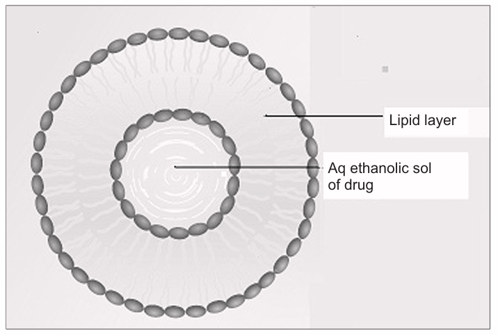
Figure 1: Proposed diagram of Ethosomes
Ethanol influences the formation of highly deformable vesicles, enabling drug application to higher epidermal layers of the skin via ethosome technology8. Ethosomes can range in size from tens of nanometers to many micrometers depending on the application requirements of the formulation. Due to their non-invasiveness, ethosomes readily facilitate the delivery of drugs through both deep dermal and systemic circulation9. Ethanol also acts as a solvent, creating flexible vesicular membranes that allow the ethosomes membranes to exhibit a more loosely packed structure; thereby enhancing the fluidity of the bilayers within the stratum corneum and allowing for better penetration of the drug into the dermal layer of the stratum corneum compared to traditional vesicular formulations. Ethosomes thus represent a new approach for the delivery of transdermal therapeutics10.
1.1.2 Type of Ethosome
On the basis of components used in the formulations, ethosomes are of two types
A) Classical ethosomes
Ethosomes are modified liposomes that contain a phospholipid-water phase and a very high percentage of ethanol (20–45% w/w). Ethosome structures also may contain additional forms of low molecular weight (molecular weight < 200g), such as PG or IPA, as well as other forms of low molecular weight alcohol11.
B) Transethosomes
The Ethosomes have been modified to enhance the delivery of drugs into the skin through the use of penetration enhancers (also known as surfactants), which make up the core of Transethosomes12. Transethosomes are a new generation of ethosomal delivery systems that retain all of the advantages offered by traditional ethosomal and transferosomal delivery systems. Before these products can be used, however, additional studies will be necessary to demonstrate their overall safety to patients because of the potential risk of skin irritation associated with combining surfactants and high levels of alcohol13.
1.1.3 Advantage of Ethosome
The skin can be penetrated readily by ethosomes, thereby allowing drugs to exert their effects topically or via transdermal means. Ethosomes can be utilized to facilitate the delivery of a variety of therapeutic agents including both small molecules and macromolecules (peptides and proteins).
Ethosomes have greater efficiency than conventional methods for delivering fluorescence probes (quantum dots) deeper into the skin while providing a greater amount of fluorescence14.
Furthermore, due to the relatively low toxicity of all components of an ethosomal system (phospholipids, ethanol, and water), there is less risk associated with drug development using ethosomal systems based upon previously available scientific information. Most ethosomal drug formulations are comprised of semisolid dosage forms (gels or creams) that are easy to apply and highly acceptable to patients15. Conversely, enhancing methods (e.g. iontophoresis, phonophoresis) tend to be much more complicated than ethosomes and therefore lead to decreased patient compliance.
Due to their proprietary technology potential, ethosomal products have significant market potential. Additionally, ethosomes have the advantage of being easily manufactured without requiring expensive and/or complicated technical infrastructure16.
2. Method of Preparation
2.1 Hot method
The drug is dissolved in a combination of ethanol and propylene glycol, which is then added to an aqueous dispersion of phospholipid kept at 40° C. After this, the mixture is sonicated for three cycles of five minutes each at 4° C with a five-minute pause in between using a sonicator, keeping it under 4° C in a refrigerated sonicator17. This ethosome dispersion is then further homogenized at 15,000 psi for three cycles using a high-pressure homogenizer in order to form nano-sized ethosomes18.
2.2 Cold method
One of the most popular methods for making ethosomes involves creating a solution of Phospholipids and your drug combined with other lipid ingredients mixed with Ethanol at room temperature. Continuous vigorous stirring will allow you to keep the mixture in solution while heating the ethanol solution using a Water Bath at 30 degrees Celsius19. Once you have reached this temperature, the Water should then be heated to 30 degrees Celsius and once the Water has reached 30 degrees Celsius add the heated water to the heated Ethanol solution and continue to stir the combined solutions in a closed container for an additional 5 minutes. If you wish to reduce the size of the Ethosomal Vesicles in the Ethosomal Formulation, you may do this by either Sonication or Extrusion. Ethosomal Formulations should be stored immediately after preparation at a refrigerated temperature20.
2.3 Classic Mechanical Dispersion Method
A round-bottomed glass vessel containing a mixture of chloroform and methanol 3:1 (volumetric) containing soya phosphatidylcholine is placed into a vacuum rotary evaporator, with the rotary evaporator maintained above the lipid's phase transition temperature. The evaporator removes all organic solvent residue, forming a thin layer of lipid on the inner surface of the vessel21. The vessel is then kept in a vacuum overnight to remove any remaining traces of organic solvent residue from the vessel walls. The thin lipid layer formed in the flask is usually formed by hydration with a variety of hydroethanol solutions of different concentrations containing the drug while rotating the flask at an appropriate temperature (generally above the drug's phase transition temperature)22.
2.4 Classic method23
Ethanol is used to dissolve the drug and phospholipid. The mixture is then placed in a water bath where it will be maintained at 30±1°C. Double distilled water is next added to the phospholipid, with stirring in a closed vessel at 700 rpm. The mixture is made into vesicles by extruding them through a polycarbonate membrane using a manual extruder for three cycles.
3. Characterization of Ethosome
3.1 Visualization of the vesicles24
To study the morphology of ethosomes, both transmission electron microscopy (TEM) and scanning electron microscopy (SEM) techniques can be utilized.
3.2 vesicle size and zeta potential25
The size and zeta potential of ethosomes can be determined by using dynamic light scattering (DLS) and photon correlation spectroscopy (pCS) methods.
3.3 Entrapment efficiency26
To measure the entrapment efficiency of ethosomes, an ultra-centrifugation technique can be employed. Following centrifugation of the formulation, the amount of unencapsulated drug that remains in the supernatant can be determined by using appropriate analytical techniques.
3.4 Transition Temperature27
The transition temperature (or melting point), enthalpy, and melting point of ethosomes are all determined through a technique called differential scanning calorimetry (DSC)
3.5 Vesicle Stability28
The stability of ethosome vesicles can be measured based upon what changes occur to the size and shape of the vesicle over an extended period of time by using both dynamic lights scattering (DLS) and transmission electron microscopy (TEM).
3.6 Penetration and permeation studies
Through the use of a confocal laser scanning microscope, one can observe how deeply into the skin ethosome penetrated after topical administration29. This can be accomplished by applying an ethosome that has been preloaded with a fluorescent dye onto the surface of an animal's skin, and measuring how far the dye moved into the skin after a specified period of time30.
4. Mechanism of ethosomes 30
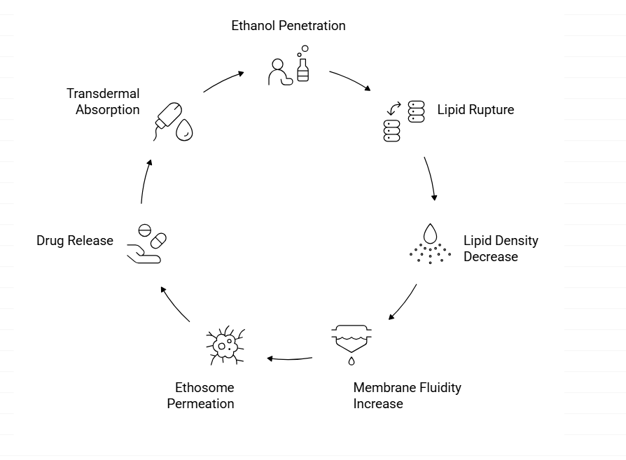
Fig: Mechanism of Ethosome
5. Therapeutic Applications of Ethosomes
5.1 Transcutaneous Immunization (Hepatitis B)
Ethosomes have been utilized in the field of transcutaneous immunization (TCI) for the delivery of Hepatitis B surface Antigen (HBsAg). The results obtained from studies using Oesophageal hborbic Tape and cabs MDC-MDiv of Mabs (EHBCT) indicate that antigen-loaded ethosomes had greater entrapment efficiency, vesicle size compared to conventional liposomes and unilamellar spherical morphology versus conventional liposomes. In vitro, spectral bioimaging and flow cytometry confirmed that HBsAg-loaded ethosomes were efficiently taken up by murine DCs after 180 minutes31. Cadaver skin permeation studies demonstrated that HBsAg-loaded ethosomes were able to penetrate cadaver skin significantly better than both conventional liposome and soluble antigen formulations32. In vivo, HBsAg-loaded ethosomes elicited a robust systemic and mucosal humoral response in mice following topical administration as compared to equivalent doses of intramuscularly administered alum-adjuvanted HBsAg, plain HBsAg solution and hydroethanolic HBsAg solution. Therefore, HBsAg-loaded ethosomes represent a viable platform with the potential to elicit a protective immune response, and may be useful as a basis for developing transcutaneous Hepatitis B vaccines33.
5.2 Transdermal Delivery of Hormones (Testosterone):
Hormonal administration via the oral route can be limited in effectiveness due to a variety of problems, including extensive first-pass metabolism, poor bioavailability associated with absorption through the digestive tract, side effects based on the amount taken (dose), and poor patient compliance with oral hormone replacement therapy34. Touitou et al. (2000) compared the amount of testosterone absorbed into the skin using ethosomes (Testosome) containing testosterone versus a commercially available testosterone transdermal patch (Testoderm®). The formulation containing ethosomes had approximately 30 times the amount absorbed into the skin compared to the commercial patch35. Pharmacokinetic data indicated the ethosome formulation produced significantly higher AUC and Cmax levels than did the patch formulation36. A follow-up study developed an ethosome formulation without the need for a patch application, which required an area ten times smaller than that of a commercial gel application (Andro Gel®) to obtain equivalent therapeutic concentrations of testosterone in plasma37.
5.3 Cannabidiol (CBD) for Rheumatoid Arthritis:
The researchers developed an ethosomal formulation containing CBD for transdermal drug delivery in treating RA. Two study designs measured drug deposition into the skin and underlying muscle via topical application using a mouse model; results demonstrated significant CBD accumulation in both tissues38. Skin permeability studies demonstrated that the CBD-loaded ethosome created a pharmacokinetic profile similar to other transdermal drug-delivery systems. Pharmacokinetic data indicated that after application of the CBD-loaded ethosome, plasma CBD concentrations were relatively constant (i.e., at steady-state) for 72 hours. Animal models (carrageenan-induced rat paw edema) exhibited enhanced anti-inflammatory effects after topical application of the CBD-loaded ethosomes, and it was concluded that CBD entrapment in ethosomes greatly improved skin penetration, tissue accumulation, and therapeutic effectiveness39.
5.4 Trihexyphenidyl Hydrochloride (THP) for Parkinson’s Disease:
In 2001, Dayan and Touitou produced an ethosomal version of trihexyphenidyl hydrochloride (THP), which is an M1 muscarinic receptor blocker indicated for the treatment of Parkinson’s disease due to its short half-life and its inability to be effectively administrated orally. Electron microscopy has confirmed the existence of permit-size phospholipid vesicles for THP in an ethosome nearby mouse dermis40. The transdermal delivery capacities of THP with ethosomes were determined to be 87-, 51-, and 4.5 times greater than the rate of THP in liposomes, phosphate buffered solution (PBS), or hydroethanol collars, respectively. In addition, after applying ethosomes, THP was retained at higher rates in the dermal tissue, confirming the advantage of ethosomes as a delivery method for THP in treatment of Parkinson's disease41.
5.5 Methotrexate for Psoriasis and Cancer Therapy:
Dubey et al. (2007) did an investigation into the properties of an ethosome formulation of methotrexate (MTX), a highly hydrophilic compound with poor transdermal permeability. Methotrexate loaded ethosomes created a more effective vehicle for deep delivery than soyate loaded ethosomes produced by other authors42. The ability to maintain this "penetration-enhancement" characteristic was confirmed by storage stability tests. The "Penetration-Enhancement" effects of ethosomes have been demonstrated in the past by Vesicle-Skin Interaction Studies, which have shown corneocyte swelling and unique pathways of penetration into the skin as well as in other areas43.
6. FUTURE PERSPECTIVE
Ethosomes are innovative vesicle-based systems with a high content of ethanol and phospholipids, making them ideal for delivering drugs through the skin. Compared to outmoded liposomes, ethosomes can reach deeper into the skin and provide both hydrophilic and lipophilic drug penetration; they have also received significant interest over the years for enhancing drug delivery via transdermal & transcutaneous methods. In the future, additional studies of ethnosometry are likely to be focused on the increasing the stability, scale, and reproducibility of formulators systems in order to aid in the establishment of commercial capabilities and the transfer of therapeutic drugs from clinical development to normal practice. By optimising the composition and processing parameters, i.e. Ethos-trans-dermal delivery systems, we can eliminate many of the hurdles currently inhibiting the sale of these products. In dermal treatments, the potential for site-specific targeting of common medications used to treat chronic skin conditions and hair loss is very promising. To achieve this effect, matures like Minoxidil, Finasteride, Dutasteride and other herbal actives may benefit tremendously from Ethosmotic Delivery by providing a higher local efficacy and fewer side effects from the treatment. In addition, a variety of emerging therapies are targeting Ethosome systems, particularly in the areas of Non-invasive immunization, Transcutaneous Immunization, Peptide/Protein Delivery via Ethosomes and Gene Therapy for Inflammatory and Neurological Disorders. Ethosome delivery systems permit efficient penetration of the dermal barrier, thereby allowing contact with immune system cells in order to create a suitable route through which future immunizations could be administered.
CONCLUSION
Ethosomes are high-performance vesicular carriers of drugs that can be absorbed through the skin and applied using conventional methods. Ethosomes differ from standard drug delivery systems in that they have a special formulation comprised of high levels of ethanol and phospholipid bilayers. The distinctive formulation of ethosomes allows them to interact with the oils and fats of the skin and, therefore, provide increased absorption of the active agent into the skin, as well as enhanced bioavailability and deeper penetration into the skin. Research over the past few years has demonstrated the successful use of ethosomes in various treatment areas, including transcutaneous vaccination, hormone replacement therapy, inflammatory and neurological diseases, and dermatological problems. It has consistently been demonstrated that ethosomes can provide superior drug entrapment, improved skin retention, sustained release, and more successful results than traditional liposomes or topical formulations. Further, ethosomes are non-invasive and easy to use, leading to greater compliance by patients in following their treatment regimen. Even though there are many encouraging results with ethosomes, more research is needed to assess the long-term stability of ethosomes, to develop a preparatory method for manufacturing ethosomes on a large scale, to validate the results from the studies, and finally, to achieve regulatory approval for the commercial sale of ethosomal products. With continued improvement in formulation techniques and advancements in nano-technology, ethosomes will likely be a major contributor to the development of safe, effective, and easy-to-use topical and transdermal delivery systems for drugs.
Compliance with ethical standards
Acknowledgments
A special thanks to Dr. Ronak Dedania for her endless support and guidance provided
during the completion of this review article.
Funding
This research did not receive any grants from funding agencies in the public or private sectors.
Disclosure of Conflict of Interest
No conflict of interest to be declared.
REFERENCE:

Palak Upadhyay, Dr. Ronak Dedaia, Vikas Maurya, “A Critical Review: Recent Advances in Ethosomal Gel for Transdermal Drug Delivery Systems’’, Int. J. of Pharm. Sci., 2026, Vol 4, Issue 1, 2425-2434. https://doi.org/10.5281/zenodo.18344838
 10.5281/zenodo.18344838