View Article

  • Moringa oleifera-Based Nanogels for Topical Management of Inflammatory Disorders: Phytochemicals, Formulation Strategies, Molecular Mechanisms and Translational Perspectives

  • Pharmacology Division, AU College of Pharmaceutical Sciences, Andhra University, Visakhapatnam, Andhra Pradesh

Abstract

Chronic inflammatory conditions like psoriasis, eczema, rheumatoid arthritis, and slow healing wounds affect millions worldwide, often leaving patients with pain, discomfort, and reduced quality of life. Current treatments such as corticosteroids and biologics can help, but they come with drawbacks from skin thinning and irritation to high costs and limited accessibility. This has sparked growing interest in safer, plant based alternatives. Moringa oleifera, often called the “miracle tree,” is packed with powerful natural compounds — flavonoids, phenolic acids, and isothiocyanates that fight oxidative stress, calm inflammation, and support tissue repair. The challenge, however, lies in delivering these delicate phytochemicals effectively through the skin, since they are often unstable, poorly soluble, and blocked by the skin’s protective barrier. Nanogels, tiny water rich polymer networks, offer a smart solution. They can encapsulate Moringa’s bioactives, protect them from degradation, and release them in a controlled way right where inflammation occurs. This review brings together the science behind Moringa’s phytochemistry, its molecular anti inflammatory mechanisms, and the latest advances in nanogel formulations. By bridging traditional herbal wisdom with modern nanotechnology, Moringa based nanogels show real promise as next generation topical therapies for chronic inflammatory disorders — safer, more effective, and patient friendly.

Keywords

Moringa oleifera, Nanogels, Phytochemicals, Chronic inflammation, Topical drug delivery, Psoriasis, Rheumatoid Arthritis, Antioxidant therapy, Herbal nanomedicine.

Introduction

× Popup Image

Chronic inflammatory disorders such as psoriasis, atopic dermatitis, rheumatoid arthritis, and chronic wounds represent a major global health burden, affecting millions of individuals and significantly impairing quality of life. Epidemiological studies estimate that psoriasis alone affects 2–3% of the world’s population, while atopic dermatitis prevalence ranges from 10–20% in children and 1–3% in adults worldwide(1,2). These conditions are characterized by persistent immune dysregulation, excessive production of pro-inflammatory cytokines (e.g., TNF-α, IL-6, IL-17), and activation of signalling pathways such as NF-κB and MAPK, leading to chronic tissue damage and impaired barrier function(3,4). Conventional topical therapies, including corticosteroids, calcineurin inhibitors, and biologics, though effective in symptom control, are associated with significant limitations. Long-term corticosteroid use often results in skin atrophy, telangiectasia, and tachyphylaxis, while biologics are costly and require parenteral administration (5,6). Moreover, many synthetic anti-inflammatory drugs suffer from poor skin penetration, instability, and systemic side effects, underscoring the need for safer, more effective alternatives.

Herbal phytochemicals have emerged as promising candidates due to their antioxidant, anti-inflammatory, and immunomodulatory properties. Moringa oleifera, a widely cultivated medicinal plant, is particularly notable for its rich phytochemical profile, including flavonoids (quercetin, kaempferol), phenolic acids, glucosinolates, and isothiocyanates, which exhibit potent anti-inflammatory and wound-healing activities (7,8). However, the clinical translation of these phytochemicals is hindered by poor solubility, instability, and limited permeability across the stratum corneum.

Nanogels crosslinked, hydrophilic polymeric networks at the nanoscale offer a novel solution to these challenges. Their high-water content, tunable porosity, and biocompatibility enable efficient encapsulation of phytochemicals, controlled release, and enhanced skin penetration (9,10). Stimuli-responsive nanogels (pH, temperature, ROS-sensitive) further allow site-specific drug delivery, minimizing systemic exposure and maximizing therapeutic efficacy. Thus, integrating Moringa oleifera phytochemicals into nanogel systems represents a rational and innovative approach for the topical management of chronic inflammatory disorders.

This review aims to comprehensively discuss the phytochemistry of Moringa oleifera, the molecular mechanisms underlying its anti-inflammatory activity, formulation strategies for nanogels, and translational perspectives in managing chronic inflammatory conditions. By synthesizing current evidence, we highlight the potential of Moringa-based nanogels as next-generation phytopharmaceuticals for topical therapy.
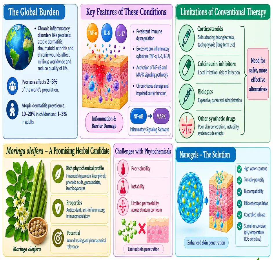
Figure No.1: Schematic overview of the rationale for Moringa oleifera–loaded nanogels in the topical management of chronic inflammatory disorders.

2. Moringa oleifera: Phytochemistry and Therapeutic Potential

Moringa oleifera Lam. (family Moringaceae), commonly known as the “drumstick tree” or “miracle tree,” is native to South Asia but widely cultivated across tropical and subtropical regions due to its nutritional and medicinal value (11). Its leaves, seeds, pods, flowers, and roots contain a diverse array of bioactive phytochemicals that contribute to its antioxidant, anti-inflammatory, and pharmaceutical relevance.

2.1 Major Phytochemicals: -The phytochemical profile of Moringa oleifera is extensive, encompassing:

  • Flavonoids: Quercetin, kaempferol, rutin (potent free radical scavengers and modulators of inflammatory signaling)  (12).
  • Phenolic acids: Chlorogenic acid, gallic acid, ferulic acid (contribute to antioxidant defense and inhibition of lipid peroxidation)(13).
  • Glucosinolates & isothiocyanates: Benzyl isothiocyanate, niazimicin (known for anti-inflammatory, anticancer, and antimicrobial properties) (14).
  • Alkaloids & saponins: Provide immunomodulatory and hepatoprotective effects (15).
  • Vitamins & micronutrients: High levels of vitamin C, β-carotene, calcium, and iron enhance its nutraceutical potential (16).

Table 1. Key Phytochemicals in Moringa oleifera and Their Biological Activities

Phytochemical

Class

Biological Activity

Pharmaceutical Relevance

Quercetin, Kaempferol

Flavonoids

Antioxidant, NF-κB inhibition

Anti-inflammatory drugs

Chlorogenic acid

Phenolic acid

Free radical scavenging, lipid regulation

Cardiovascular health

Benzyl isothiocyanate

Isothiocyanate

COX-2 inhibition, anticancer

Anti-inflammatory, oncology

Niazimicin

Glucosinolate

Antimicrobial, apoptosis induction

Antimicrobial therapy

2.2 Antioxidant and Anti-inflammatory Properties: -The antioxidant potential of Moringa oleifera is attributed to its high polyphenolic content, which enhances endogenous defense systems such as superoxide dismutase (SOD), catalase, and glutathione peroxidase (17). These compounds mitigate oxidative stress, a key driver of chronic inflammation and tissue damage.

Anti-inflammatory effects are mediated through:

  1. Suppression of NF-κB signaling, reducing TNF-α, IL-1β, and IL-6 production (18).
  2. Inhibition of COX-2 and iNOS enzymes, lowering prostaglandin and nitric oxide synthesis (19).
  3. Modulation of MAPK and Nrf2 pathways, balancing pro- and anti-inflammatory responses (20).

These mechanisms collectively position Moringa oleifera as a promising candidate for managing inflammatory disorders such as arthritis, dermatitis, and metabolic syndrome.

2.3 Pharmaceutical Relevance: -The pharmaceutical potential of Moringa oleifera lies in its ability to serve as a source of phytopharmaceuticals for novel drug delivery systems. Its bioactive compounds have demonstrated efficacy in preclinical models of:

  1. Psoriasis and dermatitis: Reduction of epidermal hyperplasia and cytokine overexpression (21).
  2. Arthritis: Attenuation of joint inflammation and oxidative stress (22).
  3. Wound healing: Acceleration of collagen deposition and angiogenesis (23).

However, challenges such as poor solubility, instability, and limited skin permeability necessitate advanced delivery strategies. Nanogels, with their ability to encapsulate and protect phytochemicals while enhancing bioavailability, represent a rational approach to harnessing Moringa oleifera’s therapeutic potential in topical applications.

3. Chronic Inflammation and Molecular Targets

Chronic inflammation is a sustained, dysregulated immune response that underlies a wide spectrum of disorders, including psoriasis, rheumatoid arthritis, atopic dermatitis, and metabolic syndrome. Unlike acute inflammation, which is protective and self-limiting, chronic inflammation persists due to continuous activation of immune pathways, leading to tissue damage, fibrosis, and impaired healing (24).

3.1 Key Inflammatory Mediators

  • Cytokines: - Pro-inflammatory cytokines such as tumor necrosis factor-alpha (TNF-α), interleukin-1 beta (IL-1β), IL-6, and IL-17 play central roles in perpetuating inflammation. They stimulate keratinocyte proliferation, synovial hyperplasia, and recruitment of immune cells (25).
  • Chemokines: - Molecules like CXCL8 (IL-8) and CCL2 drive leukocyte migration, amplifying the inflammatory cascade (26).
  • Reactive oxygen species (ROS):- Excessive ROS production contributes to oxidative stress, DNA damage, and activation of redox-sensitive transcription factors (27).

3.2 Molecular Pathways

NF-κB Pathway: The nuclear factor kappa-light-chain-enhancer of activated B cells (NF-κB) pathway is a master regulator of inflammation. Activation by cytokines, Toll-like receptors, or oxidative stress leads to nuclear translocation of NF-κB, promoting transcription of TNF-α, IL-6, COX-2, and iNOS (28). Persistent NF-κB activation is a hallmark of chronic inflammatory diseases.

MAPK Pathway: Mitogen-activated protein kinases (MAPKs), including ERK, JNK, and p38, regulate cellular responses to stress and cytokines. MAPK signaling enhances production of inflammatory mediators and contributes to keratinocyte hyperproliferation in psoriasis and synovial inflammation in arthritis (29).

Nrf2 Pathway: Nuclear factor erythroid 2–related factor 2 (Nrf2) is a transcription factor that counterbalances inflammation by upregulating antioxidant enzymes such as heme oxygenase-1 (HO-1) and glutathione peroxidase. Dysregulation of Nrf2 signaling reduces cellular resilience to oxidative stress, exacerbating chronic inflammation (30).

COX-2 and iNOS: Cyclooxygenase-2 (COX-2) and inducible nitric oxide synthase (iNOS) are key enzymes induced during inflammation. COX-2 catalyzes prostaglandin synthesis, driving pain and swelling, while iNOS generates nitric oxide, contributing to vasodilation and tissue injury (31).

3.3 Therapeutic Implications

Targeting these pathways offers multiple therapeutic opportunities. Conventional drugs such as corticosteroids and biologics act by suppressing NF-κB or cytokine signaling, but their limitations necessitate safer alternatives. Phytochemicals from Moringa oleifera including quercetin, kaempferol, and benzyl isothiocyanate have demonstrated the ability to modulate NF-κB, MAPK, and Nrf2 pathways, suggesting their potential as natural anti-inflammatory agents when delivered through advanced nanogel systems (32,33).

4. Challenges in Topical Delivery of Herbal Phytochemicals

Despite the promising pharmacological potential of herbal phytochemicals, their translation into effective topical formulations faces several critical challenges. These limitations hinder therapeutic efficacy and restrict clinical application, necessitating innovative drug delivery strategies such as nanogels.

4.1 Poor Solubility: Many phytochemicals, including flavonoids (quercetin, kaempferol) and phenolic acids, exhibit poor aqueous solubility, which reduces their ability to diffuse across the hydrophilic layers of the skin (34). Insolubility leads to low drug loading capacity in conventional creams and gels, limiting therapeutic concentrations at the target site.

4.2 Instability: Herbal compounds are prone to degradation due to environmental factors such as light, oxygen, and temperature. For example, polyphenols undergo oxidative degradation, while isothiocyanates are chemically unstable in aqueous environments (35). This instability reduces shelf life and compromises pharmacological activity.

4.3 Skin-Barrier Limitations: The stratum corneum, the outermost layer of the epidermis, is a formidable barrier to drug penetration. Its lipid-rich structure restricts the passage of hydrophilic and high-molecular-weight phytochemicals (36). Consequently, many herbal compounds fail to reach therapeutic concentrations in deeper dermal layers.

4.4 Low Permeability: Even when phytochemicals are stable and soluble, their permeability across the skin remains limited. Factors such as molecular size, polarity, and lipophilicity influence transdermal absorption. For instance, hydrophilic antioxidants like vitamin C exhibit poor penetration, reducing their clinical utility in topical formulations (37).

4.5 Bioavailability Issues: Poor solubility, instability, and restricted permeability collectively result in low bioavailability of herbal phytochemicals when applied topically. This translates into suboptimal therapeutic outcomes, requiring frequent application or higher doses, which may increase the risk of irritation or sensitization (38).

4.6 Implications for Nanogel-Based Delivery: Nanogels offer a rational solution to these challenges by:

  1. Enhancing solubility through encapsulation in hydrophilic polymeric networks.
  2. Protecting phytochemicals from degradation via crosslinked structures.
  3. Facilitating penetration across the stratum corneum through nanoscale size and stimuli-responsive release.
  4. Improving bioavailability by sustaining drug release and maintaining therapeutic concentrations.

Thus, nanogel systems represent a promising platform to overcome the inherent limitations of herbal phytochemicals in topical therapy.

5. Nanogels for Topical Drug Delivery

Nanogels are nanoscale, crosslinked hydrogel particles composed of hydrophilic polymer networks that can encapsulate bioactive compounds and deliver them in a controlled manner. Their unique physicochemical properties — high water content, tunable porosity, biocompatibility, and stimuli-responsiveness — make them particularly suitable for topical drug delivery of herbal phytochemicals, including those from Moringa oleifera (39).

5.1 Concept:

Nanogels are three-dimensional polymeric structures at the nanometer scale that swell in aqueous environments. They act as carriers for hydrophilic and hydrophobic drugs, protecting them from degradation and enabling sustained release. Their nanoscale size allows for enhanced penetration through the stratum corneum and accumulation in deeper dermal layers (40).

5.2 Types of Nanogels

Polymeric nanogels: Based on natural (chitosan, alginate, gelatin) or synthetic polymers (polyacrylamide, PEG).

  1. Stimuli-responsive nanogels: Respond to pH, temperature, redox state, or reactive oxygen species, enabling site-specific drug release.
  2. Hybrid nanogels: Incorporate inorganic nanoparticles (e.g., silica, gold) for multifunctional delivery and imaging.
  3. Lipid-polymer nanogels: Combine lipid vesicles with polymeric networks for improved stability and permeability (41).

5.3 Composition: Typical nanogels consist of:

  1. Polymeric backbone: Provides structural integrity (e.g., chitosan, PEG, polyvinyl alcohol).
  2. Crosslinking agents: Chemical or physical crosslinkers that stabilize the network.
  3. Encapsulated bioactives: Herbal phytochemicals such as quercetin, kaempferol, or benzyl isothiocyanate.
  4. Surface modifiers: PEGylation or ligand conjugation to enhance biocompatibility and targeting (42).

5.4 Advantages in Topical Delivery

  1. Enhanced solubility: Encapsulation improves aqueous solubility of poorly soluble phytochemicals.
  2. Protection from degradation: Nanogels shield sensitive compounds from oxidation, hydrolysis, and photodegradation.
  3. Improved skin penetration: Nanoscale size facilitates passage through the stratum corneum.
  4. Controlled release: Sustained and stimuli-responsive release maintains therapeutic concentrations.
  5. Reduced irritation: Lower doses and targeted delivery minimize adverse effects compared to conventional formulations (43).

5.5 Mechanisms of Skin Penetration: Nanogels penetrate the skin via multiple mechanisms:

  1. Intercellular route: Diffusion through lipid layers of the stratum corneum.
  2. Transcellular route: Passage across keratinocytes.
  3. Appendageal route: Entry through hair follicles and sweat glands. Their nanoscale size and hydrophilic nature enhance retention in epidermal and dermal layers, ensuring localized therapeutic action (44).

5.6 Controlled Release: Controlled release from nanogels is achieved through:

  1. Diffusion-controlled release: Gradual diffusion of encapsulated phytochemicals.
  2. Swelling-controlled release: Expansion of the polymeric network in response to hydration.
  3. Stimuli-responsive release: Triggered by pH changes, oxidative stress, or temperature variations in inflamed tissues (45).

Table 2. Comparison of Conventional Topical Formulations vs. Nanogels

Parameter

Conventional Formulations

Nanogels

Solubility

Limited for hydrophobic drugs

Enhanced via encapsulation

Stability

Prone to degradation

Protected by polymeric network

Skin penetration

Restricted by stratum corneum

Improved via nanoscale size

Release profile

Burst release

Sustained, controlled release

Bioavailability

Low

High, localized

Patient compliance

Moderate (frequent dosing)

High (reduced dosing frequency)

This controlled release ensures sustained therapeutic levels, reduces dosing frequency, and enhances patient compliance.

6. Moringa oleifera-Based Nanocarriers and Nanogels

The therapeutic potential of Moringa oleifera phytochemicals has prompted the development of advanced nanocarrier systems to overcome solubility, stability, and permeability challenges. Several nanocarrier platforms including liposomes, niosomes, nano-emulsions, lipid/polymeric nanoparticles, and nanogels have been explored to enhance topical delivery of Moringa-derived bioactives.

6.1 Liposomes: Liposomes are phospholipid bilayer vesicles capable of encapsulating both hydrophilic and lipophilic compounds. Moringa oleifera flavonoids such as quercetin and kaempferol have been successfully incorporated into liposomal systems, improving their stability and dermal penetration (46). Liposomes also provide controlled release and reduce irritation compared to conventional formulations.

6.2 Niosomes: Niosomes are non-ionic surfactant-based vesicles that offer improved stability over liposomes. They have been used to encapsulate Moringa polyphenols, enhancing antioxidant activity and skin retention (47). Their cost-effectiveness and ease of preparation make them attractive for herbal drug delivery.

6.3 Nanoemulsions: Nanoemulsions are kinetically stable dispersions of oil and water stabilized by surfactants. Moringa oleifera seed oil, rich in oleic acid and tocopherols, has been formulated into nanoemulsions for topical use, demonstrating enhanced hydration, antioxidant activity, and skin penetration (48). Nanoemulsions also improve sensory properties, making them suitable for cosmeceutical applications.

6.4 Lipid and Polymeric Nanoparticles: Solid lipid nanoparticles (SLNs) and polymeric nanoparticles provide a rigid matrix for encapsulating phytochemicals. Studies have shown that Moringa extracts loaded into SLNs exhibit improved stability, sustained release, and enhanced anti-inflammatory activity (49). Polymeric nanoparticles based on chitosan or carbopol further improve mucoadhesion and dermal retention.

6.5 Nanogels: Nanogels represent the most versatile platform for Moringa oleifera phytochemicals. Their hydrophilic polymeric networks encapsulate flavonoids and isothiocyanates, protecting them from degradation while enabling controlled release. Chitosan-based nanogels loaded with Moringa extracts have demonstrated superior antioxidant and anti-inflammatory effects in preclinical models (50). Stimuli-responsive nanogels further allow site-specific release in inflamed tissues, maximizing therapeutic efficacy.

6.6 Available Moringa-Based Evidence: Emerging studies highlight the potential of Moringa-based nanocarriers:

  • Liposomal quercetin from Moringa leaves showed enhanced anti-inflammatory activity in skin models (51).
  • Nano-emulsions containing Moringa seed oil improved hydration and reduced oxidative stress in dermatological applications (52).
  • Chitosan nanogels encapsulating Moringa flavonoids demonstrated sustained release and superior inhibition of pro-inflammatory cytokines(53).

Table 3. Moringa oleifera-Based Nanocarriers for Topical Delivery

Nanocarrier Type

Encapsulated Compound(s)

Key Advantages

Evidence/Applications

Liposomes

Quercetin, Kaempferol

Stability, dermal penetration

Enhanced anti-inflammatory activity (46)

Niosomes

Polyphenols

Cost-effective, skin retention

Improved antioxidant efficacy (47)

Nanoemulsions

Seed oil (oleic acid, tocopherols)

Hydration, cosmeceutical appeal

Dermatological formulations (48)

Solid lipid nanoparticles

Leaf extracts

Stability, sustained release

Anti-inflammatory activity (49)

Polymeric nanoparticles

Flavonoids, phenolics

Mucoadhesion, dermal retention

Improved topical bioavailability(49)

Nanogels

Flavonoids, isothiocyanates

Controlled release, stimuli-responsive

Superior cytokine inhibition (50)(54)

These findings underscore the translational potential of Moringa oleifera nanocarriers in topical therapy, paving the way for clinical evaluation and commercialization.

7. Therapeutic Applications in Inflammatory Disorders

The integration of Moringa oleifera phytochemicals into nanocarrier systems has demonstrated promising therapeutic outcomes in several chronic inflammatory disorders. These conditions are characterized by persistent immune dysregulation, oxidative stress, and cytokine overexpression, all of which can be modulated by Moringa-derived bioactives delivered through advanced nanogels and related nanocarriers.

7.1 Psoriasis: Psoriasis is a chronic autoimmune skin disorder marked by keratinocyte hyperproliferation and excessive cytokine release (IL-17, TNF-α). Moringa oleifera flavonoids such as quercetin and kaempferol inhibit NF-κB and MAPK signaling, reducing epidermal inflammation. Liposomal and nanogel formulations of these compounds have shown enhanced dermal penetration and sustained cytokine suppression in preclinical models (54,55).

7.2 Atopic Dermatitis: Atopic dermatitis involves barrier dysfunction, oxidative stress, and Th2-mediated inflammation. Moringa extracts rich in phenolic acids and isothiocyanates improve antioxidant defenses and reduce IL-4 and IL-13 expression. Nanoemulsion-based delivery of Moringa seed oil has demonstrated improved hydration and reduced erythema, highlighting its cosmeceutical and therapeutic relevance (56).

7.3 Chronic Wounds: Delayed wound healing is often associated with persistent inflammation and oxidative stress. Moringa oleifera extracts accelerate collagen deposition, angiogenesis, and epithelialization. Chitosan nanogels loaded with Moringa flavonoids provide controlled release, maintaining antioxidant activity and reducing pro-inflammatory cytokines, thereby promoting faster wound closure (57).

7.4 Rheumatoid Arthritis: Rheumatoid arthritis (RA) is characterized by synovial inflammation, joint destruction, and systemic oxidative stress. Moringa oleifera phytochemicals modulate TNF-α and IL-6 signaling, attenuating synovial hyperplasia. Polymeric nanoparticles and nanogels encapsulating Moringa extracts have demonstrated significant reductions in paw edema and inflammatory markers in animal models of RA (58).

7.5 Osteoarthritis: Osteoarthritis involves cartilage degradation and chronic inflammation mediated by COX-2 and matrix metalloproteinases (MMPs). Moringa flavonoids inhibit COX-2 expression and reduce oxidative damage in chondrocytes. Nanogel-based delivery enhances intra-articular retention and provides sustained anti-inflammatory effects, offering a potential alternative to conventional NSAIDs (59).

7.6 Other Relevant Conditions:  Beyond dermatological and joint disorders, Moringa oleifera nanocarriers have shown promise in:

  • Metabolic syndrome: Improving insulin sensitivity and reducing systemic inflammation (60).
  • Cancer-related inflammation: Inducing apoptosis and suppressing NF-κB-driven tumor progression (61).
  • Neuroinflammation: Modulating oxidative stress and cytokine release in neurodegenerative models (62).

Table 4. Therapeutic Applications of Moringa oleifera-Based Nanocarriers

Condition

Pathophysiology

Moringa Bio-actives

Nanocarrier System

Reported Outcomes

Psoriasis

NF-κB, IL-17, TNF-α

Quercetin, Kaempferol

Liposomes, Nanogels

Reduced cytokine expression, improved penetration(54,55)

Atopic Dermatitis

Barrier dysfunction, Th2 cytokines

Phenolic acids, Isothiocyanates

Nano-emulsions

Improved hydration, reduced erythema (33)

Chronic Wounds

Oxidative stress, delayed healing

Flavonoids, Polyphenols

Chitosan Nanogels

Accelerated collagen deposition, wound closure(57)

Rheumatoid Arthritis

TNF-α, IL-6, synovial hyperplasia

Flavonoids, Isothiocyanates

Polymeric Nanoparticles, Nanogels

Reduced edema, cytokine suppression (58)

Osteoarthritis

COX-2, MMPs, oxidative stress

Quercetin, Phenolic acids

Nanogels

Sustained anti-inflammatory effect (59)

Metabolic Syndrome

Insulin resistance, systemic inflammation

Polyphenols

Polymeric Nanoparticles

Improved insulin sensitivity (60)

Neuroinflammation

ROS, cytokine imbalance

Flavonoids

Nanogels

Reduced oxidative stress, cytokine modulation (62)

8.1 Preparation Methods

Nanogels are generally prepared using techniques such as emulsion polymerization, which allows the formation of uniform polymeric nanogels in aqueous media with controlled particle size. Ionic gelation is widely applied for natural polymers like chitosan and alginate, offering biocompatibility and biodegradability. Advanced methods such as click chemistry and photopolymerization enable the synthesis of stimuli-responsive nanogels that release phytochemicals in response to pH, temperature, or oxidative stress. For hydrophobic phytochemicals such as quercetin, solvent evaporation and nanoprecipitation are particularly effective, improving solubility and stability within the nanogel matrix.

8.2 Particle Size and Polydispersity Index (PDI): Particle size plays a decisive role in dermal penetration. Nanogels ranging between 50–300 nm have consistently demonstrated enhanced permeation through the stratum corneum and accumulation in deeper dermal layers. The polydispersity index (PDI) reflects size uniformity; values below 0.3 indicate homogeneity and stability, which are essential for reproducible therapeutic outcomes.

8.3 Zeta Potential: Zeta potential reflects the surface charge and colloidal stability of nanogels. Values above ±30 mV provide strong electrostatic repulsion, preventing aggregation and ensuring long-term stability. Chitosan-based nanogels often exhibit positive zeta potential, which enhances adhesion to negatively charged skin surfaces, thereby improving retention and localized therapeutic action.

8.4 Entrapment Efficiency (EE%): Entrapment efficiency determines the proportion of phytochemicals successfully encapsulated. High EE values (>70%) are desirable for sustained release and therapeutic efficacy. Moringa oleifera flavonoid-loaded nanogels often achieve EE values above 75–85%, ensuring that sufficient bioactive compounds are delivered to the target site. Factors influencing EE include polymer type, crosslinking density, and drug–polymer interactions.

8.5 Rheology: Rheological studies assess viscosity and spreadability. Ideal nanogels exhibit shear-thinning behavior, which allows easy application and uniform spreading across the skin. Rheology also influences drug release kinetics, patient acceptability, and compliance. Nanogels with optimal viscosity ensure both therapeutic effectiveness and cosmetic appeal.

8.6 Drug Release Studies: Drug release studies provide insight into the controlled release mechanisms of nanogels. In vitro release experiments using Franz diffusion cells have shown that Moringa-loaded nanogels achieve 2–3 fold higher drug flux compared to conventional gels, with sustained release maintained for up to 48 hours. Release kinetics are governed by diffusion, swelling of the polymeric network, and stimuli-responsive triggers such as pH, oxidative stress, or temperature variations in inflamed tissues.

8.7 Morphological Characterization: Morphological characterization using SEM and TEM consistently reveals spherical nanogels with smooth surfaces and particle sizes around 200–250 nm, confirming uniformity and absence of aggregation. This morphology ensures efficient dermal penetration and colloidal stability.

8.8 Spectroscopic and Thermal Analysis: Spectroscopic and thermal analyses validate encapsulation and stability. FTIR studies demonstrate characteristic peak shifts of flavonoids upon encapsulation, indicating hydrogen bonding with polymer backbones. DSC and TGA analyses reveal reduced crystallinity and improved thermal stability, enhancing solubility and shelf life.

8.8.1 Cytotoxicity and Biocompatibility: Biocompatibility is confirmed through MTT assays on keratinocyte and fibroblast cell lines, which show that Moringa nanogels are non-toxic at therapeutic concentrations. Silver-functionalized nanogels incorporating Moringa extracts exhibit selective cytotoxicity against cancer cells while sparing normal dermal cells, highlighting their dual potential in dermatology and oncology.

8.8.2 Ex Vivo Skin Permeation: Ex vivo permeation studies using goat or porcine skin models demonstrate deeper dermal penetration and sustained retention compared to conventional formulations. Optimized nanogels achieve higher drug flux and prolonged release, validating their superiority in topical delivery.

8.8.3 Stability Studies: Accelerated stability testing (40°C ± 2°C, 75% RH ± 5%) for three months shows >95% drug content retention, with no evidence of phase separation or microbial growth. This confirms the robustness of Moringa nanogels for long-term storage and clinical translation.

8.8.4 In Vivo Evaluation: In vivo evaluations in animal models of psoriasis, arthritis, and wound healing demonstrate superior cytokine suppression, reduced oxidative stress, and faster tissue recovery compared to conventional gels. Nanogels consistently outperform traditional formulations in reducing TNF-α, IL-6, and ROS levels.

8.8.5 Patient Compliance Studies: Nanogels reduce dosing frequency due to sustained release, while their non-greasy texture and improved spreadability enhance cosmetic acceptability. Patient compliance is significantly improved compared to conventional creams and ointments.

8.8.6 Comparative Evaluation: Comparative studies highlight that nanogels provide higher stability, controlled release, and superior cytokine inhibition compared to liposomes and nano-emulsions. This positions nanogels as the most versatile platform for Moringa phytochemicals.

8.8.7 Regulatory and Translational Perspectives: Nanogels align with FDA and EMA guidelines for topical nanocarriers. Scalable production methods such as ionic gelation and emulsion polymerization support industrial feasibility. Regulatory acceptance is facilitated by the biocompatibility of natural polymers and reproducibility of nanogel systems.

8.8.8 Future Directions: Future research should focus on hybrid nanogels that combine polymeric and inorganic components for multifunctional delivery and imaging. Personalized nanogel therapy based on patient-specific inflammatory biomarkers represents a promising frontier. Clinical trials are essential to validate efficacy in psoriasis, dermatitis, arthritis, and other chronic inflammatory disorders, paving the way for commercialization.

9. Safety, Translational Challenges, and Future Perspectives

The clinical translation of Moringa oleifera-based nanogels requires careful consideration of safety, standardization, scalability, and regulatory compliance. While preclinical evidence is promising, several challenges must be addressed before widespread clinical adoption.

    1. Toxicity Considerations: Although Moringa oleifera is generally regarded as safe, nanocarrier systems may introduce new toxicological concerns.
  1. Nanoparticle accumulation in tissues may cause long-term toxicity.
  2. Skin irritation and sensitization must be evaluated through dermatological assays.
  3. Systemic absorption of phytochemicals at high doses may lead to off-target effects. Comprehensive toxicological profiling, including cytotoxicity, genotoxicity, and immunotoxicity studies, is essential (63).
    1. Standardization of Herbal Extracts: Batch-to-batch variability in phytochemical content poses a major challenge.
  1. Factors such as cultivation conditions, extraction methods, and storage affect bioactive composition.
  2. Standardization protocols (HPLC fingerprinting, spectrophotometric assays) are required to ensure reproducibility (64).
    1. Scale-Up and Manufacturing Challenges
  1. Complex synthesis methods (e.g., emulsion polymerization, ionic gelation) may hinder large-scale production.
  2. Cost-effectiveness and industrial feasibility must be optimized.
  3. Green synthesis approaches using eco-friendly polymers and solvents are gaining attention (65).
    1. Regulatory Issues: Nanogel-based herbal formulations face regulatory hurdles:
  1. Lack of clear guidelines for herbal nanomedicines.
  2. Need for compliance with ICH stability protocols and FDA/EMA safety standards.
  3. Requirement of clinical trial data to establish efficacy and safety (66).
    1. Clinical Evidence: Currently, most studies are limited to in vitro and animal models.
  1. Human clinical trials are scarce, highlighting the need for translational research.
  2. Rigorous randomized controlled trials (RCTs) are required to validate therapeutic claims (67).

9.6 Smart Nanogels: Future nanogels are being designed with stimuli-responsive properties:

  1. pH-sensitive release in inflamed tissues.
  2. ROS-responsive systems for oxidative stress-driven disorders.
  3. Temperature-sensitive gels for localized therapy (68).

9.7 Green Nanotechnology: Eco-friendly nanogels using biodegradable polymers (e.g., chitosan, alginate) and solvent-free synthesis methods reduce environmental impact. Green nanotechnology aligns with sustainability goals and enhances patient acceptability (69).

9.8 Artificial Intelligence (AI) and Quality by Design (QbD): AI-driven predictive modeling and QbD frameworks are revolutionizing nanogel development:

  1. AI algorithms optimize polymer selection, crosslinking density, and release kinetics.
  2. QbD approaches ensure reproducibility, robustness, and regulatory compliance.
  3. Integration of AI/QbD accelerates scale-up and clinical translation (70).

Table 5. Translational Challenges and Future Directions

Challenge

Barrier

Proposed Solution

Toxicity

Nanoparticle accumulation, skin irritation

Comprehensive toxicological profiling

Standardization

Batch variability

HPLC fingerprinting, validated extraction protocols

Scale-up

Complex synthesis, cost

Green synthesis, industrial optimization

Regulatory hurdles

Lack of herbal nanomedicine guidelines

Harmonized FDA/EMA frameworks

Clinical evidence

Limited human trials

Rigorous RCTs, translational studies

Smart nanogels

Need for site-specific release

Stimuli-responsive polymers

Sustainability

Environmental impact

Biodegradable polymers, solvent-free methods

AI/QbD integration

Lack of predictive optimization

AI-driven modeling, QbD frameworks

CONCLUSION

Chronic inflammatory disorders such as psoriasis, atopic dermatitis, rheumatoid arthritis, and chronic wounds remain significant global health challenges, often inadequately managed by conventional therapies due to issues of poor solubility, instability, and systemic side effects. Herbal phytochemicals, particularly those derived from Moringa oleifera, offer a promising alternative owing to their potent antioxidant, anti-inflammatory, and immunomodulatory properties. The integration of Moringa oleifera bioactives into nanogel systems represents a transformative advancement in topical drug delivery. Nanogels overcome key limitations of herbal compounds by enhancing solubility, protecting against degradation, improving skin penetration, and enabling controlled release. Evidence from preclinical studies demonstrates that Moringa-based nanogels can effectively modulate critical inflammatory pathways such as NF-κB, MAPK, Nrf2, and COX-2, resulting in significant therapeutic benefits across dermatological and musculoskeletal disorders. Beyond efficacy, nanogels also offer patient-centric advantages, including reduced dosing frequency, improved compliance, and minimized irritation. However, translational challenges remain, particularly in the areas of toxicity profiling, phytochemical standardization, large-scale manufacturing, and regulatory approval. Addressing these barriers through green nanotechnology, AI-driven predictive modeling, and Quality by Design (QbD) frameworks will be essential for clinical adoption.

In summary, Moringa oleifera-based nanogels hold immense potential as next-generation phytopharmaceuticals for topical anti-inflammatory therapy. With continued interdisciplinary research, rigorous clinical validation, and sustainable manufacturing approaches, these systems could redefine the therapeutic landscape, offering safe, effective, and accessible solutions for chronic inflammatory disorders.

REFERENCES

  1. Parisi R, Symmons DPM, Griffiths CEM, Ashcroft DM. Global Epidemiology of Psoriasis: A Systematic Review of Incidence and Prevalence. Journal of Investigative Dermatology. 2013 Feb;133(2):377–85. doi:10.1038/jid.2012.339
  2. Nutten S. Atopic Dermatitis: Global Epidemiology and Risk Factors. Ann Nutr Metab. 2015;66(Suppl. 1):8–16. doi:10.1159/000370220
  3. Barnes PJ. Inflammatory mechanisms in patients with chronic obstructive pulmonary disease. Journal of Allergy and Clinical Immunology. 2016 Jul;138(1):16–27. doi:10.1016/j.jaci.2016.05.011
  4. Liu T, Zhang L, Joo D, Sun SC. NF-κB signaling in inflammation. Signal Transduct Target Ther. 2017 Jul 14;2(1):17023. doi:10.1038/sigtrans.2017.23
  5. Hengge UR, Ruzicka T, Schwartz RA, Cork MJ. Adverse effects of topical glucocorticosteroids. J Am Acad Dermatol. 2006 Jan;54(1):1–15. doi:10.1016/j.jaad.2005.01.010
  6. Schett G, Rahman P, Ritchlin C, McInnes IB, Elewaut D, Scher JU. Psoriatic arthritis from a mechanistic perspective. Nat Rev Rheumatol. 2022 Jun 5;18(6):311–25. doi:10.1038/s41584-022-00776-6
  7. Leone A, Spada A, Battezzati A, Schiraldi A, Aristil J, Bertoli S. Moringa oleifera Seeds and Oil: Characteristics and Uses for Human Health. Int J Mol Sci. 2016 Dec 20;17(12):2141. doi:10.3390/ijms17122141
  8. Vergara-Jimenez M, Almatrafi M, Fernandez M. Bioactive Components in Moringa Oleifera Leaves Protect against Chronic Disease. Antioxidants. 2017 Nov 16;6(4):91. doi:10.3390/antiox6040091
  9. Vinogradov S V. Nanogels in The Race for Drug Delivery. Nanomedicine. 2010 Feb 11;5(2):165–8. doi:10.2217/nnm.09.103
  10. Kabanov AV, Vinogradov SV. Nanogels as Pharmaceutical Carriers: Finite Networks of Infinite Capabilities. Angew Chem Int Ed. 2009 Jul 13;48(30):5418–29. doi:10.1002/anie.200900441
  11. Fahey J. Microbiological Monitoring of Laboratory Mice. In. 2005. p. 157–64. doi:10.1201/9781420039078.ch12
  12. Sreelatha S, Padma PR. Antioxidant Activity and Total Phenolic Content of Moringa oleifera Leaves in Two Stages of Maturity. Plant Foods for Human Nutrition. 2009 Dec 11;64(4):303–11. doi:10.1007/s11130-009-0141-0
  13. Anwar F, Latif S, Ashraf M, Gilani AH. Moringa oleifera : a food plant with multiple medicinal uses. Phytotherapy Research. 2007 Jan 6;21(1):17–25. doi:10.1002/ptr.2023
  14. Waterman C, Cheng DM, Rojas-Silva P, Poulev A, Dreifus J, Lila MA, et al. Stable, water extractable isothiocyanates from Moringa oleifera leaves attenuate inflammation in vitro. Phytochemistry. 2014 Jul;103:114–22. doi:10.1016/j.phytochem.2014.03.028
  15. M. N. Somchit. Zerumbone isolated from Zingiber zerumbet inhibits inflammation and pain in rats. Journal of Medicinal Plants Research. 2012 Jan 16;6(2). doi:10.5897/JMPR10.492
  16. Leone A, Spada A, Battezzati A, Schiraldi A, Aristil J, Bertoli S. Moringa oleifera Seeds and Oil: Characteristics and Uses for Human Health. Int J Mol Sci. 2016 Dec 20;17(12):2141. doi:10.3390/ijms17122141
  17. Verma AR, Vijayakumar M, Mathela CS, Rao C V. In vitro and in vivo antioxidant properties of different fractions of Moringa oleifera leaves. Food and Chemical Toxicology. 2009 Sep;47(9):2196–201. doi:10.1016/j.fct.2009.06.005
  18. Tiloke C, Phulukdaree A, Chuturgoon AA. The antiproliferative effect of Moringa oleifera crude aqueous leaf extract on cancerous human alveolar epithelial cells. BMC Complement Altern Med. 2013 Dec 16;13(1):226. doi:10.1186/1472-6882-13-226
  19. Sultana B, Anwar F, Przybylski R. Antioxidant activity of phenolic components present in barks of Azadirachta indica, Terminalia arjuna, Acacia nilotica, and Eugenia jambolana Lam. trees. Food Chem. 2007 Jan;104(3):1106–14. doi:10.1016/j.foodchem.2007.01.019
  20. Almatrafi M, Vergara-Jimenez M, Murillo A, Norris G, Blesso C, Fernandez M. Moringa Leaves Prevent Hepatic Lipid Accumulation and Inflammation in Guinea Pigs by Reducing the Expression of Genes Involved in Lipid Metabolism. Int J Mol Sci. 2017 Jun 22;18(7):1330. doi:10.3390/ijms18071330
  21. Waterman C, Cheng DM, Rojas-Silva P, Poulev A, Dreifus J, Lila MA, et al. Stable, water extractable isothiocyanates from Moringa oleifera leaves attenuate inflammation in vitro. Phytochemistry. 2014 Jul;103:114–22. doi:10.1016/j.phytochem.2014.03.028
  22. Abdull Razis AF, Ibrahim MD, Kntayya SB. Health Benefits of Moringa oleifera. Asian Pacific Journal of Cancer Prevention. 2014 Nov 6;15(20):8571–6. doi:10.7314/APJCP.2014.15.20.8571
  23. Amali Muhammad A, Fakurazi S, Arulselvan P, See CP, Abas F. Evaluation of wound healing properties of bioactive aqueous fraction from Moringa oleifera Lam on experimentally induced diabetic animal model. Drug Des Devel Ther. 2016 May;1715. doi:10.2147/DDDT.S96968
  24. Medzhitov R. Origin and physiological roles of inflammation. Nature. 2008 Jul 24;454(7203):428–35. doi:10.1038/nature07201
  25. Dinarello CA. Overview of the <scp>IL</scp> ‐1 family in innate inflammation and acquired immunity. Immunol Rev. 2018 Jan 16;281(1):8–27. doi:10.1111/imr.12621
  26. Zlotnik A, Yoshie O. Chemokines. Immunity. 2000 Feb;12(2):121–7. doi:10.1016/S1074-7613(00)80165-X
  27. Mittal M, Siddiqui MR, Tran K, Reddy SP, Malik AB. Reactive Oxygen Species in Inflammation and Tissue Injury. Antioxid Redox Signal. 2014 Mar 1;20(7):1126–67. doi:10.1089/ars.2012.5149
  28. Liu T, Zhang L, Joo D, Sun SC. NF-κB signaling in inflammation. Signal Transduct Target Ther. 2017 Jul 14;2(1):17023. doi:10.1038/sigtrans.2017.23
  29. Arthur JSC, Ley SC. Mitogen-activated protein kinases in innate immunity. Nat Rev Immunol. 2013 Sep 19;13(9):679–92. doi:10.1038/nri3495
  30. Ma Q. Role of Nrf2 in Oxidative Stress and Toxicity. Annu Rev Pharmacol Toxicol. 2013 Jan 6;53(1):401–26. doi:10.1146/annurev-pharmtox-011112-140320
  31. Ricciotti E, FitzGerald GA. Prostaglandins and Inflammation. Arterioscler Thromb Vasc Biol. 2011 May;31(5):986–1000. doi:10.1161/ATVBAHA.110.207449
  32. Tiloke C, Phulukdaree A, Chuturgoon AA. The antiproliferative effect of Moringa oleifera crude aqueous leaf extract on cancerous human alveolar epithelial cells. BMC Complement Altern Med. 2013 Dec 16;13(1):226. doi:10.1186/1472-6882-13-226
  33. Almatrafi M, Vergara-Jimenez M, Murillo A, Norris G, Blesso C, Fernandez M. Moringa Leaves Prevent Hepatic Lipid Accumulation and Inflammation in Guinea Pigs by Reducing the Expression of Genes Involved in Lipid Metabolism. Int J Mol Sci. 2017 Jun 22;18(7):1330. doi:10.3390/ijms18071330
  34. Singh BN, Shankar S, Srivastava RK. Green tea catechin, epigallocatechin-3-gallate (EGCG): Mechanisms, perspectives and clinical applications. Biochem Pharmacol. 2011 Dec;82(12):1807–21. doi:10.1016/j.bcp.2011.07.093
  35. Waterman C, Cheng DM, Rojas-Silva P, Poulev A, Dreifus J, Lila MA, et al. Stable, water extractable isothiocyanates from Moringa oleifera leaves attenuate inflammation in vitro. Phytochemistry. 2014 Jul;103:114–22. doi:10.1016/j.phytochem.2014.03.028
  36. Prausnitz MR, Langer R. Transdermal drug delivery. Nat Biotechnol. 2008 Nov 7;26(11):1261–8. doi:10.1038/nbt.1504
  37. Pinnell SR. Cutaneous photodamage, oxidative stress, and topical antioxidant protection. J Am Acad Dermatol. 2003 Jan;48(1):1–22. doi:10.1067/mjd.2003.16
  38. Peng Y, Yang Z, Li J, Liu S. Research progress on nanotechnology of traditional Chinese medicine to enhance the therapeutic effect of osteoarthritis. Drug Deliv Transl Res. 2024 Jun 15;14(6):1517–34. doi:10.1007/s13346-024-01517-w
  39. Vinogradov S V. Nanogels in The Race for Drug Delivery. Nanomedicine. 2010 Feb 11;5(2):165–8. doi:10.2217/nnm.09.103
  40. Kabanov AV, Vinogradov SV. Nanogels as Pharmaceutical Carriers: Finite Networks of Infinite Capabilities. Angew Chem Int Ed. 2009 Jul 13;48(30):5418–29. doi:10.1002/anie.200900441
  41. Nayak S, Lyon LA. Soft Nanotechnology with Soft Nanoparticles. Angew Chem Int Ed. 2005 Dec 2;44(47):7686–708. doi:10.1002/anie.200501321
  42. Oh JK, Drumright R, Siegwart DJ, Matyjaszewski K. The development of microgels/nanogels for drug delivery applications. Prog Polym Sci. 2008 Apr;33(4):448–77. doi:10.1016/j.progpolymsci.2008.01.002
  43. Hamidi M, Azadi A, Rafiei P. Hydrogel nanoparticles in drug delivery. Adv Drug Deliv Rev. 2008 Dec;60(15):1638–49. doi:10.1016/j.addr.2008.08.002
  44. Prausnitz MR, Mitragotri S, Langer R. Current status and future potential of transdermal drug delivery. Nat Rev Drug Discov. 2004 Feb;3(2):115–24. doi:10.1038/nrd1304
  45. Peppas NA, Hilt JZ, Khademhosseini A, Langer R. Hydrogels in Biology and Medicine: From Molecular Principles to Bionanotechnology. Advanced Materials. 2006 Jun 6;18(11):1345–60. doi:10.1002/adma.200501612
  46. Samad A, Sultana Y, Aqil M. Liposomal Drug Delivery Systems: An Update Review. Curr Drug Deliv. 2007 Oct 1;4(4):297–305. doi:10.2174/156720107782151269
  47. Uchegbu IF, Vyas SP. Non-ionic surfactant based vesicles (niosomes) in drug delivery. Int J Pharm. 1998 Oct;172(1–2):33–70. doi:10.1016/S0378-5173(98)00169-0
  48. Salvia-Trujillo L, Rojas-Graü A, Soliva-Fortuny R, Martín-Belloso O. Physicochemical characterization and antimicrobial activity of food-grade emulsions and nanoemulsions incorporating essential oils. Food Hydrocoll. 2015 Jan;43:547–56. doi:10.1016/j.foodhyd.2014.07.012
  49. Müller RH, Radtke M, Wissing SA. Solid lipid nanoparticles (SLN) and nanostructured lipid carriers (NLC) in cosmetic and dermatological preparations. Adv Drug Deliv Rev. 2002 Nov;54:S131–55. doi:10.1016/S0169-409X(02)00118-7
  50. Hamidi M, Azadi A, Rafiei P. Hydrogel nanoparticles in drug delivery. Adv Drug Deliv Rev. 2008 Dec;60(15):1638–49. doi:10.1016/j.addr.2008.08.002
  51. Khogta S, Patel J, Barve K, Londhe V. Herbal nano-formulations for topical delivery. J Herb Med. 2020 Apr;20:100300. doi:10.1016/j.hermed.2019.100300
  52. Leone A, Spada A, Battezzati A, Schiraldi A, Aristil J, Bertoli S. Moringa oleifera Seeds and Oil: Characteristics and Uses for Human Health. Int J Mol Sci. 2016 Dec 20;17(12):2141. doi:10.3390/ijms17122141
  53. Tiloke C, Phulukdaree A, Chuturgoon AA. The antiproliferative effect of Moringa oleifera crude aqueous leaf extract on cancerous human alveolar epithelial cells. BMC Complement Altern Med. 2013 Dec 16;13(1):226. doi:10.1186/1472-6882-13-226
  54. Tiloke C, Phulukdaree A, Chuturgoon AA. The antiproliferative effect of Moringa oleifera crude aqueous leaf extract on cancerous human alveolar epithelial cells. BMC Complement Altern Med. 2013 Dec 16;13(1):226. doi:10.1186/1472-6882-13-226
  55. Nickoloff BJ, Nestle FO. Recent insights into the immunopathogenesis of psoriasis provide new therapeutic opportunities. Journal of Clinical Investigation. 2004 Jun 15;113(12):1664–75. doi:10.1172/JCI22147
  56. Vergara-Jimenez M, Almatrafi M, Fernandez M. Bioactive Components in Moringa Oleifera Leaves Protect against Chronic Disease. Antioxidants. 2017 Nov 16;6(4):91. doi:10.3390/antiox6040091
  57. Amali Muhammad A, Fakurazi S, Arulselvan P, See CP, Abas F. Evaluation of wound healing properties of bioactive aqueous fraction from Moringa oleifera Lam on experimentally induced diabetic animal model. Drug Des Devel Ther. 2016 May;1715. doi:10.2147/DDDT.S96968
  58. Abdull Razis AF, Ibrahim MD, Kntayya SB. Health Benefits of Moringa oleifera. Asian Pacific Journal of Cancer Prevention. 2014 Nov 6;15(20):8571–6. doi:10.7314/APJCP.2014.15.20.8571
  59. Gavane VS, Gojare SA, Bandawane DD. Evaluation of anti-osteoarthritic activity of hydroalcoholic extract of Moringa oleifera by using papain-inducing model in experimental animals. J Complement Integr Med. 2026 Aug 28. doi:10.1515/jcim-2026-0236
  60. Vergara-Jimenez M, Almatrafi M, Fernandez M. Bioactive Components in Moringa Oleifera Leaves Protect against Chronic Disease. Antioxidants. 2017 Nov 16;6(4):91. doi:10.3390/antiox6040091
  61. Waterman C, Cheng DM, Rojas-Silva P, Poulev A, Dreifus J, Lila MA, et al. Stable, water extractable isothiocyanates from Moringa oleifera leaves attenuate inflammation in vitro. Phytochemistry. 2014 Jul;103:114–22. doi:10.1016/j.phytochem.2014.03.028
  62. Mittal M, Siddiqui MR, Tran K, Reddy SP, Malik AB. Reactive Oxygen Species in Inflammation and Tissue Injury. Antioxid Redox Signal. 2014 Mar 1;20(7):1126–67. doi:10.1089/ars.2012.5149
  63. Nel A, Xia T, Mädler L, Li N. Toxic Potential of Materials at the Nanolevel. Science (1979). 2006 Feb 3;311(5761):622–7. doi:10.1126/science.1114397
  64. Mukherjee PK, Harwansh RK, Bahadur S, Banerjee S, Kar A, Chanda J, et al. Development of Ayurveda – Tradition to trend. J Ethnopharmacol. 2017 Feb;197:10–24. doi:10.1016/j.jep.2016.09.024
  65. Jeevanandam J, Barhoum A, Chan YS, Dufresne A, Danquah MK. Review on nanoparticles and nanostructured materials: history, sources, toxicity and regulations. Beilstein Journal of Nanotechnology. 2018 Apr 3;9:1050–74. doi:10.3762/bjnano.9.98
  66. Ventola CL. Progress in Nanomedicine: Approved and Investigational Nanodrugs. P T. 2017 Dec;42(12):742–55. PubMed PMID: 29234213.
  67. Gupta N, Shaik AR, Chettupalli AK, Bukke SPN, Goruntla N. Nanomedicine for the management of rheumatoid arthritis: current status and prospective therapeutic approaches. Discover Medicine. 2025 Apr 28;2(1):111. doi:10.1007/s44337-025-00316-7
  68. Nayak S, Lyon LA. Soft Nanotechnology with Soft Nanoparticles. Angew Chem Int Ed. 2005 Dec 2;44(47):7686–708. doi:10.1002/anie.200501321
  69. Kotnala S, Tiwari S, Nayak A, Bhushan B, Chandra S, Siyadatpanah A, et al. Green nanotechnology: Eco-friendly approaches, applications and future prospects. Sustainable Chemistry One World. 2026 Jun;10:100231. doi:10.1016/j.scowo.2026.100231
  70. Yu LX, Amidon G, Khan MA, Hoag SW, Polli J, Raju GK, et al. Understanding Pharmaceutical Quality by Design. AAPS J. 2014 Jul 23;16(4):771–83. doi:10.1208/s12248-014-9598-3    

Reference

  1. Parisi R, Symmons DPM, Griffiths CEM, Ashcroft DM. Global Epidemiology of Psoriasis: A Systematic Review of Incidence and Prevalence. Journal of Investigative Dermatology. 2013 Feb;133(2):377–85. doi:10.1038/jid.2012.339
  2. Nutten S. Atopic Dermatitis: Global Epidemiology and Risk Factors. Ann Nutr Metab. 2015;66(Suppl. 1):8–16. doi:10.1159/000370220
  3. Barnes PJ. Inflammatory mechanisms in patients with chronic obstructive pulmonary disease. Journal of Allergy and Clinical Immunology. 2016 Jul;138(1):16–27. doi:10.1016/j.jaci.2016.05.011
  4. Liu T, Zhang L, Joo D, Sun SC. NF-κB signaling in inflammation. Signal Transduct Target Ther. 2017 Jul 14;2(1):17023. doi:10.1038/sigtrans.2017.23
  5. Hengge UR, Ruzicka T, Schwartz RA, Cork MJ. Adverse effects of topical glucocorticosteroids. J Am Acad Dermatol. 2006 Jan;54(1):1–15. doi:10.1016/j.jaad.2005.01.010
  6. Schett G, Rahman P, Ritchlin C, McInnes IB, Elewaut D, Scher JU. Psoriatic arthritis from a mechanistic perspective. Nat Rev Rheumatol. 2022 Jun 5;18(6):311–25. doi:10.1038/s41584-022-00776-6
  7. Leone A, Spada A, Battezzati A, Schiraldi A, Aristil J, Bertoli S. Moringa oleifera Seeds and Oil: Characteristics and Uses for Human Health. Int J Mol Sci. 2016 Dec 20;17(12):2141. doi:10.3390/ijms17122141
  8. Vergara-Jimenez M, Almatrafi M, Fernandez M. Bioactive Components in Moringa Oleifera Leaves Protect against Chronic Disease. Antioxidants. 2017 Nov 16;6(4):91. doi:10.3390/antiox6040091
  9. Vinogradov S V. Nanogels in The Race for Drug Delivery. Nanomedicine. 2010 Feb 11;5(2):165–8. doi:10.2217/nnm.09.103
  10. Kabanov AV, Vinogradov SV. Nanogels as Pharmaceutical Carriers: Finite Networks of Infinite Capabilities. Angew Chem Int Ed. 2009 Jul 13;48(30):5418–29. doi:10.1002/anie.200900441
  11. Fahey J. Microbiological Monitoring of Laboratory Mice. In. 2005. p. 157–64. doi:10.1201/9781420039078.ch12
  12. Sreelatha S, Padma PR. Antioxidant Activity and Total Phenolic Content of Moringa oleifera Leaves in Two Stages of Maturity. Plant Foods for Human Nutrition. 2009 Dec 11;64(4):303–11. doi:10.1007/s11130-009-0141-0
  13. Anwar F, Latif S, Ashraf M, Gilani AH. Moringa oleifera?: a food plant with multiple medicinal uses. Phytotherapy Research. 2007 Jan 6;21(1):17–25. doi:10.1002/ptr.2023
  14. Waterman C, Cheng DM, Rojas-Silva P, Poulev A, Dreifus J, Lila MA, et al. Stable, water extractable isothiocyanates from Moringa oleifera leaves attenuate inflammation in vitro. Phytochemistry. 2014 Jul;103:114–22. doi:10.1016/j.phytochem.2014.03.028
  15. M. N. Somchit. Zerumbone isolated from Zingiber zerumbet inhibits inflammation and pain in rats. Journal of Medicinal Plants Research. 2012 Jan 16;6(2). doi:10.5897/JMPR10.492
  16. Leone A, Spada A, Battezzati A, Schiraldi A, Aristil J, Bertoli S. Moringa oleifera Seeds and Oil: Characteristics and Uses for Human Health. Int J Mol Sci. 2016 Dec 20;17(12):2141. doi:10.3390/ijms17122141
  17. Verma AR, Vijayakumar M, Mathela CS, Rao C V. In vitro and in vivo antioxidant properties of different fractions of Moringa oleifera leaves. Food and Chemical Toxicology. 2009 Sep;47(9):2196–201. doi:10.1016/j.fct.2009.06.005
  18. Tiloke C, Phulukdaree A, Chuturgoon AA. The antiproliferative effect of Moringa oleifera crude aqueous leaf extract on cancerous human alveolar epithelial cells. BMC Complement Altern Med. 2013 Dec 16;13(1):226. doi:10.1186/1472-6882-13-226
  19. Sultana B, Anwar F, Przybylski R. Antioxidant activity of phenolic components present in barks of Azadirachta indica, Terminalia arjuna, Acacia nilotica, and Eugenia jambolana Lam. trees. Food Chem. 2007 Jan;104(3):1106–14. doi:10.1016/j.foodchem.2007.01.019
  20. Almatrafi M, Vergara-Jimenez M, Murillo A, Norris G, Blesso C, Fernandez M. Moringa Leaves Prevent Hepatic Lipid Accumulation and Inflammation in Guinea Pigs by Reducing the Expression of Genes Involved in Lipid Metabolism. Int J Mol Sci. 2017 Jun 22;18(7):1330. doi:10.3390/ijms18071330
  21. Waterman C, Cheng DM, Rojas-Silva P, Poulev A, Dreifus J, Lila MA, et al. Stable, water extractable isothiocyanates from Moringa oleifera leaves attenuate inflammation in vitro. Phytochemistry. 2014 Jul;103:114–22. doi:10.1016/j.phytochem.2014.03.028
  22. Abdull Razis AF, Ibrahim MD, Kntayya SB. Health Benefits of Moringa oleifera. Asian Pacific Journal of Cancer Prevention. 2014 Nov 6;15(20):8571–6. doi:10.7314/APJCP.2014.15.20.8571
  23. Amali Muhammad A, Fakurazi S, Arulselvan P, See CP, Abas F. Evaluation of wound healing properties of bioactive aqueous fraction from Moringa oleifera Lam on experimentally induced diabetic animal model. Drug Des Devel Ther. 2016 May;1715. doi:10.2147/DDDT.S96968
  24. Medzhitov R. Origin and physiological roles of inflammation. Nature. 2008 Jul 24;454(7203):428–35. doi:10.1038/nature07201
  25. Dinarello CA. Overview of the <scp>IL</scp> ?1 family in innate inflammation and acquired immunity. Immunol Rev. 2018 Jan 16;281(1):8–27. doi:10.1111/imr.12621
  26. Zlotnik A, Yoshie O. Chemokines. Immunity. 2000 Feb;12(2):121–7. doi:10.1016/S1074-7613(00)80165-X
  27. Mittal M, Siddiqui MR, Tran K, Reddy SP, Malik AB. Reactive Oxygen Species in Inflammation and Tissue Injury. Antioxid Redox Signal. 2014 Mar 1;20(7):1126–67. doi:10.1089/ars.2012.5149
  28. Liu T, Zhang L, Joo D, Sun SC. NF-κB signaling in inflammation. Signal Transduct Target Ther. 2017 Jul 14;2(1):17023. doi:10.1038/sigtrans.2017.23
  29. Arthur JSC, Ley SC. Mitogen-activated protein kinases in innate immunity. Nat Rev Immunol. 2013 Sep 19;13(9):679–92. doi:10.1038/nri3495
  30. Ma Q. Role of Nrf2 in Oxidative Stress and Toxicity. Annu Rev Pharmacol Toxicol. 2013 Jan 6;53(1):401–26. doi:10.1146/annurev-pharmtox-011112-140320
  31. Ricciotti E, FitzGerald GA. Prostaglandins and Inflammation. Arterioscler Thromb Vasc Biol. 2011 May;31(5):986–1000. doi:10.1161/ATVBAHA.110.207449
  32. Tiloke C, Phulukdaree A, Chuturgoon AA. The antiproliferative effect of Moringa oleifera crude aqueous leaf extract on cancerous human alveolar epithelial cells. BMC Complement Altern Med. 2013 Dec 16;13(1):226. doi:10.1186/1472-6882-13-226
  33. Almatrafi M, Vergara-Jimenez M, Murillo A, Norris G, Blesso C, Fernandez M. Moringa Leaves Prevent Hepatic Lipid Accumulation and Inflammation in Guinea Pigs by Reducing the Expression of Genes Involved in Lipid Metabolism. Int J Mol Sci. 2017 Jun 22;18(7):1330. doi:10.3390/ijms18071330
  34. Singh BN, Shankar S, Srivastava RK. Green tea catechin, epigallocatechin-3-gallate (EGCG): Mechanisms, perspectives and clinical applications. Biochem Pharmacol. 2011 Dec;82(12):1807–21. doi:10.1016/j.bcp.2011.07.093
  35. Waterman C, Cheng DM, Rojas-Silva P, Poulev A, Dreifus J, Lila MA, et al. Stable, water extractable isothiocyanates from Moringa oleifera leaves attenuate inflammation in vitro. Phytochemistry. 2014 Jul;103:114–22. doi:10.1016/j.phytochem.2014.03.028
  36. Prausnitz MR, Langer R. Transdermal drug delivery. Nat Biotechnol. 2008 Nov 7;26(11):1261–8. doi:10.1038/nbt.1504
  37. Pinnell SR. Cutaneous photodamage, oxidative stress, and topical antioxidant protection. J Am Acad Dermatol. 2003 Jan;48(1):1–22. doi:10.1067/mjd.2003.16
  38. Peng Y, Yang Z, Li J, Liu S. Research progress on nanotechnology of traditional Chinese medicine to enhance the therapeutic effect of osteoarthritis. Drug Deliv Transl Res. 2024 Jun 15;14(6):1517–34. doi:10.1007/s13346-024-01517-w
  39. Vinogradov S V. Nanogels in The Race for Drug Delivery. Nanomedicine. 2010 Feb 11;5(2):165–8. doi:10.2217/nnm.09.103
  40. Kabanov AV, Vinogradov SV. Nanogels as Pharmaceutical Carriers: Finite Networks of Infinite Capabilities. Angew Chem Int Ed. 2009 Jul 13;48(30):5418–29. doi:10.1002/anie.200900441
  41. Nayak S, Lyon LA. Soft Nanotechnology with Soft Nanoparticles. Angew Chem Int Ed. 2005 Dec 2;44(47):7686–708. doi:10.1002/anie.200501321
  42. Oh JK, Drumright R, Siegwart DJ, Matyjaszewski K. The development of microgels/nanogels for drug delivery applications. Prog Polym Sci. 2008 Apr;33(4):448–77. doi:10.1016/j.progpolymsci.2008.01.002
  43. Hamidi M, Azadi A, Rafiei P. Hydrogel nanoparticles in drug delivery. Adv Drug Deliv Rev. 2008 Dec;60(15):1638–49. doi:10.1016/j.addr.2008.08.002
  44. Prausnitz MR, Mitragotri S, Langer R. Current status and future potential of transdermal drug delivery. Nat Rev Drug Discov. 2004 Feb;3(2):115–24. doi:10.1038/nrd1304
  45. Peppas NA, Hilt JZ, Khademhosseini A, Langer R. Hydrogels in Biology and Medicine: From Molecular Principles to Bionanotechnology. Advanced Materials. 2006 Jun 6;18(11):1345–60. doi:10.1002/adma.200501612
  46. Samad A, Sultana Y, Aqil M. Liposomal Drug Delivery Systems: An Update Review. Curr Drug Deliv. 2007 Oct 1;4(4):297–305. doi:10.2174/156720107782151269
  47. Uchegbu IF, Vyas SP. Non-ionic surfactant based vesicles (niosomes) in drug delivery. Int J Pharm. 1998 Oct;172(1–2):33–70. doi:10.1016/S0378-5173(98)00169-0
  48. Salvia-Trujillo L, Rojas-Graü A, Soliva-Fortuny R, Martín-Belloso O. Physicochemical characterization and antimicrobial activity of food-grade emulsions and nanoemulsions incorporating essential oils. Food Hydrocoll. 2015 Jan;43:547–56. doi:10.1016/j.foodhyd.2014.07.012
  49. Müller RH, Radtke M, Wissing SA. Solid lipid nanoparticles (SLN) and nanostructured lipid carriers (NLC) in cosmetic and dermatological preparations. Adv Drug Deliv Rev. 2002 Nov;54:S131–55. doi:10.1016/S0169-409X(02)00118-7
  50. Hamidi M, Azadi A, Rafiei P. Hydrogel nanoparticles in drug delivery. Adv Drug Deliv Rev. 2008 Dec;60(15):1638–49. doi:10.1016/j.addr.2008.08.002
  51. Khogta S, Patel J, Barve K, Londhe V. Herbal nano-formulations for topical delivery. J Herb Med. 2020 Apr;20:100300. doi:10.1016/j.hermed.2019.100300
  52. Leone A, Spada A, Battezzati A, Schiraldi A, Aristil J, Bertoli S. Moringa oleifera Seeds and Oil: Characteristics and Uses for Human Health. Int J Mol Sci. 2016 Dec 20;17(12):2141. doi:10.3390/ijms17122141
  53. Tiloke C, Phulukdaree A, Chuturgoon AA. The antiproliferative effect of Moringa oleifera crude aqueous leaf extract on cancerous human alveolar epithelial cells. BMC Complement Altern Med. 2013 Dec 16;13(1):226. doi:10.1186/1472-6882-13-226
  54. Tiloke C, Phulukdaree A, Chuturgoon AA. The antiproliferative effect of Moringa oleifera crude aqueous leaf extract on cancerous human alveolar epithelial cells. BMC Complement Altern Med. 2013 Dec 16;13(1):226. doi:10.1186/1472-6882-13-226
  55. Nickoloff BJ, Nestle FO. Recent insights into the immunopathogenesis of psoriasis provide new therapeutic opportunities. Journal of Clinical Investigation. 2004 Jun 15;113(12):1664–75. doi:10.1172/JCI22147
  56. Vergara-Jimenez M, Almatrafi M, Fernandez M. Bioactive Components in Moringa Oleifera Leaves Protect against Chronic Disease. Antioxidants. 2017 Nov 16;6(4):91. doi:10.3390/antiox6040091
  57. Amali Muhammad A, Fakurazi S, Arulselvan P, See CP, Abas F. Evaluation of wound healing properties of bioactive aqueous fraction from Moringa oleifera Lam on experimentally induced diabetic animal model. Drug Des Devel Ther. 2016 May;1715. doi:10.2147/DDDT.S96968
  58. Abdull Razis AF, Ibrahim MD, Kntayya SB. Health Benefits of Moringa oleifera. Asian Pacific Journal of Cancer Prevention. 2014 Nov 6;15(20):8571–6. doi:10.7314/APJCP.2014.15.20.8571
  59. Gavane VS, Gojare SA, Bandawane DD. Evaluation of anti-osteoarthritic activity of hydroalcoholic extract of Moringa oleifera by using papain-inducing model in experimental animals. J Complement Integr Med. 2026 Aug 28. doi:10.1515/jcim-2026-0236
  60. Vergara-Jimenez M, Almatrafi M, Fernandez M. Bioactive Components in Moringa Oleifera Leaves Protect against Chronic Disease. Antioxidants. 2017 Nov 16;6(4):91. doi:10.3390/antiox6040091
  61. Waterman C, Cheng DM, Rojas-Silva P, Poulev A, Dreifus J, Lila MA, et al. Stable, water extractable isothiocyanates from Moringa oleifera leaves attenuate inflammation in vitro. Phytochemistry. 2014 Jul;103:114–22. doi:10.1016/j.phytochem.2014.03.028
  62. Mittal M, Siddiqui MR, Tran K, Reddy SP, Malik AB. Reactive Oxygen Species in Inflammation and Tissue Injury. Antioxid Redox Signal. 2014 Mar 1;20(7):1126–67. doi:10.1089/ars.2012.5149
  63. Nel A, Xia T, Ma?dler L, Li N. Toxic Potential of Materials at the Nanolevel. Science (1979). 2006 Feb 3;311(5761):622–7. doi:10.1126/science.1114397
  64. Mukherjee PK, Harwansh RK, Bahadur S, Banerjee S, Kar A, Chanda J, et al. Development of Ayurveda – Tradition to trend. J Ethnopharmacol. 2017 Feb;197:10–24. doi:10.1016/j.jep.2016.09.024
  65. Jeevanandam J, Barhoum A, Chan YS, Dufresne A, Danquah MK. Review on nanoparticles and nanostructured materials: history, sources, toxicity and regulations. Beilstein Journal of Nanotechnology. 2018 Apr 3;9:1050–74. doi:10.3762/bjnano.9.98
  66. Ventola CL. Progress in Nanomedicine: Approved and Investigational Nanodrugs. P T. 2017 Dec;42(12):742–55. PubMed PMID: 29234213.
  67. Gupta N, Shaik AR, Chettupalli AK, Bukke SPN, Goruntla N. Nanomedicine for the management of rheumatoid arthritis: current status and prospective therapeutic approaches. Discover Medicine. 2025 Apr 28;2(1):111. doi:10.1007/s44337-025-00316-7
  68. Nayak S, Lyon LA. Soft Nanotechnology with Soft Nanoparticles. Angew Chem Int Ed. 2005 Dec 2;44(47):7686–708. doi:10.1002/anie.200501321
  69. Kotnala S, Tiwari S, Nayak A, Bhushan B, Chandra S, Siyadatpanah A, et al. Green nanotechnology: Eco-friendly approaches, applications and future prospects. Sustainable Chemistry One World. 2026 Jun;10:100231. doi:10.1016/j.scowo.2026.100231
  70. Yu LX, Amidon G, Khan MA, Hoag SW, Polli J, Raju GK, et al. Understanding Pharmaceutical Quality by Design. AAPS J. 2014 Jul 23;16(4):771–83. doi:10.1208/s12248-014-9598-3    

Photo
Rahul Punia
Corresponding author

School of Pharmaceutical Sciences, MVN University, Palwal, Haryana.

Photo
Manisha Sharma
Co-author

School of Pharmaceutical Sciences, MVN University, Palwal, Haryana.

Photo
Dheeraj
Co-author

PhD Scholar, SGT University, Gurugram

Photo
Pooja
Co-author

Guru Jambheshwar University, Hisar

Photo
Vanshika Dabur
Co-author

School of Pharmaceutical Sciences, MVN University, Palwal, Haryana.

Rahul Punia, Manisha Sharma, Dheeraj, Pooja, Vanshika Dabur, Moringa oleifera-Based Nanogels for Topical Management of Inflammatory Disorders: Phytochemicals, Formulation Strategies, Molecular Mechanisms and Translational Perspectives, Int. J. of Pharm. Sci., 2026, Vol 4, Issue 9, 3710-3727. https://doi.org/10.5281/zenodo.23022450

More related articles
A Comprehensive Review on Chlorhexidine-Aloe Vera ...
Sumanth N, Punith Gowda L, Harish Gowda S M, Pallavi A, Beny Baby...
Post-Expiry Salbutamol: Chemical Stability, Degrad...
Pratik Patel, Nimita Manocha, Praveen Sharma, Kashish Verma, Yash...
Formulation And Evaluation Of Herbal Microcapsules Containing Amla (Emblica offi...
Sivaranjani, P.Gopi Arun, S.Logeshwari, D.Madhumitha, S.Delsiya, Dr.M.Rajesh...
Formulation And Evaluation Of Resveratrol Nanoparticle-Loaded Nasal In-Situ Gel ...
Thilagavathi S., Dharun Dexit .T, Sakthi Sundar. M., Rokesh S., Vishwadharshan B., Parameshwari P., ...
Related Articles
Inflammation and Carcinogenesis: Molecular Targets and Small Molecule Interventi...
K. Malleswari, A. Lakshmi Prasanna Sahithi, Dr. D. Rama Brahma Reddy...
Comparative Study of Different Samples of Chandamarutha sindoora for Neuro-Regen...
Dr. Samarth Ballyaya D, Dr. Rachana C, Dr. Savitha K Bhat...
Nanofabrication of Stimuli-Responsive Nanocarriers for Targeted Overcoming of Ch...
Balakoti Erothi, Chunduru Venkata Ragha Vinay, Ummadisetty Manoj Kumar, K Eswar Kumar, Routhu Pratyu...
Plasmodium Adaptation and the Emerging Pharmacology of Precision Malaria Elimina...
Biswa Bhusan Sarangi, Seyam Sundar, Mohammed Mundhir, Omm Bhusan Sarangi...
A Comprehensive Review on Chlorhexidine-Aloe Vera Nanogels for Antimicrobial Act...
Sumanth N, Punith Gowda L, Harish Gowda S M, Pallavi A, Beny Baby ...
More related articles
A Comprehensive Review on Chlorhexidine-Aloe Vera Nanogels for Antimicrobial Act...
Sumanth N, Punith Gowda L, Harish Gowda S M, Pallavi A, Beny Baby ...
Post-Expiry Salbutamol: Chemical Stability, Degradation Pathways, and the Case f...
Pratik Patel, Nimita Manocha, Praveen Sharma, Kashish Verma, Yashraj Sondhiya, Aman Patel, Preeti Sa...
A Comprehensive Review on Chlorhexidine-Aloe Vera Nanogels for Antimicrobial Act...
Sumanth N, Punith Gowda L, Harish Gowda S M, Pallavi A, Beny Baby ...
Post-Expiry Salbutamol: Chemical Stability, Degradation Pathways, and the Case f...
Pratik Patel, Nimita Manocha, Praveen Sharma, Kashish Verma, Yashraj Sondhiya, Aman Patel, Preeti Sa...