

We use cookies to ensure our website works properly and to personalise your experience. Cookies policy
M Pharm Pharmaceutics, Karnataka College of Pharmacy.
Type 2 diabetes mellitus (T2DM) has become pandemic as an increasingly prevalent metabolic disorder that is characterized by insulin resistance, progressive ?-cell failure and chronic hyperglycemia with serious microvascular and?macrovascular complications. Although there are numerous traditional antidiabetic treatments, they are commonly associated with drawbacks of long-term use including poor patient compliance, systemic effects, requirement for multiple dosing and extensive?first pass metabolism. Berberine hydrochloride, as an isoquinoline alkaloid extracted from various medicinal plants like Berberis species, has shown a potential in the treatment of diabetes mellitus by targeting on the activation of AMP-activated protein kinase (AMPK), improvement of insulin sensitivity and inhibition of gluconeogenesis and?regulation lipid metabolism. And high oral bioavailability of hesperidin can hardly be achieved due to extremely low oral and intestinal absorption and a considerable?first-pass effect in the liver.The limitation can be addressed by resorting to mucoadhesive buccal drug delivery systems, which offer?the possibility of direct absorption through the buccal mucosa with avoidance of first-pass hepatic metabolism and enhancing systemic bioavailability. Buccal film have drawn interest due to their unique benefits of comfort in administration, fast onset of action, long?duration of time at the application site, enhanced patient compliance and drug release control. This review is an attempt to summarize the epidemiology and pathophysiology of?T2DM, conventional as well as novel antidiabetic drug delivery systems and pharmacological profile of berberine hydrochloride. Attention is given to the justification for delivering a product by the buccal route, anatomy and physiology of oral cavity, mucoadherent forces, ingredients of formulations and?manufacturing methods and assessment parameters of mucoadhesive films.
Diabetes mellitus (DM), or simply diabetes that was perceived as diseases associated with “sweety urine” and muscle wasting. The pancreas produces a hormone that keeps blood sugar levels where they should be. As these level rises, the pancreases create insulin and continue the glucose level. In diabetics, insulin is released either not at all or in reduced level that results in hyperglycemia.(1)
Diabetes mellitus (DM) is a chronic metabolic disorder characterized by elevated blood glucose levels resulting from inadequate insulin production, impaired insulin action, or both. Type 1 diabetes occurs when the pancreas produces little or no insulin, whereas Type 2 diabetes develops when the body becomes resistant to insulin or fails to produce sufficient amounts to meet physiological needs.(2)Globally, more than 400 million people are affected by diabetes, making it a major public health challenge. If not adequately controlled, the condition can progress to significant and sometimes life-threatening consequences, including microvascular, macrovascular, and neuropathic problems.(3)
This metabolic syndrome progressively causes potentially deadly chronic microvascular, macrovascular, and neuropathic effects. Insufficient insulin secretion, damage to pancreatic β-cells, or decreased sensitivity of bodily tissues to insulin are the main causes of diabetes. Unhealthy eating habits and physical inactivity are two lifestyle factors that are strongly associated with the increased incidence of diabetes. The number of diabetic patients among the old population (those over 65) is expected to reach over 366 million by 2030 due to rising urbanization and sedentary lifestyles.(4)
There is increasing interest in natural bioactive compounds because of their potential to promote health and prevent disease, mainly due to their strong antioxidant activity. Oxidative stress occurs when the production of reactive oxygen species (ROS) exceeds the body’s ability to neutralize them with antioxidant defenses. This imbalance contributes significantly to the development of many chronic conditions, such as cardiovascular diseases, diabetes, and neurodegenerative disorders. (5)
Among these natural compounds, berberine has been widely studied for their potent antioxidant activities and mechanisms of action. Berberine, an isoquinoline alkaloid, is derived from several medicinal plants, including Berberis aristata and Coptis chinensis. (6)
Berberine's effects on improving the glycemic profile have also been attributed to a number of different mechanisms, including increased insulin sensitivity, adenosine monophosphate 2 (AMP2) activation of protein kinase (AMPK) 1, inhibition of gluconeogenesis, stimulation of glycogenesis, GLP-13 secretion, and expression of LDL receptor mRNA secretion.(7)
Stimulating AMPK can also increase the translocation of the glucose transporter type 4 (GLUT4) by increasing the phosphorylation of Acetyl-CoA carboxylase (ACC). This will indirectly speed up the uptake of glucose in the blood and free fatty acids to the mitochondria, both of which cause the reduction of glucose and lipids.(8)
Mucoadhesion refers to the ability of a formulation to adhere to mucus or a mucosal surface, allowing the two materials to remain in close contact for a prolonged period. (9)
The buccal region has emerged as an attractive alternative to conventional oral dosage forms because it is highly vascularized, lacks significant enzymatic activity, and minimizes drug degradation. This region enables rapid onset of action and allows patients to administer medication easily. Currently, several commercial buccal drug delivery systems are available on the market, including buccal tablets, sprays, mucoadhesive systems, sublingual lozenges, chewing gums, films, and oral mucosal solutions. (10)
EPIDEMOLOGY
Diabetes has become one of the most serious global health challenges of this century and is now ranked among the top ten causes of death worldwide, along with cardiovascular diseases, respiratory disorders, and cancer. (11)
In the past, type 2 diabetes—which makes up about 90% of all instances of the disease—was thought to be primarily found in wealthy Western nations. However, it has already spread around the world and become a significant contributor to disability and early mortality, progressively impacting younger populations. In many developing nations, especially China and India, the disease has spread to pandemic proportions. The World Health Organization (WHO) reports that 74% of fatalities worldwide in 2019 were caused by non-communicable diseases, with diabetes alone accounting for around 1.6 million deaths, making it the tenth biggest cause of death globally. (11)
Additionally, it is predicted that approximately 592 million individuals may have diabetes by 2035, underscoring the critical need for efficient preventative and control techniques. (12)
RISK FACTORS OF TYPE 2 DIABETES: (13)(14)(15)(16)
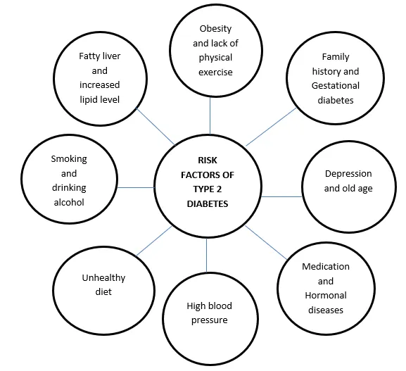
Figure 1: Risk factor of Type-II Diabetes
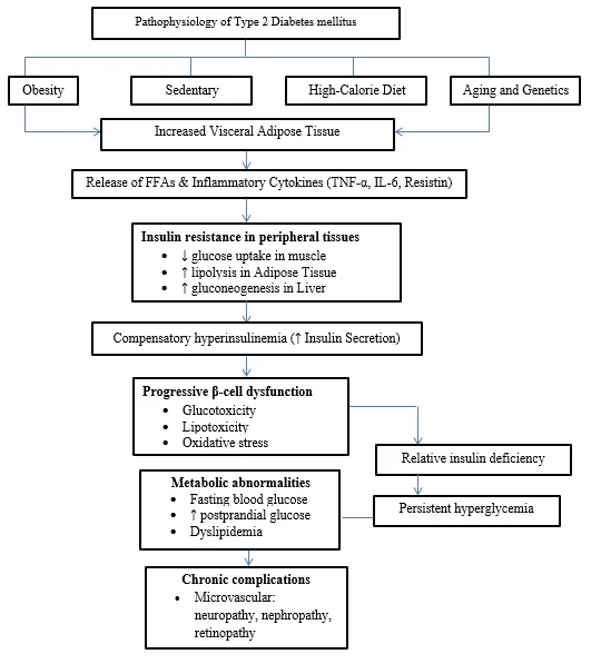
PATHOPHYSIOLOGY OF TYPE 2 DIABETES: (17)(18)(19)
Table 1: Pathophysiology of Type-II Diabetes
CONVENTIAL DRUG DELIVERY SYSTEM FOR TYPE 2 DIABETES: (20)(21)
Table 2: CONVENTIAL DRUG DELIVERY SYSTEM FOR TYPE 2 DIABETES
CONVENTIAL DRUG DELIVERY SYSTEM FOR TYPE 2 DIABETES: (20)(21)
Table 2: CONVENTIAL DRUG DELIVERY SYSTEM FOR TYPE 2 DIABETES
|
Sulfonylureas |
Glipizide, Glimepiride |
Stimulate insulin release from β-cells |
1–20 mg/day |
Oral tablet |
Effective, inexpensive |
Hypoglycemia, weight gain |
|
Thiazolidinediones (TZDs) |
Pioglitazone |
↑ Insulin sensitivity (PPAR-γ activation) |
15–45 mg/day |
Oral tablet |
Improves insulin resistance |
Weight gain, edema, heart failure risk |
|
DPP-4 inhibitors |
Sitagliptin, Saxagliptin |
↑ Endogenous GLP-1 levels |
5–100 mg/day |
Oral tablet |
Weight neutral, low hypoglycemia |
Pancreatitis risk, modest efficacy |
|
GLP-1 receptor agonists |
Exenatide, Liraglutide |
↑ Insulin, ↓ glucagon, slow gastric emptying |
0.6–1.8 mg/day |
SC injection |
Weight loss, CV benefit |
Injectable, nausea, pancreatitis |
|
SGLT-2 inhibitors |
Canagliflozin, Empagliflozin |
↑ Urinary glucose excretion |
10–300 mg/day |
Oral tablet |
Weight loss, CV & renal benefit |
UTI, dehydration, ketoacidosis |
|
α-Glucosidase inhibitors |
Acarbose, Miglitol |
Delay carbohydrate absorption |
25–100 mg TID |
Oral tablet |
Controls post-prandial glucose |
Flatulence, diarrhea |
|
Meglitinides |
Repaglinide |
Short-acting insulin secretagogue |
0.5–4 mg |
Oral tablet |
Flexible meal dosing |
Hypoglycemia, weight gain |
|
Amylin analogues |
Pramlintide |
↓ Glucagon, ↓ gastric emptying |
0.06–0.12 mg |
SC injection |
Post-meal glucose control |
Nausea, injections |
|
Insulin |
Glargine, Lispro |
Replaces endogenous insulin |
Dose in IU |
SC injection |
Most effective therapy |
Hypoglycemia, weight gain |
NOVEL DRUG DELIVERY SYSTEM FOR ANTI DIABETES DRUGS FOR T2DM:
Novel Drug Delivery Systems (NDDSs) have emerged as an important area of research in recent years because of their ability to reduce dosing frequency, improve drug bioavailability, protect drugs from degradation in the acidic gastric environment, and deliver medications more effectively to targeted sites while minimizing side effects. In contrast, conventional drug delivery systems have several limitations, including reduced therapeutic effectiveness due to inappropriate or inefficient dosing, decreased drug potency caused by metabolism, and poor target specificity. (22)(23)
Although many NDDSs have been developed and studied for the treatment of various diseases, only a limited number have been reported for the management of type 2 diabetes mellitus (T2DM). These systems can be broadly classified as:
Table 3: Novel Drug Delivery System for Anti Diabetes Drugs for T2DM
|
System |
Key Component |
Primary Advantage |
Diabetes drugs |
References |
|
Nanoparticles |
Polymers/Lipids |
High surface area; targeted delivery. |
Metfromin |
(24) |
|
Glipizide |
(25) |
|||
|
Liposomes |
Phospholipids |
Biocompatible; carries both drug types. |
Metfromin |
(26) |
|
Glicazide |
(27) |
|||
|
Niosomes |
Non-ionic surfactant and cholesterol |
Stable, targeted delivery |
Pioglitazone |
(28) |
|
Repaglinide |
(29) |
|||
|
SNEDDS |
Oil/Surfactant mix |
Enhances oral bioavailability of oily drugs. |
Glimepiride |
(30) |
|
Glibenclamide |
(31) |
|||
|
Transdermal |
Adhesives/Membranes |
Non-invasive; avoids hepatic metabolism. |
Metformin |
(32) |
|
Glipizide |
(33) |
BERBERINE HYDROCHLORIDE AS AN ANTI-DIABETIC DRUG
SOURCE AND CHEMICAL CHARACTERISTICS:
Berberine is primarily obtained from the roots and rhizomes of several medicinal plants, including Berberis vulgaris (barberry), Berberis aristata (tree turmeric), Berberis thunbergii, Berberis aquifolium (Oregon grape or holly-leaved barberry), Fibraurea tinctoria, Hydrastis canadensis (goldenseal), Xanthorhiza simplicissima (yellow root), Phellodendron amurense (Amur cork tree), Coptis chinensis (Chinese goldthread), Tinospora cordifolia, and Argemone mexicana (prickly poppy). These plant sources are widely used for the production of various berberine-based nutraceutical formulations.(34)
PHYSIOCHEMICAL PROPERTIES OF BERBERINE: (35)
Table 4: Physiochemical Properties of Berberine
|
Property |
Description |
|
Chemical structure |
|
|
Chemical formula |
C₂₀H₁₈NO₄⁺ |
|
Chemical name |
5,6-dihydro-9,10-dimethoxybenzo[g]-1,3-benzodioxolo[5,6-a]quinolizinium |
|
Chemical class |
Quaternary benzylisoquinoline alkaloid |
|
Melting point |
145 °C |
|
Color |
Yellow |
|
Physical state |
Solid |
|
Color index (standard dye identifier) |
75160 |
|
Industry use |
Dyeing wool, leather, and wood; histological staining |
|
Taste |
Bitter |
|
Natural sources |
Berberis spp. and some other plants |
|
Modern application |
Nutraceuticals |
MECHANISM OF ANTIDIABETIC ACTIVITY OF BERBERINE HYDROCHLORIDE:
Numerous clinical and animal studies have demonstrated that berberine (BBR) plays an important role in improving insulin resistance. Research indicates that BBR can lower the levels of inflammatory mediators, thereby protecting pancreatic β-cells from apoptosis. (36)
Recent studies have also shown that BBR activates AMP-activated protein kinase (AMPK) phosphorylation in 3T3-L1 adipocytes. This activation enhances the activity of glucose transporter 1 (GLUT1), leading to improved glucose transport and increased glucose uptake by body tissues. (37)
AMP-activated protein kinase (AMPK), a crucial cellular energy-sensing and signaling system that reacts to variations in the AMP/ATP ratio, is known to be triggered by berberine. In addition to enhancing insulin sensitivity, AMPK activation is crucial for controlling mitochondrial activity. (38)(39)

Figure 2: Pharmacological mechanism and Molecular targets of Berberine in
Type 2 diabetes mellitus (7)
PHARMACOKINETICS AND BIOAVAILABILITY:
Berberine hydrochloride shows complicated pharmacokinetic behavior and is known for its very low oral bioavailability, which is usually reported to be less than 1%.(40) After oral administration, its absorption in the intestine is poor because of its quaternary ammonium structure and high polarity, making it difficult for the drug to passively diffuse across biological membranes. (41)In addition, berberine acts as a substrate for P-glycoprotein (P-gp), a transport protein that actively pumps it back into the intestinal lumen, further reducing its absorption into the bloodstream.(42)
Moreover, berberine undergoes extensive first-pass metabolism in both the intestine and the liver, which significantly lowers its plasma concentration. During this process, it is converted into several major metabolites, such as berberrubine, demethyleneberberine, and jatrorrhizine, through phase I and phase II metabolic pathways. As a result of these combined factors, only a very small amount of orally administered berberine reaches systemic circulation. (40)(43)
After being absorbed, berberine is widely distributed throughout the body, with higher concentrations found in organs such as the liver, kidneys, muscles, and adipose tissue. This broad tissue distribution helps explain why berberine remains therapeutically effective despite having low levels in the bloodstream. (40)(44) The liver serves as a major target organ, where berberine accumulates and plays an important role in regulating glucose and lipid metabolism by activating AMP-activated protein kinase (AMPK). (44) Berberine is extensively metabolized in the liver, mainly by cytochrome P450 enzymes, including CYP2D6, CYP1A2, and CYP3A4. This is followed by conjugation processes such as glucuronidation, which further modify the drug for elimination.(43) Berberine has a relatively short elimination half-life, and both the parent compound and its metabolites are mainly excreted through bile and feces, with only a small amount eliminated through the kidneys.(40)(43)
As a result, alternative drug delivery systems, such as mucoadhesive buccal films, have been suggested to overcome these limitations. This approach helps bypass hepatic first-pass metabolism, enhances drug absorption through the oral mucosa, and improves overall systemic bioavailability. Buccal delivery therefore represents a promising strategy for maintaining more stable plasma levels of berberine while minimizing dose-related side effects. By improving drug availability and tolerability, this method can enhance the clinical usefulness of berberine in the management of Type 2 Diabetes Mellitus.
CLINICAL EVIDENCE ON METABOLIC AND THERAPEUTIC PROPERTIES OF BERBERINE AND ITS DERIVATIVES:
Table 5: Clinical Evidence on Metabolic and Therapeutic Properties Of Berberine And Its Derivatives
|
Therapeutic Area |
Major Biological Actions |
Key Mechanisms Involved |
Reference Numbers |
|
Cardiovascular |
Cardioprotection, antihypertensive |
Regulation of blood pressure and heart rate; improvement of endothelial function; antioxidant activity; anti-inflammatory effects |
(45)(46)(47) |
|
Anti-diabetic |
Glycemic control, metabolic regulation |
Enhancement of insulin secretion; suppression of gluconeogenesis enzymes; improvement of fatty acid metabolism; antioxidant |
(48)(49) |
|
Renal |
Nephroprotection |
Enhanced endogenous antioxidant defense; reduction of inflammatory mediators |
(50)(51) |
|
Gastrointestinal & Liver |
Hepatoprotection, gut health |
Antioxidant activity; anti-inflammatory effects |
(51)(52)(53) |
|
Periodontitis |
Oral anti-infective activity |
Antimicrobial action; antioxidant enhancement; |
(54) |
|
Reproductive System |
Reproductive protection |
Antioxidant activity; anti-inflammatory mechanisms |
(55) |
|
Central Nervous System |
Neuroprotection |
Enhancement of endogenous antioxidant system; anti-inflammatory effects |
(56) |
CLINICAL TRIALS OF BERBERINE (BBR) IN METABOLIC DISORDERS: (8)
Table 6: Clinical Trials of Berberine In Metabolic Disorders
|
Disease Status |
Trial Status |
Phase |
Intervention Measures |
Dose |
Identifier |
|
Prediabetes |
Completed |
III |
BBR + Bifidobacterium / Bifidobacterium / Placebo |
0.5 g, BID |
NCT03330184 |
|
Prediabetes |
– |
IV |
BBR / Metformin |
0.5 g, TID |
NCT03029390 |
|
Diabetes Mellitus |
Completed |
I |
BBR / Placebo |
– |
NCT03972215 |
|
T2DM |
Completed |
I, II |
BBR / Metformin |
– |
NCT00425009 |
|
T2DM |
Active, not recruiting |
III |
BBR + Probiotics / Placebo + Probiotics / BBR + Placebo / Placebo + Placebo |
0.6 g, BID |
NCT02861261 |
|
T2DM with Dyslipidemia |
Completed |
III |
BBR / Placebo |
1.0 g/day |
NCT00462046 |
|
Dyslipidemia |
Terminated |
IV |
BBR + Statins / Statins |
0.5 g, BID |
NCT01697735 |
|
Dyslipidemia |
– |
II |
BBR / Bezafibrate / BBR + Bezafibrate |
0.5 g, TID |
NCT02548832 |
|
NAFLD |
Recruiting |
IV |
BBR / Placebo |
0.5 g, TID |
NCT03198572 |
|
NAFLD |
Completed |
II |
LSI / LSI + Pioglitazone / LSI + BBR |
0.5 g, TID |
NCT00633282 |
|
NAFLD |
Completed |
– |
BBR / LSI |
6.25 g/day |
NCT04049396 |
|
Metabolic Syndrome |
Recruiting |
– |
BBR / Placebo |
1.5 g/day |
NCT03976336 |
BUCCAL DRUG DELIVERY SYSTEM:
The buccal region is considered an effective site for delivering drugs directly to the mucosa for both local and systemic effects, as medicines can be absorbed through the mucosal membrane lining the oral cavity. Compared to conventional oral drug delivery, the buccal mucosa offers several unique advantages. It is richly supplied with blood vessels and has relatively low enzymatic activity, which helps reduce drug degradation. This region is less sensitive, easy to access, and allows patients to remove the dosage form easily if any discomfort or adverse effects occur. Buccal delivery also avoids drug breakdown in the acidic environment of the stomach and bypasses hepatic first-pass metabolism. As a result, this route improves drug bioavailability, allowing lower doses to be used and reducing dose-related side effects. Furthermore, buccal administration generally provides better patient compliance than many other non-oral drug delivery methods. (57)(58)(59)
However, buccal drug delivery also has certain limitations. The available surface area is relatively small (about 50 cm²), and continuous saliva secretion (approximately 0.5–2 L per day) can dilute the drug. Swallowing saliva may reduce drug absorption, while accidental ingestion of the dosage form may cause choking, especially in children, elderly individuals, and patients with swallowing difficulties. In addition, the presence of the dosage form can interfere with eating and drinking, leading to discomfort and inconvenience for some patients. (60)
ANATOMY AND PHYSIOLOGY OF ORAL CAVITY:
Figure3: Schematic representation of Buccal area (9)
The oral cavity is a complex structure made up of different regions that vary in their epithelial type, thickness, and ability to allow drug passage, all of which are important for drug delivery.(58) It is generally divided into two main types: keratinized tissues, such as the gingiva and hard palate, and non-keratinized tissues, including the buccal mucosa, sublingual area, soft palate, underside of the tongue, and inner lips.(61)Keratinized regions mainly provide mechanical protection and are less permeable because they contain a tough outer layer called the stratum corneum. In contrast, non-keratinized regions have a thinner and more hydrated epithelial layer, which makes them more favorable for drug absorption through the mucosa. As a result, these areas are considered more suitable for effective transmucosal drug delivery. (62)
The buccal mucosa, located on the inner lining of the cheeks, is made up of stratified squamous non-keratinized epithelium supported by connective tissue and a dense network of blood vessels. This rich blood supply helps promote efficient absorption of drugs into the systemic circulation. (63)The sublingual mucosa, found beneath the tongue, is the thinnest and most permeable region in the oral cavity, which makes it especially suitable for rapid drug absorption and quick onset of action. (64)The tongue has a rough upper surface containing specialized papillae responsible for taste, while the lower surface is smooth and highly vascularized, supporting effective transport of substances into the bloodstream. (65)
Overall, the structural and histological differences within the oral cavity play a major role in determining how long a drug remains in place, how easily it passes through the tissue, and how well it is absorbed. Therefore, these features are crucial factors in the design and effectiveness of buccal drug delivery systems.
AN OVERVIEW OF MUCOADHESIVE FILM TECHNOLOGY:
Mucoadhesive buccal drug delivery systems represent a promising approach for systemic drug administration, as they help bypass first-pass metabolism and improve drug bioavailability. These systems, especially mucoadhesive films, are designed to attach firmly to the buccal mucosa and remain in place for prolonged periods.(66)(67)In practice, such films may also adhere to other regions of the oral cavity, including the sublingual and gingival mucosa. Therefore, it is often more appropriate e to classify these mucoadhesive films based on the specific site of adhesion, referring to them as sublingual films, gingival films, or buccal films, depending on their location of application. (68)(69)
MECHANISM OF MUCOADHESION:(70)
Mucoadhesion generally occurs in two main stages:
Contact stage: In this initial phase, the mucoadhesive formulation comes into contact with the mucous layer. Upon contact, the material begins to absorb moisture, swell, and spread over the mucosal surface, creating close physical interaction with the membrane.
Consolidation stage: In this stage, moisture further activates and softens the mucoadhesive material, making the system more flexible. This allows the adhesive molecules to rearrange and interact with the mucosal surface through weak forces such as van der Waals interactions and hydrogen bonding, resulting in stronger and more stable adhesion.
THEORIES OF MUCOADHESION:
Table 7: Theories of Mucoadhesion
|
Theory |
Basic Concept |
Key Contribution to Mucoadhesion |
Major Limitation |
References |
|
Electronic Theory |
Adhesion occurs due to electron transfer between the mucoadhesive polymer and mucosal surface, leading to formation of an electrical double layer |
Explains electrostatic attraction between oppositely charged surfaces |
Does not account for polymer chain interpenetration or mucus dynamics |
(70) |
|
Wetting Theory |
Mucoadhesion depends on the ability of a polymer to spread and wet the mucosal surface effectively |
Useful for liquid and semi-solid formulations; emphasizes contact angle and surface energy |
Limited applicability to solid dosage forms and long-term adhesion |
(71) |
|
Adsorption Theory |
Adhesion results from secondary chemical bonds such as hydrogen bonding, van der Waals forces, and hydrophobic interactions |
Explains strong interfacial bonding at polymer–mucus interface |
Bond strength may be affected by environmental conditions like pH and moisture |
(71)(72) |
|
Diffusion Theory |
Polymer chains diffuse and interpenetrate with mucin glycoprotein chains forming a semi-permanent bond |
Widely accepted theory for polymeric mucoadhesive systems |
Assumes ideal conditions and neglects continuous mucus turnover |
(70)(72) |
|
Fracture Theory |
Focuses on the force required to detach the mucoadhesive system from the mucosal surface |
Useful for quantifying adhesive strength mechanically |
Difficult to apply accurately to soft, hydrated biological tissues |
(73) |
|
Mechanical Theory |
Adhesion occurs due to mechanical interlocking of polymer into surface irregularities of mucus |
Explains adhesion in rough or porous mucosal surfaces |
Less relevant for smooth mucosal membranes like buccal or sublingual mucosa |
(70)(72) |
FACTORS AFFECTING MUCOADHESION: (74)(75)(76)(10)
The mucoadhesive strength of a polymer generally increases when its molecular weight exceeds 100,000. In particular, polyoxyethylene polymers show a clear relationship between molecular weight and adhesive strength, with higher mucoadhesion observed in the range of about 200,000 to 7,000,000. This indicates that polymers with larger molecular chains tend to form stronger interactions with the mucosal surface, leading to improved adhesion.
The average pore size, the number and molecular weight of cross-linked polymer chains, and the degree of cross-linking are three important and closely related structural features of a polymer network. These factors together influence how the polymer behaves when it comes in contact with moisture.
As the density of cross-linking increases, the movement of water into the polymer network becomes slower. This reduced water penetration leads to limited swelling of the polymer, which in turn decreases the ability of the polymer chains to interpenetrate with mucin. As a result, higher cross-linking density can weaken the overall mucoadhesive interaction.
Mucoadhesion begins when polymer chains spread and diffuse into the contact area between the dosage form and the mucus layer. For this process to occur effectively, the polymer chains must be sufficiently flexible so that they can intertwine and form strong bonds with the mucus. In general, the mobility and flexibility of polymers are linked to their viscosity and diffusion properties. More flexible polymers can move more easily and penetrate deeper into the mucus network, leading to better interpenetration and stronger mucoadhesive interaction.
Hydrogen bonding also plays a key role in the mucoadhesive behavior of polymers. For effective adhesion, polymers should contain suitable functional groups that can readily form hydrogen bonds with mucus. In addition, the flexibility of the polymer chains enhances their ability to interact closely with the mucus layer, thereby strengthening these bonds. Polymers such as polyvinyl alcohol, hydroxylated methacrylate, and poly(methacrylic acid), along with their copolymers, are known for their strong hydrogen bonding ability and therefore show good mucoadhesive properties.
Hydration is essential for a mucoadhesive polymer to swell and form a suitable network structure of adequate size. This process also increases the mobility of polymer chains, allowing them to move freely and penetrate into the mucin layer more effectively. When the polymer swells, it exposes bioadhesive sites that can participate in hydrogen bonding and electrostatic interactions with the mucus, leading to stronger adhesion. However, there is an optimal level of hydration at which swelling and mucoadhesion are maximized, as too little or too much water can reduce adhesive strength.
In general, the electrical charge of bioadhesive polymers plays an important role in their mucoadhesive behavior. Studies have shown that non-ionic polymers usually exhibit weaker adhesion when compared to anionic polymers. A strong negative charge on the polymer is often considered a key feature for effective mucoadhesion. In addition, some positively charged (cationic) polymers may also show excellent mucoadhesive properties due to their strong interactions with the mucus layer.
This factor is important because it influences the formation of a strong adhesive bond with the mucus layer, which depends on how effectively polymer chains can penetrate into it. When the polymer concentration is too low, only a small number of chains are available to enter the mucus network. As a result, the interaction between the polymer and mucus becomes weak and unstable, leading to poor mucoadhesion.
Saliva, which acts as the dissolution medium in the oral cavity, plays an important role in influencing the behavior of mucoadhesive polymers and their adhesive properties. Factors such as the rate of salivary flow and the pH of saliva can significantly affect polymer hydration, swelling, and adhesion. In addition, the continuous movement of buccal tissues during activities like eating, drinking, and speaking may disturb the contact between the polymer and the mucosal surface, thereby reducing its bioadhesive effectiveness.
The pH of the surrounding medium plays a crucial role in the hydration and mucoadhesive behavior of polymers. A pH range of 6.5 to 7.5 is generally considered optimal for achieving strong mucoadhesion. When the pH moves away from this range, especially under more alkaline conditions or with changes in ionic strength, the mucoadhesive ability of the polymer tends to decrease. As a result, maintaining suitable pH and ionic conditions is important for ensuring effective adhesion to the mucosal surface.
The physicochemical properties of mucus can change during certain disease conditions, such as the common cold, gastric ulcers, ulcerative colitis, and bacterial or fungal infections. These changes may modify the structure and composition of the mucus layer. As a result, alterations in the normal physiological state of the body can influence the bioadhesive behavior of drug delivery systems and may affect their ability to adhere effectively to mucosal surfaces.
Adequate initial contact between the polymer and the mucus layer is essential for proper swelling and effective interpenetration of polymer chains. This close interaction allows the polymer to absorb moisture and expand. As a result, stronger entanglement is formed, leading to improved mucoadhesive strength and stability.
BASIC COMPONENTS OF MUCOADHESIVE DRUG DELIVERY SYSTEM:
The selection of a suitable active pharmaceutical ingredient should be based on its pharmacokinetic characteristics. Ideally, the drug should be effective at a low dose, preferably not exceeding 25 mg per administration. It should also have a short biological half-life, typically between 2 and 8 hours. In addition, drugs that undergo significant first-pass metabolism are good candidates for buccal drug delivery, as this route helps bypass hepatic metabolism and improves systemic availability. (10)
The choice of a bioadhesive polymer plays a key role in determining important factors such as mucoadhesive strength, film thickness, in-vitro drug release, and the residence time of the delivery system. Polymers with higher molecular weight are generally preferred because they provide better control over drug release. An ideal polymer should be chemically inert, compatible with both the drug and the surrounding environment, and capable of rapidly adhering to the mucosal surface while maintaining strong and prolonged adhesion for the required duration. (10)(77)
Examples of bioadhesive polymers used are: (58)
Table 8: Examples of bioadhesive polymers
|
Polymer Category |
Examples of Bioadhesive Polymers |
Key Features Relevant to Mucoadhesion |
|
Synthetic polymers |
Carbopol (Polyacrylic acid) |
Strong hydrogen bonding, high mucoadhesive strength, good swelling and controlled release |
|
Polycarbophil, Polyvinylpyrrolidone (PVP) |
||
|
Poly(methacrylic acid) |
||
|
Semi-synthetic polymers |
Hydroxypropyl Cellulose (HPC) |
Flexible polymer chains, good film-forming ability, moderate to good mucoadhesion |
|
Hydroxypropyl Methylcellulose (HPMC) |
||
|
Sodium Carboxymethyl Cellulose |
||
|
(CMC) Hydroxyethyl Cellulose (HEC) |
||
|
Natural polymers |
Chitosan |
Biocompatible, biodegradable, electrostatic interaction with mucus, safe for oral use |
|
Hyaluronic acid |
||
|
Xanthan gum |
||
|
Pectin |
||
|
Locust bean gum |
Penetration enhancers improve drug transport by interacting with epithelial cell components such as keratin, intercellular lipids, and proteins, thereby temporarily modifying the barrier properties of the tissue. They may increase the drug’s diffusion coefficient, enhance its thermodynamic activity in the formulation, or promote greater partitioning into the buccal epithelium. (78)Various types of absorption enhancers are used for this purpose, including surfactants, bile salts, fatty acids, complexing agents, polymers, cyclodextrins, and other compounds such as azole analogues. (9)
Plasticizers are added to drug delivery systems to enhance their folding endurance and mechanical flexibility. They help make the dosage form more flexible and less brittle, which improves patient comfort, acceptability, and overall compliance. By providing better elasticity, plasticizers also support the durability of the formulation during handling and use. Commonly used plasticizers include PEG-400, PEG-600, dibutyl phthalate, and propylene glycol. (10)
METHOD OF PREPARATION OF MUCOADHESIVE BUCCAL FILM:
Various techniques have been explored for the preparation of mucoadhesive buccal films, including solvent casting, hot melt extrusion, inkjet printing, and 3D printing. Each of these methods has its own benefits and limitations in terms of cost, scalability, precision, and product quality. Therefore, the choice of manufacturing technique depends on the desired characteristics of the final dosage form and the intended application.
Solvent casting is the most commonly used method for preparing buccal films because of its simplicity and low production cost. In this process, water-soluble polymers are dissolved to form a uniform viscous solution, followed by the addition of the active pharmaceutical ingredient and other excipients. The solution is then poured, dried, and cut into films of desired size containing a specific drug dose. This technique offers several advantages, including good mechanical properties, easy processing, low cost, and uniform film thickness. However, it also has limitations. Films prepared by solvent casting may become brittle during storage due to gradual loss of residual solvent, leading to reduced flexibility and elongation. (79)(80)
Hot Melt Extrusion (HME) is a continuous, reproducible, and easily automated manufacturing technique widely used in pharmaceutical production.(81) In this process, a hot melt extruder is employed to convert polymer blends into films through controlled heating. A dry mixture of the active pharmaceutical ingredient and excipients is fed into the hopper, where it is conveyed, mixed, heated, and melted before being extruded as a molten mass. This molten material is then cast into films and allowed to solidify. The casting and drying stages are critical for product quality. HME offers several advantages, including shorter processing times, lower operating temperatures, elimination of organic solvents, reduced material wastage, precise process control, and good scalability for large-scale production. (79)(82)
Inkjet printing is a pharmaceutical manufacturing technique similar to conventional printing, but it uses specially formulated drug-containing “inks” instead of regular ink. In this method, the active pharmaceutical ingredient is dissolved or dispersed in a suitable liquid medium and loaded into the printer. The formulation is then deposited onto a substrate in the form of fine droplets through a nozzle. (83)
Inkjet printing is often combined with other buccal film fabrication methods such as solvent casting, hot melt extrusion, and 3D printing. It is mainly used to deposit the drug onto an already prepared substrate rather than incorporating it directly into the film matrix. (84)(85)
Compared to solvent casting and HME, inkjet printing offers several advantages, including improved mechanical strength, better long-term stability, and the possibility of personalized dosing. However, challenges include nozzle clogging, which may affect dose accuracy, the need for drug solubility and stability in the ink, and limited suitability for low-potency drugs. (86)
Three-dimensional (3D) printing has emerged as a promising approach for overcoming formulation challenges in the production of buccal films. Currently, most buccal dosage forms are limited to potent drugs due to their low drug-loading capacity, and 3D printing offers opportunities to improve this limitation. (9)
Fused Deposition Modeling (FDM) is one of the most widely used 3D printing techniques. In this method, thermoplastic filaments are melted or softened and extruded through a nozzle to build objects layer by layer according to a computer-aided design (CAD) model. The material is heated slightly above its melting point and rapidly solidifies after deposition, forming a stable three-dimensional structure. (87)
Semi-Solid Extrusion (SSE) is another important 3D printing technique that involves the layer-by-layer deposition of semi-solid materials using a syringe-based system. These materials are prepared by blending polymers with suitable solvents to achieve the viscosity required for smooth and accurate printing. (87)
EVALUATION OF MUCOADHESIVE BUCCAL FILM: (9)(88)(89)
Table 9: Evaluation of Mucoadhesive Buccal Film
|
Evaluation parameter |
Method / Instrument |
Purpose / Outcome |
|
Surface morphology |
Scanning Electron Microscopy (SEM) |
To observe surface texture, porosity, and drug distribution in plain polymer film and drug-loaded buccal film |
|
Drug–polymer interaction |
Fourier Transform Infrared Spectroscopy (FTIR) |
To detect possible chemical interactions between drug and film excipients |
|
Thermal behavior |
Differential Scanning Calorimetry (DSC) |
To evaluate crystallinity and thermal stability of drug within the film matrix |
|
Surface pH |
Digital pH meter |
To ensure non-irritancy and compatibility with buccal mucosa (acceptable pH ~6–7) |
|
Folding endurance |
Manual folding method |
To assess mechanical strength and flexibility of films |
|
Swelling ratio (%) |
Gravimetric method |
To determine hydration capacity and mucoadhesive potential |
|
Film thickness |
Digital micrometer |
To ensure uniform distribution of formulation components |
|
In-vitro disintegration time |
USP method |
Time required for the film to start breaking |
|
In-vitro dissolution time |
USP method |
Time required for complete dissolution of the film |
|
Tensile strength (TS) |
Santam Testing Machine (STM-20) |
To determine maximum stress the film can withstand |
|
Percentage elongation at break (%EB) |
Santam Testing Machine |
To assess elasticity and deformability of films |
|
Young’s modulus |
Stress–strain analysis |
To evaluate stiffness and brittleness of buccal films |
|
Drug content uniformity |
UV–Visible spectrophotometry |
To ensure uniform drug distribution (acceptable limit: 85–115%) |
|
In-vitro drug release |
UV–Visible spectrophotometry |
To study release kinetics and extent of drug release from films |
FUTURE PERSPECTIVES AND DIRECTIONS:
The delivery of macromolecules through the buccal mucosa has been explored less extensively compared to other routes of administration. Solvent casting remains the most commonly used method for incorporating active substances into biocompatible polymeric films. However, there is increasing interest in advanced manufacturing techniques such as hot melt extrusion, fused deposition modeling, and inkjet printing. Although buccal films can improve the stability and permeability of macromolecules compared to oral formulations, their limited surface area makes it difficult to achieve high drug loading. Encapsulation of drugs into nanoparticles and their incorporation into mucoadhesive polymer matrices may help overcome this limitation. Ongoing research is focused on developing nanoparticle-based buccal films and functionalization strategies to enhance mucosal permeation and enable effective systemic targeting.(9)
Mucoadhesive buccal films containing berberine hydrochloride offer a promising and innovative strategy for the management of Type 2 Diabetes Mellitus. However, additional research and development are still needed to clearly establish their clinical effectiveness and commercial feasibility.
Future studies should concentrate on optimizing key formulation factors such as polymer combinations, drug loading, film thickness, and mechanical strength to improve stability, enhance mucoadhesive properties, and ensure controlled drug release. Moreover, the use of novel biodegradable and stimuli-responsive polymers may further increase residence time in the oral cavity while improving patient comfort and acceptance. Advanced drug delivery approaches, such as incorporating nanocarriers, lipid-based systems, and cyclodextrin complexes into buccal films, can greatly improve the solubility, permeability, and bioavailability of berberine. In particular, buccal films loaded with nanoparticles show strong potential to overcome challenges related to low drug loading and poor mucosal penetration, thereby enhancing therapeutic effectiveness.
Future research should also investigate combination therapy strategies in which berberine is delivered together with other antidiabetic drugs, probiotics, or bioenhancers through buccal films to produce synergistic effects and achieve better glycemic control. Overall, with ongoing progress in material science, nanotechnology, and pharmaceutical engineering, mucoadhesive buccal films containing berberine hydrochloride have great potential to develop into an effective, patient-friendly, and clinically dependable treatment option for the long-term management of Type 2 Diabetes Mellitus.
CONCLUSION
Type 2 diabetes mellitus requires innovative treatment strategies to address the limitations of conventional oral antidiabetic therapies, such as low bioavailability, frequent dosing, and unwanted systemic side effects. Berberine hydrochloride has shown strong antidiabetic potential through multiple mechanisms, including activation of AMP-activated protein kinase, enhancement of insulin sensitivity, and regulation of glucose and lipid metabolism. However, its clinical use is limited by poor oral absorption and extensive first-pass metabolism. Mucoadhesive buccal film delivery systems provide a promising alternative by allowing direct absorption through the oral mucosa, improving bioavailability, enabling sustained drug release, and enhancing patient compliance. Incorporating berberine hydrochloride into mucoadhesive buccal films offers a novel, convenient, and patient-friendly approach for managing Type 2 diabetes mellitus. Further formulation refinement and clinical studies are needed to confirm its therapeutic effectiveness and practical applicability.
REFERENCES

Pallavi A, Beny Baby, Bhuvana, Tejashree K C, Navya N, Mucoadhesive Buccal Film Infused with Berberine Hydrochloride for Management of Type 2 Diabetes, Int. J. of Pharm. Sci., 2026, Vol 4, Issue 9, 3790-3813, https://doi.org/10.5281/zenodo.23031708
 10.5281/zenodo.23031708