View Article

Abstract

Microneedle drug delivery system is an effective approach for the delivery of the drugs by the transdermal route. The microneedles create the micro sized pores on the skin surface and the drug enters to the dermis through cuticle layer. Stratum corneum is a barrier of skin, sclera and cornea that challenges the topical administration of the drugs and entry of the exogenous materials. Also, for transdermal delivery the drug should obey specific physicochemical properties like, the drug should have a molecular size less than 500 D, it should have a low melting point, as well as adequate lipophilicity. Ideally log P should be within 2-3. Thus the drugs which are hydrophilic in nature, or macromolecular agents like proteins, peptides, DNA, small interfering RNA, etc. are difficult to administer via transdermal route. Therefore, microneedle system can be incorporated to increase the drug permeation through subcutaneous layer. It also bypasses the hepatic circulation and first pass metabolism, increasing the bioavailability of the drugs. In this review article we have discussed about the microneedle drug delivery system including its types, applications, and newer approaches in the field of microneedles.

Keywords

Microneedles, transdermal drug delivery, microfabrication, injection, skin permeation.

Introduction

× Popup Image

One of the most practical ways to administer medications is orally however occasionally this is not possible since medications may be poorly absorbed or destroyed as a result of enzymatic breakdown in the liver or gastrointestinal tract over the past century injection using a hypodermic needle has become a popular substitute for oral drug delivery which are more painful and invasive also create the biohazardous waste and require trained personnel Hypodermic needles are inserted to muscles where there is a lower immune response than there in the skin  so TDDS is used as an alternative method to the hypodermic needles TDDS also bypasses the hepatic first pass metabolism and it is non-invasive painless alternative and it can be self-administered stratum corneum  is the outermost layer of the skin made up of the dead corneocytes that acts as a primary barrier for transdermal administration it causes significantly reduced effectiveness of the drug delivery and restricts the different kinds of the drugs that are transported via skin Microneedle technology was proposed in year 1976 to overcome the problems associated with old transdermal delivery technique The microneedles are about 25 um to 2000um long and are much sharper than the usual hypodermic needles so that they can cross stratum corneum and create micro scale drug delivery channels without disturbing the nerve fibers and blood vessels present in the epidermis and dermis layers of the skin therefore the efficacy of the drug delivery can be markedly improved and a wide range of drugs delivered by TDDS can be significantly expanded in a painless and least invasive way.

2. CLASSIFICATION OF MICRONEEDLES:

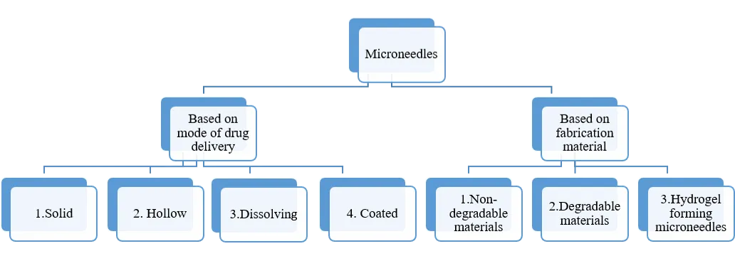
Figure 1: Classification of microneedles

2.1 Based on mode of drug delivery:

There are four types of microneedles: Solid, hollow, dissolving and coated microneedles.

2.1.1 Solids microneedles:

Since solid microneedles are introduced and removed to create pores on the skin’s surface that are micro in size, they can be utilized in the skin pre-treatments They function by creating micro channels using the “poke and patch” method. These micro channels increase the permeability of the medication by permitting diffusion straight into the dermal layer from a formulation. An experiments on rat skin has demonstrated that when placed under occlusive conditions such as occlusive tape, the micro pores created by the microneedles persisted for at least 72 hours following microneedle therapy. Without applying occlusive tape, the micro pores quickly closed. It was found that the solid MN-created micro channels healed quickly in just 2 hours, without causing secondary infection. The “scrape and patch” method is modification of the standard solid microneedle technique in which microneedle, micro projections, or micro blades are scraped over the skin to create micro-abrasions. These microprojection are subsequently covered with drug solution in patch. An additional alternative is to utilize a roller with solid microneedles that repeatedly puncture the stratum corneum as the roller rotates across the skin pores. A study by Ita et al (2015) used Microneedle Systems with a cylindrical surface to administer medications for high blood pressure. Transdermal flow values have been observed to rise 5-6 times following microneedle roller treatment using pig skin.

2.1.2 Hollow microneedles:

The microneedles are a tiny version of the traditional hypodermic needle systems. A pressure-driven liquid formulation flow is used to administer drugs. This method can deliver significant dosages into the dermal layer. It is challenging to construct hollow microneedles because of their fragility and structure. The hollow microneedle work on the principle of ‘puncture & flow’. Davis et al (2005) created a series of hollow metal microneedles for one such study. In diabetic rats, the administration of insulin using these microneedles was investigated. The study found that the pharmacodynamics profiles of hollow microneedles are similar to those of a conventional hypodermic injection.

 2.1.3 Dissolving microneedles:

These types of microneedles are governed by “poke & release” principle. Compared to other microneedles, they are simple to use and manufacture, which is why they have attracted a lot of interest recently. The biodegradable elements used to make dissolving Microneedles include sugars and biodegradable polymers loaded with medical compounds. The conjugated therapeutic agent is released when the microneedles dissolve into the skin after applications. Because of their single-step application approach and ease of manufacture, these microneedles offer an advantage over convenient. Microarrays made of dissolving hyaluronic acid were created by Liu et al. When used as microneedle array, the cumulative penetrated amount of dextran rose by an astonishing 610 times with minimal lag drug penetration time. Researchers are looking into using dissolving microneedles to deliver vaccines via the skin. It has been demonstrated that carboxy methyl cellulose and hyaluronan microneedle totally disintegrate in rat skin, 5minutes after applications, releasing the IgG payload specific to the antigen.      

2.1.4 Coated microneedles:

Coated microneedles are those that operate on the “coat and poke” principle. These microneedles are made out of a solid microneedle foundation covered in medication solutions or dispersions. Numerous studies have been investigated for coating microneedles. Dip coating is the most used technique, but it is challenging because it requires exact control to ensure that the microneedles are introduced into the dipping solution precisely. Another technique for drug deposition on microneedles is spray coating; however, this approach is unable to prevent drug loss due to spraying on the array of microneedles’ substrate, which is inaccessible for drug penetration.

Although aerosol coating techniques are more effective, the usage of thermolabile medications may be restricted due to the requirement for temperatures as high as 200°C. Ma et al (2018) used a polyethylene glycol matrix to cover solid microneedles with suspended lidocaine that demonstrated a noticeably greater delivery than marketed product. Since they provide a comparatively modest surface area for medication absorption, coated microneedles are employed less frequently than other microneedles.

Following is a comprehensive overview of the benefits, drawbacks, and mode of drug delivery of different microneedles:

Table 1 : An overview of microneedles

Type of microneedles

Benefits

Drawbacks

Mode of drug delivery

Solid microneedles

These can be fabricated with different types of materials

1.Can break beneath the dermal layers.

2.Minimum surface area for drug absorption

By making micropores to the skin through which the drug is delivered

Hollow Microneedles

High doses of drugs can be delivered

Require a strong material of manufacturing to resist flow pressure.

Pressure driven flow through the needles

Dissolving microneedles

Simple manufacturing

Can be made up of only biodegradable substances

Dissolves  beneath skin to release drug payload

Coated microneedles

Potent drugs requiring low dose can be delivered

1.Drug loss during fabrication

2.Temperature control is required

Drug is released through the coating

2.2 Based on fabrication material:

Microneedles are manufactured using a various types of material including metals, ceramics, silicones, and polymers. These materials used for microneedles manufacture are classified as non- degradable and degradable materials. Materials such as metals, ceramics and silicones are non- degradable materials while the polysaccharides and polymers are the degradable materials that are employed in microneedle manufacturing.

2.2.1 Non degradable materials:

  1. Metals: One of the first metal based microneedle product to be introduced was Soluvia®. Microjet®, a silicone- based product was another that arrived on the market. The two metals that are most frequently utilized to create microneedles are titanium and stainless steel. Metal microneedles can be manufactured as hollow, solid, or microneedles with coatings. Wet etching, metal electroplating, laser cutting, and three- dimensional laser ablation are examples of fabrication methods. The AdmiPen® is a type of hollow microneedle that has been released commercially. Its length ranges from 600 to 1500 µm. Titanium or stainless steel derma rollers are used professionally for cosmetic applications.
  2. Silicone: For more than 20 years, silicones had been studied as one of the most popular material for microneedle devices. In micromoulding, silicone microneedle arrays could serve as the main mould. Silicones make it possible to create microneedles with a variety of shapes. Using an etching process, Narayanan et al created silicon microneedles. Etching factors including time, different concentrations of tetra methyl ammonium hydroxide (TMAH) , sharper microneedles with greater aspect ratio were created by adjusting the temperature, etching rate, and other factors. According to Krieger et al, a 3D printer can also be used to create high aspect ratio microneedle silicone master mould.
  3. Ceramic: One of the most popular materials used to make microneedles is alumina i.e., (Al2O3). Because of its porosity, alumina can hold a specific volume of active ingredient. Microneedles have also been created using gypsum and brushite. Microneedles have been created using Ormocer® which is a new material composed of organically modified silicon alkoxides and organic monomers. It creates a three-dimensional network that expands the region where the drug solution can be absorbed.
  4. Synthetic polymers: A number of synthetic polymers, including poly vinyl alcohol (PVA) and polyvinyl pyrrolidone (PVP), have been studied for usage I microneedles. For microneedle arrays, poly (methyl methacrylate) i.e., PMMA is a biocompatible polymer.

2.2.2 Degradable materials:

  1. Natural materials: For the production of microneedles, carbohydrates are a good substitute. The affordability and safety of carbohydrates is one of their most significant qualities. They have low toxicity in addition to having great biocompatibility. It is recognized that certain carbohydrates have a notable degree of toughness or strength. These materials are inexpensive and readily biodegradable. Using master templates, it is simple to shape carbohydrates into microneedles. A combination of active substance and carbohydrates can be created. After that, this mixture can be poured into molds that dissolve when inserted into the skin, releasing the pharmacological payload. Maltose is one of the sugars most frequently utilized in the preparation of microneedle arrays. In parenteral formulations that have received FDA approval, maltose is a frequently used excipient. FDA approved parenteral preparations use maltose as an excipient.
  2. Synthetic materials: Microneedles have been created using a wide range of synthetic macromolecular materials. These consist of poly lactic acid (PLA), poly lactic co-glycolic acid (PLGA), polyglycolic acid (PGA). Microneedles control the drug release for these kinds of materials, which is dependent on the microneedle materials’s composition.
  3. Hydrogel forming microneedles: These are called as in – situ microneedles and fabricated with a polymer is cross-linked to form dry hydrogels. They absorb in water from interstitial fluid when injected into the skin, which causes microneedles to swell.  Conduits that serve as drug reservoirs and regulate dug distribution are created by swelling hydrogel network. Additionally, it keeps the microneedles in skin intact. The molding method, which also involves a cross-linked phase, is mostly used to create these microneedles. Numerous materials have been investigated for hydrogel forming microneedles. Polysaccharides are the most prevalent. Otherv polymers have been investigated for hydrogel forming microneedles, including Poly (ethylene glycol) diacrylate i.e., PEGDA and PVA, polyacrylic acid- co- maleic acid (PAMA), and methylvinylether-co- maliec acid. Because of microneedle base’s capacity to swell, the primary benefit of hydrogel forming microneedles is the delivery of large quantities of medication.

Microneedles have been made from a variety of materials. They can be made from either biodegradable or non-biodegradable materials. Although there are many options for both biodegradable and non-biodegradable materials, it is important to take into account supporting factors that may affect the building materials. Several types of materials have been used for fabrication of microneedles. The choice of material is influenced by a number of factors, including the available manufacturing method, the drugs and excipients of choice, the drug’s compatibility with the material and fabrication process, the eventual usage, and the mode of delivery.

3. FABRICATION TECHNIQUES OF MICRONEEDLES:

Skin is an elastic organ that has considerablevtensile strength. Microneedles need to be as strong as possible to penetrate without breaking or cracking during insertion. As a result, creating microneedles is difficult. The required skin depth, microneedles geometry, and preferred material all influence the construction process. For example, solvent- casting might work well for polymeric microneedles, whereas metal microneedles are best designed using microelectromechanical system. The manufacturing technology has been used to categorize the fabrication procedures.

  1. Fabrication of polymeric microneedles using solvent:
  1. Micromolding: Micromolding is used to create a variety of polymers, from PVA and PVP (synthetic or semisynthetic polymers) to pectin (natural polymer). The method uses many methods to evenly pour a liquid polymeric solution onto the mold, then removes air gaps. Air spaces can be eliminated by using a vacuum (or, alternately, a centrifuge), drying in an oven, and then being removed from the molds.
  2. Atomized spraying to fill molds: Because of the centrifugation or vacuuming process involved, continuous production of microneedles using micromolding techniques can be difficult. This difficulty can be solved by atomized spraying the polymer solution into the molds. As demonstrated by McGrath et al., atomization of aqueous solution might effectively cover the whole surface of the mould without creating any voids. Using atomized spraying, the authors created dissolving microneedles (5% solids concentration) in silicon micromolds.
  3. Droplet born air blowing (DAB): This technique uses air blowing to create a polymer droplet the shape of microneedle, enabling mild conditions without the requirement for harsh external conditions like heat or UV radiations. The first step in the procedure is to dispense the solution onto the upper and lower plates. The viscous solution is extended by keeping the two plates in contact and gradually pulling them apart. After that, the extended viscous solution is subjected to controlled blowing air. This drying stage provides the necessary form. Kim et al suggested the manufacturing process. The droplet’s size, concentration, and regulated drug loading without drug loss are all managed using a single polymer drop per microneedle. The authors successfully lowered blood glucose levels in diabetic rats by using this method to create insulin loaded dissolving microneedles.
  4. Pulling pipettes: The pulling pipettes method is only useful for creating hollow glass microneedles. Glass is heated to a high temperaturebefore being pulled with a micropipette puller to form microneedle arrays. Programmable pullers can be used to make this method more reliable.
  1. Fabrication of microneedles using electromagnetic/ microsystem/ laser technology:
  1. Microelectromechanical system (MEMS): Integrated circuit technology has been used for developing MESM. The three primary processes of MEMS are:

(i) Deposition, (ii) patterning, and (iii) etching.

  1. Deposition:

This process involves the creation of films on substrate, which is a base material. A chemical or physical process creates the films. While the chemical vapour deposition (CVD) technique involves a chemical reaction between the hot substrate and the carrier gases in the chamber, the physical vapour deposition (PVD) technique involves removing the materials from the source and depositing them on the substrate.

  1. Patterning:

The microneedle geometry pattern, such as height, base width, tip angle, and bore size details is transferred onto the film during the patterning process. One popular method for patterning is lithography. In short, a microneedle pattern is transferred when a radiation source, like light strikes a photosensitive substrate. Radiation can come from a variety of sources, including x-ray, ion, or electron beam lithography. The mask transfer onto the photosensitive coated substrate typically involves the following successive steps:

The first stage is heating in the presence of steam or humidified oxygen to generate an oxide layer. A thin layer of an organic polymer known as a photosensitive/ photoresist or resist substances (sensitive to UV radiation) is then spin coated. The resist layer is then heated in order to eliminate the solvent. After that a mask is exposed to a UV lamp, which enables an almost flawless transfer or to put it another way, ‘printing’ of the mask image onto the resist-coated wafers.

  1. Etching:

In order to create an interesting microneedle design, the unprotected portions of the substrate are etched out using a strong caustic solution. Both wet and dry methods can be used for etching.

  • Dry process: Ion beam milling or reactive ion etching techniques may be used in the dry process. Solid and hollow microneedles are produed by the dry etching method.
  • Wet process: This method involves immersing the substrate in a chemical liquid to assist removal of extra material. Wet etching can occur at distinct (anisotropic) or the same (isotropic) rates. Silicon and metallic arrays are made with it.
  1. Laser cutting: This technique involves cutting metallic sheets into the shapes of microneedles using an infrared laser. The microneedle’s geometry is designed using AutoCAD software. The metal sheet is subsequently removed by the laser beam in accordance with the design, creating in-plane Microneedles. After being cleaned, the manufactured microneedles are bent at a 90° angle. After that, electropolishing is used to adjust the tips. Metallic microneedles are produced using this technique. Stainless steel is the most commonly used material.
  2. Laser ablation: This technique creates a bulge by impinging a laser on a metal plate, as the name implies. To create a microneedle, light pulses are employed to give the bulge to distinct shape. Laser ablation is also used to create solid metallic arrays, much like laser cutting. Additionally, metals such as tantalum are manufactured.
  3. Deep X-ray lithography: This method involves coating and curing a polymer solution on a silicon wafer. The silicon wafer is exposed to a vertical deep x-ray, creating an array of triangular columns and channels. To achieve the required microneedle geometry, the substrate and the x-ray mask are continually aligned and modified. This method is well known for producing hollow microneedles with high precision. Microneedle array can be made up of glass, ceramic, polymeric and metallic materials.
  1. Additive manufacturing:
  1. Two photon polymerization: This is a low-cost, high throughput methods of creating 3- dimensional structures layer by layer. Resins are polymerized into microneedle structures using femtosecond or picosecond laser pulse. Two-photon absorption, which produces an energy centered at laser focal point, starts the polymerization. A computer-aided design (CAD) model is used to first slice the required structure. After that, the laser’s contour in the photosensitive resin. Resin is cleaned with a solvent once the microneedle is made, and ultraviolet radiation is used to cure it.
  2. Microstereolithography (µSL): A high- throughput Fabrication method is called microstereolithography (µSL). It uses a bottom-up addition method to create 3D microstructures. Tissue engineering and biomedicine have made extensive use of this method. The creation of a liquid’s spatially controlled solidification using photopolymerization. A laser beam produces an microneedle pattern on a resin’s surface, giving it a distinct depth. Following support platform is made easier by this. The first layer is photo- polymerized, separated, and realigned, and then the first built layer is recoated. The second layer cures the pattern. To create a solid, 3- dimensional object, continue these processes.
  3. Continuous liquid interface (CLIP): Microneedles are computationally designed using CLIP. In contrast to the above-mentioned conventional layer-by-layer procedure, this innovative approach uses a continuous additive process. The primary benefit of CLIP over the conventional method is that it produces microneedles with a much higher resolution. By doing away with separation and realignement, efficiency is increased and microneedles are produced more quickly. Johnson et al created microneedle arrays utilizing CLIP employing triethylol propane triacrylate, polyethylene glycol dimethacrylate, polycaprolactone trimethacrylate, and polyacrylic acid. It was demonstrated that prepared microneedles could pierce the skin of mice and release rhodamine dye. In 2 -10 minutes, any microneedle patch may be designed using the CLIP technique. The CLIP approach’s great versatility for microneedle generation and rapid prototyping allows for the examination of a wide range of parameters, such as size, shape, and composition, in a throughput process.
  4. Microneedle pain and effectiveness of drug delivery: The primary goal of microneedles is to effectively and painlessly administer medications into the skin. Skin permeability is improved for solid microneedles by flat tips, longer needle length, greater pressing power, and longer skin residence time. Permeability is also improved by increasing microneedle number, but the “bed of nails” effect may make very high density less effective. Drug delivery effectiveness is greatly impacted by microneedle geometry, although insertion speed yields inconsistent findings. Drug release rates are enhanced by separating dissolvable microneedle designs and flexible or self-swelling substrates. Fluid flow through needle conduits is necessary for drug delivery in hollow microneedles. Poisuille’s law governs the resistance, however skin tissue adds additional resistance that lowers flow rate. Deeper insertion, the use of bevel tips, microneedle vibration or retraction, and co-injection of hyaluronidase can all enhance flow. Due to strong cutaneous resistance, tip opening size has minimal impact. Conduit designs that have been modified enhance flow and lessen blockage.

When compared to hypodermic needles, microneedles have a significant benefit in terms of pain. Microneedles’ number has a minor impact on pain, whereas microneedles’ length has a greater influence. Microneedles that are less than 400µm often cause little pain. Pain for hollow microneedles diminishes with reduced flow and hyaluronidase use, but increases with increased pressure and flow rate. In general, microneedles are painless, and their design is not significantly hampered by discomfort.

4. APPLICATIONS OF MICRONEEDLES:

In contrast to traditional patches, microneedle systems frequently need outside help to penetrate biological tissues consistently and reliably. While applicator devices allow for consistent microneedle insertion with less inter-individual variability, manual application may result in varying insertion depth and applied pressure. Microneedle arrays provide regulated penetration into stratum corneum and can be applied to the skin before device is activated or pre-mounted with applicator. Both single use and multiple use setups are supported by reported applicator design.

The ability of a molecule to enter the skin is a major factor in the effectiveness of transdermal medication delivery. Due to their enormous molecular size, many compounds particularly peptides and vaccines have faced challenges during formulation and application. When compared to traditional delivery methods, microneedles have shown efficacy in delivering these molecules intradermally and, in certain instances, have been able to achieve the same therapeutic impact at lower doses. For instance, employing microneedles rather than conventional intramuscular administration resulted in a similar increase in hemagglutinin inhibition antibody titer with less than half the medication dose.

Several regulatory bodies have approved microneedle devices for the delivery of seasonal influenza vaccinations, further demonstrating their efficacy. Microneedles have also been investigated for the treatment of psoriasis, dermatitis, viral warts, and even skin cancer. Microneedles are being studied for ocular medication administration in addition to intradermal applications, and they have shown encouraging outcomes in the treatment of disorders such glaucoma, macular degeneration, uveitis, and retinal vascular occlusion. Currently, the majority of commercially available microneedles are used in cosmetic applications; dissolving microneedles are frequently used to treat acne, while dermarollers improve the general health of the skin.

Yun Liu et al described various applications of polymeric microneedles as follows:

  1. Diabetes:

Insulin delivery devises, which are known for their rapid in vivo glucose responsiveness, usually have limited insulin carrying capacity and pose manufacturing issues. Therefore, the rapid in vivo glucose release response seen in pancreatic β-cells is being replicated in microneedle devices for blood glucose management, while also emphasizing excellent biocompatibility to address acute and long term toxicity concerns. By avoiding the epidermal barrier and enabling the direct administration of macromolecules using a variety of microneedles, the microneedle patch provides a clear advantage. This characteristic increases their effectiveness in reducing pain, making them favoured choice for people with long term medical conditions like diabetes tat requires ongoing treatment. Dopamine nanoparticles (PDA NPs) are incorporated into the exterior layer of microneedle system in a unique method devised by Ma and colleagues. By improving the microneedles’ adherence to skin tissue, this design helps diabeteic wounds heal more quickly. Synthetic nanovescicles ( NV) generated from mesenchymal stem cells (MSCs) are produced by extrusion via a porous membrane showed significant improvements in mRNA and protein expression levels, as well as a noteworthy 250- fold increase in manufacturing yield. The PDA microneedle patch’s powerful therapeutic growth factor offers significant benefits in boosting collagen regeneration, decreasing inflammation, and encouraging angiogenesis. Additionally, the potential benefits of polymeric microneedles medication delivery devices for wound healing management were shown.

  1. Cancer:

The medicine can reach the stimulation site and the associated subcutaneous tissues and be quickly dissolved or destroyed with excellent bioavailability thanks to the microneedle drug delivery system, which completely encapsulates the drug within the microarrays. One of the most successful medication delivery techniques for skin cancer and other cancer treatments is microneedling. Compared to conventional metal and glass micro and nanomaterials, polymer- based micro and nanocomposited provide benefits such as high drug loading, structural durability, and the elimination of the requirement for external drug storage. The very hydrophobic and biocompatible polycaprolactone (PCL) enhaces the solubility and bioavailability of low molecular weight lipid soluble medications.

Drugs are delivered at a more effective drug release rate using a hydrophilic crosslinked PVP polymer microneedle device. In the 500 µg/mL concentration range, PCL and PNVP are both highly biodegradable and non-toxic, which enhances the effectiveness of low molecular weight drug delivery and guarantees long-term safety. Additionally, both of them work well with blood, which facilitate the systematic distribution of medications. The polymer based hydrogel made from carboxy methyl cellulose can mimic the pharmacokinetic process of cellular DOX release in vivo, hence enhancing the therapy of melanoma cancer cells. Polymer microneedles insertion site’s long term durability guarantees the steady blood pressure and long lasting pharmacological benefits that improve performance and lower the risk of infection. In conclusion, compared to traditional drug release techniques, polymer microneedle architectures exhibit significant potential for enhancing drug bioavailability, extending circulation time, regulating drug release and tumor targeting.

  1. Pain management:

In order to avoid systemic toxicity or infection, the long term management of chronic pain restricts the use of delivery methods like parenteral injections. Treatment resistance brought on by patient’s skin nociceptive sensitivity to pain is effectively reduced by microneedle device reacts to biophysical cues gathered in subcutaneous tissue for potential physiological monitoring in case of secondary pain brought on by complex disorders like diabetes.

Therapeutic agents must be delivered locally and specifically to treat problems connected to chronic pain. For the treatment of chronic pain, transdermal delivery devices such as microneedles offer a slow- release mechanism that guarantees sustained plasma delivery and medication efficacy. The microneedle administration devices prevented substance misuse and dramatically decreases the frequency of treatment programs for managing chronic pain. The physicochemical characteristics of the medication (such as solubility, partition coefficient, hydrophobicity), loading dose, molecular diffusivity, and polymer layer all affect the therapeutic efficacy of traditional transdermal patches. Although oral meloxicam effectively reduces osteoarthritis pain in individuals, it has side effects such as ulceration and perforation.

  1. Rheumatoid arthritis:

Currently, corticosteroids, non-steroidal anti-inflammatory agents (NSAIDs), and disease modifying antirheumatic drugs (DMARDs) including Etanercept and methotrexate are the main treatments for rheumatoid arthritis. These drugs are often administered orally or by subcutaneous and intramuscular injections. A viable alternative for transdermal drug delivery is microneedle arrays, which may avoid first pass metabolism and its dangerous side effects, including hepatotoxicity, myelosuppression and gastrointestinal bleeding. Compared to traditional delivery methods, this strategy also reduces the risk of infection and improves patient compliance.

Drug delivery vehicles are made of nanoscale synthetic and natural polymers with diameters under 200 mm. These polymers encapsulation of therapeutic substances enables tailored administration to inflammatory areas, encouraging localized drug accumulation and reducing medication waste overall. According to a new study, delivering antigens to antigen presenting cells (APCs) via polymer microneedles promotes a crucial interaction with antigen specific CD4+T cells. Significant anti-inflammatory qualities are conferred with a living organism by this interaction, which successfully controls T cell development into regulatory T cells. Its therapeutic potential has been demonstrated by a notable reduction in the anti-inflammatory response. Additionally, a variety of therapeutic agents, such as genes, nucleic acids and antigen-specific modified cells and proteins, can be delivered more easily. For rheumatoid arthritis, microneedle system offers a very promising substitute for traditional drug delivery techniques, with the possibility for affordable, secure, and long lasting therapeutic treatments.

  1. Vaccine delivery:

The most effective method of controlling infectious diseases is preventive vaccination, and the way is administered is crucial to its effectiveness. An important development that offers a viable substitute for the conventional approaches is the introduction of microneedle system-based vaccination administration. The need for cryogenic storage and transportation, which limits accessibility, is a significant obstacle in the delivery of the BCG vaccination. This invention offers a potential remedy for the cold-chain dependency by preserving the vaccine’s immunogenicity and antigenic qualities without sacrificing its efficacy or integrity.

While significantly preventing skin irritation and the development of post-vaccination scars, the BCG vaccine administered using microneedle powder (BCG-MNP) shows efficacy comparable to the conventional intradermal delivery. By effectively introducing antigens to the skin’s dendritic cells (DCs), this microneedle-based vaccination approach produces a strong local immune response that is not seen with intramuscular injections. The choice of microneedle matrix biomaterials is important since they can enhance the immune response and modify the vaccine’s immunogenicity.

5. CONCLUSION:

By successfully getting over the stratum corneum barrier, microneedle-based medication delivery systems mark a substantial breakthrough in the field of transdermal therapies. Microneedles provide a less intrusive, painless, and patient-friendly option with better bioavailability and fewer systemic side effects than traditional delivery methods like oral administration and hypodermic injections.

Depending on the therapeutic need, a variety of microneedle types, such as solid, hollow, dissolving, and coated systems, offer flexible platforms for drug administration. Additionally, the development of microneedles with improved mechanical strength, accuracy, and drug-loading capacity has been made possible by the variety of fabrication materials and sophisticated production procedures.

Microneedles have a wide range of therapeutic applications, including vaccine administration, diabetes management, cancer therapy, pain management, and dermatological diseases. Notwithstanding these encouraging benefits, issues with cost-effectiveness, long-term safety, regulatory approval, and large-scale manufacturing still need to be resolved.

All things considered, microneedle technology has enormous potential as a cutting-edge medication delivery method that could transform contemporary treatments and enhance patient compliance.

6. FUTURE OUTLOOK:

Microneedle drug delivery systems have a bright future thanks to continuous developments in biomedical engineering, materials science, and microfabrication technologies. It is anticipated that new techniques such hydrogel-forming systems, stimuli-responsive drug delivery platforms, and smart microneedles would further improve therapeutic accuracy and efficiency. Personalized medicine may be made possible by the integration of microneedles with digital health technology, such as wearables and biosensors, which could allow for regulated drug delivery and real-time monitoring. Furthermore, the creation of self-administrable microneedle patches has the potential to greatly increase healthcare accessibility, particularly in environments with low resources.

Since microneedles reduce the requirement for cold-chain storage and skilled staff, more study is also expected in the field of vaccine delivery, especially for quick immunization during pandemics. It is anticipated that developments in 3D printing and additive manufacturing would enable affordable mass production and customizable designs.

Future research, however, must concentrate on guaranteeing long-term safety, drug formulation stability, regulatory consistency, and patient acceptance in order to achieve broad clinical adoption. Microneedle technologies have the potential to be a key component of targeted and non-invasive medication delivery in the future with further development and interdisciplinary cooperation.

REFERENCES

  1. Henry S, McAllister DV, Allen MG, Prausnitz MR. Microfabricated microneedles: A novel approach to transdermal drug delivery. J Pharm Sci. 1998;87(8):922–925. doi:10.1021/js980042+
  2. Prausnitz MR. Microneedles for transdermal drug delivery. Adv Drug Deliv Rev. 2004;56(5):581–587. doi:10.1016/j.addr.2003.10.023
  3. Kim YC, Park JH, Prausnitz MR. Microneedles for drug and vaccine delivery. Adv Drug Deliv Rev. 2012;64(14):1547–1568. doi:10.1016/j.addr.2012.04.005
  4. Ita K. Transdermal delivery of drugs with microneedles—Potential and challenges. Pharmaceutics. 2015;7(3):90–105. doi:10.3390/pharmaceutics7030090
  5. Donnelly RF, Singh TRR, Woolfson AD. Microneedle-based drug delivery systems: Microfabrication, drug delivery, and safety. Drug Deliv. 2010;17(4):187–207. doi:10.3109/10717541003667798
  6. Bariya SH, Gohel MC, Mehta TA, Sharma OP. Microneedles: An emerging transdermal drug delivery system. J Pharm Pharmacol. 2012;64(1):11–29. doi:10.1111/j.2042-7158.2011.01369.x
  7. Larrañeta E, Lutton REM, Woolfson AD, Donnelly RF. Microneedle arrays as transdermal and intradermal drug delivery systems: Materials science, manufacture and commercial development. Mater Sci Eng R Rep. 2016;104:1–32. doi:10.1016/j.mser.2016.03.001
  8. Gill HS, Prausnitz MR. Coated microneedles for transdermal delivery. J Control Release. 2007;117(2):227–237. doi:10.1016/j.jconrel.2006.10.017
  9. Davis SP, Landis BJ, Adams ZH, Allen MG, Prausnitz MR. Insertion of microneedles into skin: Measurement and prediction of insertion force and needle fracture force. J Biomech. 2004;37(8):1155–1163. doi:10.1016/j.jbiomech.2003.12.010
  10. Davis SP, Martanto W, Allen MG, Prausnitz MR. Hollow metal microneedles for insulin delivery to diabetic rats. IEEE Trans Biomed Eng. 2005;52(5):909–915. doi:10.1109/TBME.2005.845240
  11. Martanto W, Davis SP, Holiday NR, Wang J, Gill HS, Prausnitz MR. Transdermal delivery of insulin using microneedles in vivo. Pharm Res. 2004;21(6):947–952. doi:10.1023/B:PHAM.0000029282.44140.22
  12. Donnelly RF, Raj Singh TR, Garland MJ, Migalska K, Majithiya R, McCrudden CM, et al. Hydrogel-forming microneedle arrays for enhanced transdermal drug delivery. Adv Funct Mater. 2012;22(23):4879–4890. doi:10.1002/adfm.201201864
  13. Lee K, Lee CY, Jung H. Dissolving microneedles for transdermal drug administration prepared by stepwise controlled drawing of maltose. Biomaterials. 2011;32(11):3134–3140. doi:10.1016/j.biomaterials.2011.01.040
  14. Kolli CS, Banga AK. Characterization of solid maltose microneedles and their use for transdermal delivery. Pharm Res. 2008;25(1):104–113. doi:10.1007/s11095-007-9393-4
  15. Gill HS, Söderholm J, Prausnitz MR, Sällberg M. Cutaneous vaccination using microneedles coated with hepatitis C DNA vaccine. Gene Ther. 2010;17(6):811–814. doi:10.1038/gt.2010.22
  16. Kim YC, Quan FS, Compans RW, Kang SM, Prausnitz MR. Formulation and coating of microneedles with inactivated influenza virus to improve vaccine stability and immunogenicity. J Control Release. 2010;142(2):187–195. doi:10.1016/j.jconrel.2009.10.013
  17. Sullivan SP, Koutsonanos DG, del Pilar Martin M, Lee JW, Zarnitsyn V, Choi SO, et al. Dissolving polymer microneedle patches for influenza vaccination. Nat Med. 2010;16(8):915–920. doi:10.1038/nm.2182
  18. Kim YC, Jarrahian C, Zehrung D, Mitragotri S, Prausnitz MR. Delivery systems for intradermal vaccination: Current status and future progress. Drug Deliv Transl Res. 2012;2(3):193–212. doi:10.1007/s13346-012-0085-3
  19. Norman JJ, Arya JM, McClain MA, Frew PM, Meltzer MI, Prausnitz MR. Microneedle patches: Usability and acceptability for self-vaccination against influenza. Vaccine. 2014;32(16):1856–1862. doi:10.1016/j.vaccine.2014.01.076
  20. Prausnitz MR, Langer R. Transdermal drug delivery. Nat Biotechnol. 2008;26(11):1261–1268. doi:10.1038/nbt.1504
  21. Ita K. Dissolving microneedles for transdermal drug delivery: Advances and challenges. Biomed Pharmacother. 2017;93:1116–1127. doi:10.1016/j.biopha.2017.07.019
  22. McCrudden MTC, Alkilani AZ, Courtenay AJ, McCrudden CM, McCloskey B, Walker C, et al. Considerations in the sterile manufacture of polymeric microneedle arrays. Drug Deliv Transl Res. 2015;5(1):3–14. doi:10.1007/s13346-014-0211-2
  23. Lee JW, Park JH, Prausnitz MR. Dissolving microneedles for transdermal drug delivery. Biomaterials. 2008;29(13):2113–2124. doi:10.1016/j.biomaterials.2007.12.048
  24. Park JH, Allen MG, Prausnitz MR. Polymer microneedles for controlled-release drug delivery. Pharm Res. 2006;23(5):1008–1019. doi:10.1007/s11095-006-0028-9
  25. Kim M, Jung B, Park JH. Hydrogel swelling as a trigger to release drugs from polymer microneedles. Acta Biomater. 2012;8(3):1080–1087. doi:10.1016/j.actbio.2011.11.012
  26. He X, Sun J, Zhuang J, Xu H, Liu Y, Wu D. Microneedle system for transdermal drug and vaccine delivery: Devices, safety, and future prospects. Drug Deliv. 2019;26(1):186–201. doi:10.1080/10717544.2018.1552043
  27. Chen X, Prow TW, Crichton ML, Jenkins DWK, Roberts MS, Frazer IH, et al. Dry-coated microprojection array patches for targeted delivery of vaccine to the skin. J Control Release. 2009;139(3):212–220. doi:10.1016/j.jconrel.2009.06.026
  28. Chen MC, Huang SF, Lai KY, Ling MH. Fully embeddable chitosan microneedles as a sustained release depot for intradermal vaccination. Biomaterials. 2013;34(12):3077–3086. doi:10.1016/j.biomaterials.2013.01.021
  29. Ma G, Wu C, Wu Y, Li J, Wang Y. Dissolving microneedles for transdermal delivery of lidocaine with enhanced therapeutic efficacy. Eur J Pharm Sci. 2018;121:177–188. doi:10.1016/j.ejps.2018.05.019
  30. Lutton REM, Moore J, Larrañeta E, Ligett S, Woolfson AD, Donnelly RF. Microneedle characterisation: The need for universal acceptance criteria and test methods. Eur J Pharm Biopharm. 2015;93:152–163. doi:10.1016/j.ejpb.2015.03.016
  31. Narayanan SP, Raghavan S. Fabrication and characterization of silicon microneedles for transdermal drug delivery. Sens Actuators A Phys. 2013;201:246–255. doi:10.1016/j.sna.2013.07.012
  32. Krieger KJ, Bertollo N, Dangol M, Sheridan JT, Lowery MM, O’Cearbhaill ED. Simple and customizable fabrication of high-aspect-ratio microneedle molds using low-cost stereolithography 3D printing. Micromachines. 2019;10(1):42. doi:10.3390/mi10010042
  33. McGrath MG, Vucen S, Vrdoljak A, Kelly A, O’Mahony C, Crean AM, et al. Production of dissolvable microneedles using an atomized spray process. Int J Pharm. 2014;466(1–2):83–94. doi:10.1016/j.ijpharm.2014.03.009
  34. Kim JD, Kim M, Yang H, Lee K, Jung H. Droplet-born air blowing: Novel dissolving microneedles for efficient transdermal drug delivery. J Control Release. 2013;170(3):430–436. doi:10.1016/j.jconrel.2013.06.005
  35. Roxhed N, Samel B, Nordquist L, Griss P, Stemme G. Painless drug delivery through microneedle-based transdermal patches featuring active infusion. IEEE Trans Biomed Eng. 2008;55(3):1063–1071. doi:10.1109/TBME.2007.909639
  36. Li J, Zeng M, Shan H, Tong C. Microneedle patches as drug and vaccine delivery platform. Curr Med Chem. 2017;24(22):2413–2422. doi:10.2174/0929867324666170307122935
  37. Ullah A, Khan H, Ullah I, Khan M, Khan S. Biodegradable microneedles for transdermal drug delivery: Challenges and opportunities. Int J Pharm. 2020;586:119465. doi:10.1016/j.ijpharm.2020.119465
  38. Ullah I, Choi HJ, Kim GM. Smart microneedles for drug delivery and biomedical applications. Pharmaceutics. 2021;13(11):1773. doi:10.3390/pharmaceutics13111773
  39. Johnson AR, Caudill CL, Tumbleston JR, Bloomquist CJ, Moga KA, Ermoshkin N, et al. Single-step fabrication of computationally designed microneedles by continuous liquid interface production. PLoS One. 2016;11(10):e0162518. doi:10.1371/journal.pone.0162518
  40. Donnelly RF, Morrow DIJ, Singh TRR, Woolfson AD. Microneedle-mediated transdermal and intradermal drug delivery. John Wiley & Sons. 2012. doi:10.1002/9781118293188
  41. Arya J, Henry S, Kalluri H, McAllister DV, Pewin WP, Prausnitz MR. Tolerability, usability and acceptability of dissolving microneedle patch administration in human subjects. Biomaterials. 2017;128:1–7. doi:10.1016/j.biomaterials.2017.03.021
  42. Kochhar JS, Quek TC, Soon WJ, Choi J, Zou S, Kang L. Effect of microneedle geometry and supporting substrate on skin penetration and efficacy. J Pharm Sci. 2013;102(11):4100–4112. doi:10.1002/jps.23724
  43. Birchall JC, Clemo R, Anstey A, John DN. Microneedles in clinical practice—An exploratory study into pain perception and patient acceptability. J Drug Target. 2011;19(8):586–592. doi:10.3109/1061186X.2010.530080
  44. Kim YC, Prausnitz MR. Enabling skin vaccination using new delivery technologies. Drug Deliv Transl Res. 2011;1(1):7–12. doi:10.1007/s13346-010-0001-5
  45. Courtenay AJ, McCrudden MTC, McAvoy KJ, McCarthy HO, Donnelly RF. Microneedle-mediated transdermal delivery of therapeutic agents: Advances in materials and methods. Drug Deliv Transl Res. 2018;8(6):1473–1488. doi:10.1007/s13346-018-0561-3
  46. Liu S, Jin MN, Quan YS, Kamiyama F, Katsumi H, Sakane T, et al. The development and characteristics of novel microneedle arrays fabricated from hyaluronic acid for transdermal drug delivery. J Control Release. 2012;161(3):933–941. doi:10.1016/j.jconrel.2012.05.030

Reference

  1. Henry S, McAllister DV, Allen MG, Prausnitz MR. Microfabricated microneedles: A novel approach to transdermal drug delivery. J Pharm Sci. 1998;87(8):922–925. doi:10.1021/js980042+
  2. Prausnitz MR. Microneedles for transdermal drug delivery. Adv Drug Deliv Rev. 2004;56(5):581–587. doi:10.1016/j.addr.2003.10.023
  3. Kim YC, Park JH, Prausnitz MR. Microneedles for drug and vaccine delivery. Adv Drug Deliv Rev. 2012;64(14):1547–1568. doi:10.1016/j.addr.2012.04.005
  4. Ita K. Transdermal delivery of drugs with microneedles—Potential and challenges. Pharmaceutics. 2015;7(3):90–105. doi:10.3390/pharmaceutics7030090
  5. Donnelly RF, Singh TRR, Woolfson AD. Microneedle-based drug delivery systems: Microfabrication, drug delivery, and safety. Drug Deliv. 2010;17(4):187–207. doi:10.3109/10717541003667798
  6. Bariya SH, Gohel MC, Mehta TA, Sharma OP. Microneedles: An emerging transdermal drug delivery system. J Pharm Pharmacol. 2012;64(1):11–29. doi:10.1111/j.2042-7158.2011.01369.x
  7. Larrañeta E, Lutton REM, Woolfson AD, Donnelly RF. Microneedle arrays as transdermal and intradermal drug delivery systems: Materials science, manufacture and commercial development. Mater Sci Eng R Rep. 2016;104:1–32. doi:10.1016/j.mser.2016.03.001
  8. Gill HS, Prausnitz MR. Coated microneedles for transdermal delivery. J Control Release. 2007;117(2):227–237. doi:10.1016/j.jconrel.2006.10.017
  9. Davis SP, Landis BJ, Adams ZH, Allen MG, Prausnitz MR. Insertion of microneedles into skin: Measurement and prediction of insertion force and needle fracture force. J Biomech. 2004;37(8):1155–1163. doi:10.1016/j.jbiomech.2003.12.010
  10. Davis SP, Martanto W, Allen MG, Prausnitz MR. Hollow metal microneedles for insulin delivery to diabetic rats. IEEE Trans Biomed Eng. 2005;52(5):909–915. doi:10.1109/TBME.2005.845240
  11. Martanto W, Davis SP, Holiday NR, Wang J, Gill HS, Prausnitz MR. Transdermal delivery of insulin using microneedles in vivo. Pharm Res. 2004;21(6):947–952. doi:10.1023/B:PHAM.0000029282.44140.22
  12. Donnelly RF, Raj Singh TR, Garland MJ, Migalska K, Majithiya R, McCrudden CM, et al. Hydrogel-forming microneedle arrays for enhanced transdermal drug delivery. Adv Funct Mater. 2012;22(23):4879–4890. doi:10.1002/adfm.201201864
  13. Lee K, Lee CY, Jung H. Dissolving microneedles for transdermal drug administration prepared by stepwise controlled drawing of maltose. Biomaterials. 2011;32(11):3134–3140. doi:10.1016/j.biomaterials.2011.01.040
  14. Kolli CS, Banga AK. Characterization of solid maltose microneedles and their use for transdermal delivery. Pharm Res. 2008;25(1):104–113. doi:10.1007/s11095-007-9393-4
  15. Gill HS, Söderholm J, Prausnitz MR, Sällberg M. Cutaneous vaccination using microneedles coated with hepatitis C DNA vaccine. Gene Ther. 2010;17(6):811–814. doi:10.1038/gt.2010.22
  16. Kim YC, Quan FS, Compans RW, Kang SM, Prausnitz MR. Formulation and coating of microneedles with inactivated influenza virus to improve vaccine stability and immunogenicity. J Control Release. 2010;142(2):187–195. doi:10.1016/j.jconrel.2009.10.013
  17. Sullivan SP, Koutsonanos DG, del Pilar Martin M, Lee JW, Zarnitsyn V, Choi SO, et al. Dissolving polymer microneedle patches for influenza vaccination. Nat Med. 2010;16(8):915–920. doi:10.1038/nm.2182
  18. Kim YC, Jarrahian C, Zehrung D, Mitragotri S, Prausnitz MR. Delivery systems for intradermal vaccination: Current status and future progress. Drug Deliv Transl Res. 2012;2(3):193–212. doi:10.1007/s13346-012-0085-3
  19. Norman JJ, Arya JM, McClain MA, Frew PM, Meltzer MI, Prausnitz MR. Microneedle patches: Usability and acceptability for self-vaccination against influenza. Vaccine. 2014;32(16):1856–1862. doi:10.1016/j.vaccine.2014.01.076
  20. Prausnitz MR, Langer R. Transdermal drug delivery. Nat Biotechnol. 2008;26(11):1261–1268. doi:10.1038/nbt.1504
  21. Ita K. Dissolving microneedles for transdermal drug delivery: Advances and challenges. Biomed Pharmacother. 2017;93:1116–1127. doi:10.1016/j.biopha.2017.07.019
  22. McCrudden MTC, Alkilani AZ, Courtenay AJ, McCrudden CM, McCloskey B, Walker C, et al. Considerations in the sterile manufacture of polymeric microneedle arrays. Drug Deliv Transl Res. 2015;5(1):3–14. doi:10.1007/s13346-014-0211-2
  23. Lee JW, Park JH, Prausnitz MR. Dissolving microneedles for transdermal drug delivery. Biomaterials. 2008;29(13):2113–2124. doi:10.1016/j.biomaterials.2007.12.048
  24. Park JH, Allen MG, Prausnitz MR. Polymer microneedles for controlled-release drug delivery. Pharm Res. 2006;23(5):1008–1019. doi:10.1007/s11095-006-0028-9
  25. Kim M, Jung B, Park JH. Hydrogel swelling as a trigger to release drugs from polymer microneedles. Acta Biomater. 2012;8(3):1080–1087. doi:10.1016/j.actbio.2011.11.012
  26. He X, Sun J, Zhuang J, Xu H, Liu Y, Wu D. Microneedle system for transdermal drug and vaccine delivery: Devices, safety, and future prospects. Drug Deliv. 2019;26(1):186–201. doi:10.1080/10717544.2018.1552043
  27. Chen X, Prow TW, Crichton ML, Jenkins DWK, Roberts MS, Frazer IH, et al. Dry-coated microprojection array patches for targeted delivery of vaccine to the skin. J Control Release. 2009;139(3):212–220. doi:10.1016/j.jconrel.2009.06.026
  28. Chen MC, Huang SF, Lai KY, Ling MH. Fully embeddable chitosan microneedles as a sustained release depot for intradermal vaccination. Biomaterials. 2013;34(12):3077–3086. doi:10.1016/j.biomaterials.2013.01.021
  29. Ma G, Wu C, Wu Y, Li J, Wang Y. Dissolving microneedles for transdermal delivery of lidocaine with enhanced therapeutic efficacy. Eur J Pharm Sci. 2018;121:177–188. doi:10.1016/j.ejps.2018.05.019
  30. Lutton REM, Moore J, Larrañeta E, Ligett S, Woolfson AD, Donnelly RF. Microneedle characterisation: The need for universal acceptance criteria and test methods. Eur J Pharm Biopharm. 2015;93:152–163. doi:10.1016/j.ejpb.2015.03.016
  31. Narayanan SP, Raghavan S. Fabrication and characterization of silicon microneedles for transdermal drug delivery. Sens Actuators A Phys. 2013;201:246–255. doi:10.1016/j.sna.2013.07.012
  32. Krieger KJ, Bertollo N, Dangol M, Sheridan JT, Lowery MM, O’Cearbhaill ED. Simple and customizable fabrication of high-aspect-ratio microneedle molds using low-cost stereolithography 3D printing. Micromachines. 2019;10(1):42. doi:10.3390/mi10010042
  33. McGrath MG, Vucen S, Vrdoljak A, Kelly A, O’Mahony C, Crean AM, et al. Production of dissolvable microneedles using an atomized spray process. Int J Pharm. 2014;466(1–2):83–94. doi:10.1016/j.ijpharm.2014.03.009
  34. Kim JD, Kim M, Yang H, Lee K, Jung H. Droplet-born air blowing: Novel dissolving microneedles for efficient transdermal drug delivery. J Control Release. 2013;170(3):430–436. doi:10.1016/j.jconrel.2013.06.005
  35. Roxhed N, Samel B, Nordquist L, Griss P, Stemme G. Painless drug delivery through microneedle-based transdermal patches featuring active infusion. IEEE Trans Biomed Eng. 2008;55(3):1063–1071. doi:10.1109/TBME.2007.909639
  36. Li J, Zeng M, Shan H, Tong C. Microneedle patches as drug and vaccine delivery platform. Curr Med Chem. 2017;24(22):2413–2422. doi:10.2174/0929867324666170307122935
  37. Ullah A, Khan H, Ullah I, Khan M, Khan S. Biodegradable microneedles for transdermal drug delivery: Challenges and opportunities. Int J Pharm. 2020;586:119465. doi:10.1016/j.ijpharm.2020.119465
  38. Ullah I, Choi HJ, Kim GM. Smart microneedles for drug delivery and biomedical applications. Pharmaceutics. 2021;13(11):1773. doi:10.3390/pharmaceutics13111773
  39. Johnson AR, Caudill CL, Tumbleston JR, Bloomquist CJ, Moga KA, Ermoshkin N, et al. Single-step fabrication of computationally designed microneedles by continuous liquid interface production. PLoS One. 2016;11(10):e0162518. doi:10.1371/journal.pone.0162518
  40. Donnelly RF, Morrow DIJ, Singh TRR, Woolfson AD. Microneedle-mediated transdermal and intradermal drug delivery. John Wiley & Sons. 2012. doi:10.1002/9781118293188
  41. Arya J, Henry S, Kalluri H, McAllister DV, Pewin WP, Prausnitz MR. Tolerability, usability and acceptability of dissolving microneedle patch administration in human subjects. Biomaterials. 2017;128:1–7. doi:10.1016/j.biomaterials.2017.03.021
  42. Kochhar JS, Quek TC, Soon WJ, Choi J, Zou S, Kang L. Effect of microneedle geometry and supporting substrate on skin penetration and efficacy. J Pharm Sci. 2013;102(11):4100–4112. doi:10.1002/jps.23724
  43. Birchall JC, Clemo R, Anstey A, John DN. Microneedles in clinical practice—An exploratory study into pain perception and patient acceptability. J Drug Target. 2011;19(8):586–592. doi:10.3109/1061186X.2010.530080
  44. Kim YC, Prausnitz MR. Enabling skin vaccination using new delivery technologies. Drug Deliv Transl Res. 2011;1(1):7–12. doi:10.1007/s13346-010-0001-5
  45. Courtenay AJ, McCrudden MTC, McAvoy KJ, McCarthy HO, Donnelly RF. Microneedle-mediated transdermal delivery of therapeutic agents: Advances in materials and methods. Drug Deliv Transl Res. 2018;8(6):1473–1488. doi:10.1007/s13346-018-0561-3
  46. Liu S, Jin MN, Quan YS, Kamiyama F, Katsumi H, Sakane T, et al. The development and characteristics of novel microneedle arrays fabricated from hyaluronic acid for transdermal drug delivery. J Control Release. 2012;161(3):933–941. doi:10.1016/j.jconrel.2012.05.030

Photo
Srushti Devkule
Corresponding author

Womens College of Pharmacy, Peth Vadgaon, Maharashtra, India

Photo
Shruti Patil
Co-author

Womens College of Pharmacy, Peth Vadgaon, Maharashtra, India

Photo
Vaishnavi Mane
Co-author

Womens College of Pharmacy, Peth Vadgaon, Maharashtra, India

Photo
Lija Mujawar
Co-author

Womens College of Pharmacy, Peth Vadgaon, Maharashtra, India

Photo
Sindhu Shedbale
Co-author

Womens College of Pharmacy, Peth Vadgaon, Maharashtra, India

Photo
Dhanraj Jadge
Co-author

Womens College of Pharmacy, Peth Vadgaon, Maharashtra, India

Srushti Devkule, Shruti Patil, Vaishnavi Mane, Lija Mujawar, Sindhu Shedbale, Dhanraj Jadge, A Review on Microneedle Drug Delivery Systems: Classification, Fabrication, and Biomedical Applications, Int. J. of Pharm. Sci., 2026, Vol 4, Issue 6, 81-96. https://doi.org/10.5281/zenodo.20483349

More related articles
Waste-to-Value Approach for Herbal Mouthwash Formu...
Omkar Dhanawade, Yogesh Kolekar, Shraddha Gulve, Nitish Majgaonka...
Nanotechnology in Medicine...
Mahek Shikalgar, Dr. Dhanraj Jadge, Pooja Koli, Sakshi Powar, Mu...
Artificial Intelligence In Healthcare: Application...
Siddhi Karande, Dr. Dhanraj Jadge , Pooja Koli, Sakshi Powar, Mub...
To formulation and Evaluation of Herbal Sunscreen Lotion Containing Centella asi...
Kirti Gadhari, Vaibhavi Pawar, Ishwar Chandane, Yash Shimpi, Raindra Rathod, Amrapali Pagare...
Review on Antibiotic Resistance...
Sakshi More, Dr. Dhanraj Jadge, Shraddha Lakambare...
Carbon Nanotubes in Ferulic Acid of Cancer Treatment...
Amruta Bhat, Dr. Dhanraj Jadge, Samiksha Gaikwad, Pragati Patil, Madhura Khot, Arpita Bhatane, Saksh...
Related Articles
Encephalitis Disease: Pathophysiology, Clinical Manifestations, Diagnosis, Treat...
Shruti Bongale, Dhanraj Jadge, Sushilkumar Sarvagod, Srushti Barwade...
Clinical Trials and Future Perspectives of Stem Cell Therapy: A Comprehensive Re...
Ankita Kadam, Dhanraj Jadge, Ravina Kamble, Sanika Charne, Sadiya Jamdar...
Challenges and Opportunities in Tele Pharmacy...
Sakshi Powar, Dr. Dhanraj Jadge, Pooja Koli, Siddhi Karande, Mahek Shikalgar...
Waste-to-Value Approach for Herbal Mouthwash Formulation Using Clove and Mint Re...
Omkar Dhanawade, Yogesh Kolekar, Shraddha Gulve, Nitish Majgaonkar, Stuti Ganbote, Pooja Khade, Devy...
More related articles
Waste-to-Value Approach for Herbal Mouthwash Formulation Using Clove and Mint Re...
Omkar Dhanawade, Yogesh Kolekar, Shraddha Gulve, Nitish Majgaonkar, Stuti Ganbote, Pooja Khade, Devy...
Nanotechnology in Medicine...
Mahek Shikalgar, Dr. Dhanraj Jadge, Pooja Koli, Sakshi Powar, Mubina Shaikh, Siddhi Karande...
Artificial Intelligence In Healthcare: Applications In Disease Diagnosis, Screen...
Siddhi Karande, Dr. Dhanraj Jadge , Pooja Koli, Sakshi Powar, Mubina Shaikh, Mahek Shikalgar...