
View Article

Abstract

Human malignancies are genetically and epigenetically diverse, and they can take over a number of cellular functions to support their development, survival, and resistance to treatment. Overexpressing proteins that inhibit apoptosis, including the Bcl-2 family protein Mcl-1, is one tactic. A number of human malignancies exhibit overexpression of the Mcl-1 protein, which is essential for preventing cell death. Using genetic and pharmacological methods to target Mcl-1 for extinction in these tumors offers a potentially effective way to create novel, effective cancer treatments. Here, we go over the various approaches that have been used to target this essential protein and the substantial potential that these targeting medicines offer in terms of both inhibiting the growth of cancer and reversing resistance to traditional cancer treatments.

Keywords

apoptosis, BCL-2 family, MCL-1 protein, BCL-2 family proteins, MCL-1 inhibitors and cancer treatment

Introduction

× Popup Image

Cellular homeostasis depends on the process of programd cell death, or apoptosis, whose dysregulation has been linked to chemoresistance and carcinogenesis. Apoptosis is traditionally induced either extrinsically by the death ligand-receptor route or internally by the mitochondrial system. The BCL-2 family of proteins, which includes proapoptotic and antiapoptotic molecules, regulates the former. [1,2] An anti-apoptotic protein called myeloid cell leukemia-1 MCL-1 heterodimerizes with proapoptotic components to stop apoptotic cell death. MCL-1 mediates resistance to apoptosis induced by chemotherapy and targeted therapy and is overexpressed in a number of malignancies [3, 4]. MCL-1i inhibitor development (MCL-1i). Under both normal and pathological circumstances, cells use the orderly and sensitive management of apoptosis as an independent clearance mechanism to preserve their own homeostasis. In order to preserve cellular homeostasis and regular physiological function, cell growth, proliferation, and death normally maintain a dynamic balance. Cell proliferation may surpass apoptosis when an organism is exposed to specific stressors that upset this equilibrium, maybe even resulting in cancer. [5] Myeloid leukemia 1 (MCL-1) is an antiapoptotic BCL-2 family protein that binds to pro-apoptotic BCL-2 proteins to stop apoptosis. MCL-1 overexpression is commonly seen in a variety of tumor types and is strongly linked to treatment resistance, poor prognosis, and tumorigenesis [6].

MCL-1 is a desirable target for cancer treatment due to its crucial function in controlling the mitochondrial apoptotic pathway. MCL-1 inhibitors have advanced significantly, and some of them are now undergoing clinical trials. Here, we examine the development of MCL-1 small molecule inhibitors and their use in cancer treatment, as well as the mechanism by which MCL-1 controls cancer cell death [6,7].

Sale and colleagues' related study used complimentary methods to reach comparable findings. They found that the MCL-1:BCL-XL ratio is significantly greater in melanoma cell lines and tumors than in colorectal, lung, and pancreatic cancers.Therefore, compared to inhibitors of BCL-2/BCL-XL and ERK pathway-driven colorectal cancer cell lines, MCL-1 inhibitors significantly sensitized BRAF and NRAS-driven melanoma cell lines to inhibition of the RAF-MEKERK pathway [8,9].

1.1       BCL-2 family proteins regulate apoptosis

Apoptosis is a genetically programmed cell death that is important for development, tissue homeostasis, and immunity. Its dysregulation has been linked to various ailments in which uncontrolled apoptosis can contribute to neurodegenerative diseases, and decreased apoptosis can promote cancer and autoimmune diseases. Apoptosis can be triggered by 2 distinct routes: the mitochondrial and death receptor pathways. Both pathways eventually activate the effector caspases (caspases 3, 7, and 6) to initiate apoptosis [10].

1.2       MCL-1 protein: structure; regulation; and function

1.3       The MCL-1 protein is a distinct protein belonging to the BCL-2 family. Compared to its other prosurvival cousins, it has 350 amino acid residues, whereas BCL-2 and BCL-xL have 239 and 233 residues, respectively. The transmembrane region of the MCL-1 protein's C-terminus (170–350 aa) is crucial for the protein's mitochondrial localization.
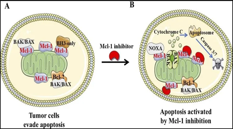


Fig 1 : Small molecule Mcl-1 inhibitor


2.         Biological Roles of MCL-1 in Cancer

i.          The Canonical Anti-Apoptotic Function

By attaching to the BH3 domains of pro-apoptotic proteins and preventing cytochrome c release and mitochondrial membrane permeabilization, MCL-1 inhibits apoptosis [11,12,13]. The sequestering of pro-apoptotic partners maintains mitochondrial integrity by directly binding pro-apoptotic Bcl-2 family members in combination with cell survival factors. This prevents caspase action and triggers apoptotic cell death. Additionally, MCL-1 has been demonstrated to have distinct interactions due to its binding affinities and kinetics with pro-apoptotic family members, which might lead to selective reliance on MCL-1 in certain cancer lines, even tho it shares some important characteristics with other anti-apoptotic paralogs

ii.         Regulation and Post-Translational Modifications

ii.         The accumulation of MCL-1 is regulated by its phosphorylation, ubiquitination, and degradation by the proteasome[14,15,16]. Reversible post-translational modifications, specifically phosphorylation by kinases such as ERK and GSK- 3β at specific amino acid residues, modulate MCL-1 interaction with E3 ligases, such as MULE and β-TRCP, reducing its half-life[18,22]. For example, it has been shown that phosphorylation of MCL-1 at Thr163 by ERK promotes stabilization of the protein in response to growth factor signaling, while phosphorylation by GSK-3β at the same location targets MCL-1 for turnover by ubiquitin- mediated proteolysis[17]. Kinase dysregulation and hyper stabilization of MCL-1 in cancer cells can enhance its anti-apoptotic function.

iii.       Non-Apoptotic Cellular Programs for Tumor Growth

iv.        MCL-1 is involved in non-apoptotic cellular processes that promote tumor growth, according to recent research. MCL-1 interacts with voltage-dependent anion channels (VDAC) in the mitochondrial matrix to improve oxidative phosphorylation and regulate calcium homeostasis [18, 19]. When there is a lack of nutrients, MCL-1 encourages energy reprogramming for growth. Cell survival and metastatic potential can be significantly impacted by MCL-1's regulation of mitochondrial processes, such as cristae construction, metabolism, and ROS signaling [20,21].

v.         Beyond the mitochondria, MCL-1 can context-dependently regulate DNA damage responses, autophagy initiation, and hypoxia stress adaptability, expanding its carcinogenic role beyond apoptosis regulation [22,23,24].

vi.        Immune Modulation

CL-1 has been linked in recent research to the preservation of immune cell survival and function characteristics [29, 30]. Despite promoting lymphocyte survival, overexpression of MCL-1 in tumor-infiltrating immune cells might paradoxically increase immunosuppressive microenvironments in malignancies.M Tumor-associated macrophages (TAMs) can be reprogrammed by selectively targeting MCL-1 to

serve as a dual treatment strategy to eradicate tumor cells and boost antitumor immunity by increasing effector T-cell persistence and inducing tumor cell death [25, 26]. An immunosuppressive tumor microenvironment is promoted by MCL-1 overexpression in TAMs and myeloid-derived suppressor cells (MDSCs), despite the fact that MCL-1 enhances lymphocyte persistence and immune cell survival [27, 28 In this sense, preventingIn addition to causing tumor cells to undergo apoptosis, MCL-1 has the ability to reprogram populations of suppressive immune cells to enhance T-cell-mediated anti-tumor immunity activity.Role of Mcl-1 in Cancer

Numerous human cancer cell lines, including those from the breast, CNS, colon, lung, ovarian, prostate, renal, and melanoma, have high expression of Mcl-1. Mcl-1 protein is expressed by human melanoma cell lines, but not at a higher level than in normal melanocyte controls. However, research on paraffin sections of original melanoma, metastatic melanoma, and benign nevi revealed an increase in Mcl-1 expression that was associated with the advancement of melanoma. [29, 30]

Cancer cells use Mcl-1 overexpression as a strategy to build resistance to various chemotherapeutic drugs and to avoid cell death, i.e., to live. Although they won't be included in this study, the other antiapoptotic members of the Bcl-2 protein family are also commonly overexpressed in malignancies. Mcl-1's capacity to inhibit apoptosis plays a significant role in the altered state even though overexpression of the protein does not lead to increased cell proliferation. As previously mentioned, the equilibrium between pro- and anti-apoptotic proteins within cells is the basis for the regulation of apoptosis. A cancer cell has a significant edge in defending itself against harmful substances because of its capacity to tip the scales in favor of survival. Chemoresistance may be a clinical manifestation of this condition. For instance, several chemotherapeutic drugs are thot to act by initiating apoptosis in a cell, tho they do so thru a number of methods. Therefore, protection from apoptosis thru Mcl-1 overexpression could be a major obstacle to chemotherapeutic drugs' efficacy. [31]

Techniques used to target Mcl-1

Many strategies have been developed to target Mcl-1. While most of these tactics target several anti-apoptotic proteins in the Bcl-2 family, some of them concentrate mainly on Mcl-1. The most important inhibitors created to date are reviewed here: downregulation Many medications exhibit a mechanism of action that involves downregulating Mcl-1, even tho they are not made to explicitly target Mcl-1. Research using these


Drugs continue to show how crucial this protein is to the upkeep and spread of cancer.

3.         Cyclin-dependent kinase inhibitors

4.1.1    Flavopiridol

By vying for the kinase active site, the semisynthetic flavonoid flavopiridol inhibits cyclin-dependent kinases. It causes G1/S and G2/S cell cycle arrest and can decrease the activity of certain CDKs [32]. Additionally, flavopiridol can bind double-stranded DNA and substantially reduce transcription levels in the cell by inhibiting P-TEFb [38]. Flavopiridol's capacity to reduce Mcl-1 protein levels in B-cell chronic lymphocytic leukemia cells, however, was one of the more intriguing findings [33]. Notably, patients with high-risk CLL on a hybrid infusional flavopiridol schedule showed notable activity. A 30-minute loading dosage was administered every week for four to six weeks before a four-hour infusion. Of the 42 patients with refractory CLL in the study, 45% had a partial response. [34]

SNS-032

SNS-032, another cyclin-dependent kinase inhibitor, was first found in a screen designed to find particular Cdk2 inhibitors. Since then, it has been demonstrated that this substance also inhibits Cdk7 and Cdk9. Similar to flavopiridol, SNS-032 exhibits potent cytotoxic effects in vitro and reduces anti-apoptotic proteins like Mcl-1. Unfortunately, when this chemical was tested in clinical studies, the findings were unsatisfactory. [35]

Originally created as a B-Raf inhibitor, sorafenib is a multi-kinase inhibitor that has been proven to inhibit several other kinases, including PDGFR, FLT, Kit, and VEGFR. Sorafenib at pharmacologically attainable concentrations caused apoptosis in human leukemia cells via downregulating Mcl-1. It has been discovered that sorafenib-induced ER stress is the cause of this translation inhibition. [36]WP1130

Deubiquitinate Inhibitors

Mcl-1 is quickly broken down in the cell by a proteasome-dependent process, as was previously covered in this article. Mcl-1 protein levels may rise when the proteasome is blocked. Deubiquitinases (DUBs), which can extract ubiquitin from ubiquitinated Mcl-1, can also save Mcl-1 from destruction. Schwickart et al. discovered that the DUB USP9X is one of the proteins that co-immunoprecipitates with Mcl-1 in a study examining interactions between Mcl-1 and other proteins in the cell. This team postulated that this Mcl-1 may become more stable as a result of contact, and it may be crucial for malignancies that overexpress Mcl-1. In fact, they demonstrated that samples of ductal adenocarcinoma, colon adenocarcinoma, and small cell lung cancer had overexpressed USP9X in addition to Mcl-1. Furthermore, a poor prognosis for multiple myeloma was observed to be correlated with USP9X overexpression. The idea that USP9X knockdown leads to enhanced Mcl-1 degradation is supported by the fact that siRNA knockdown of USP9X reduced Mcl-1 protein levels but not RNA levels. Additionally, this knockdown made leukemia and colon cancer cell lines—which are often resistant to the drug—more sensitive to ABT-737, a substance covered later in this study. [37]

A tiny chemical with several anti-cancer properties is WP1130. It has been demonstrated to cause Bcr-Abl to be removed from the cytoplasm and placed in aggresomes, where it cannot support oncogenic signaling. It also directly inhibits USP9X, which is the most intriguing aspect of this review. WP1130 induces apoptosis, compartmentalizes Bcr-Abl, and lowers Mcl-1 levels when administered as a therapy for CML. [38]

4.2       Antisense oligonucleotide (ASO) treatment

There have been numerous research examining the effects of Mcl-1 ASO treatment on cancer in vitro and in vivo in animal models, but no successful translation of a Mcl-1 ASO treatment into the clinic has been reported. This is perhaps because people may experience problems when using ASO therapy. Because ASO has a very short half-life in the human body, it is challenging to achieve adequate blood concentrations. They can also be broken down by DNases, which exacerbates stability issues. Another problem with ASO delivery is that it is challenging to guide ASO to its precise cellular target.. [39]

4.3       BH3 Mimetics

One of the most promising translational approaches has been the idea of BH3 mimetics. As previously mentioned, physical interactions between the anti-apoptotic Bcl-2 protein and the BH3 proteins ultimately result in pro-apoptotic or anti-apoptotic effects. The BH1, BH2, and BH3 domains of the antiapoptotic proteins fold to form a hydrophobic binding pocket. This The BH3 domain of BH3-only proteins occupies and binds the hydrophobic pocket. [40]. Small compounds that resemble the BH3 domain have been created based on this modeling assumption. These molecules can fit into the hydrophobic pocket of anti-apoptotic proteins and prevent them from binding pro-apoptotic proteins, so impairing their function. Screening experiments have produced some of these mimetics, while structural studies have produced others. Regardless of how these medications were found, they offer a fresh and intriguing approach to the development of cancer treatments. [41].


4.3.1    ABT-737

ABT-737, one of the most effective and well researched BH3 mimetics, was found thru NMR-based screening and showed a high binding affinity to the anti-apoptotic proteins Bcl-2, Bcl-XL, and Bcl-w. Mcl-1 and Bfl-1 are not bound by ABT-737. As was previously mentioned in this review, this variation in affinity is caused by variations in the architectures of these proteins. According to preliminary research, ABT-737 proved successful in causing cytotoxicity in small cell lung cancer (SCLC) and follicular lymphoma when used alone.

lines of cells. Furthermore, it was discovered to increase paclitaxel's lethality in non-small cell lung cancer (NSCLC).)[42ABT-737 has since been extensively researched in a range of malignancies. It has been demonstrated to cause cytotoxicity in glioblastoma, multiple myeloma, leukemia and lymphoma, and SCLC both in vitro and in vivo [43, 44]. There are still many tumors that are resistant to ABT-737, despite numerous encouraging trials in these cancer types. It has been demonstrated that the overexpression of Mcl-1, an anti-apoptotic protein that ABT-737 does not bind to, is the cause of this resistance. Certain cell lines that express larger amounts of Mcl-1 show enhanced resistance to ABT-737, even among malignancies that are sensitive to the drug. [45]. The literature has repeatedly demonstrated Mcl-1-dependent ABT-737 resistance, and numerous studies reveal that downregulating Mcl-1 via a range of pathways can result in sensitivity to ABT-737 [46,47]. Notably, thru a mechanism involving Mcl-1 down-regulation, the CDK inhibitor roscovitine significantly boosted ABT-737 lethality in human leukemia cells.. [48]

4.3.2. Obatoclax (GX15)

Another BH3 mimic is obatoclax. Unlike ABT-737, this mimetic binds to and inhibits Mcl-1 and Bfl-1 in addition to targeting Bcl-2, Bcl-XL, and Bcl-w. Research has demonstrated that this substance causes cytotoxicity in several cancer forms, such as NSCLC and several leukemias. Furthermore, it has demonstrated synergy with a variety of traditional chemotherapeutic drugs [49,50,51]. The significance of Mcl-1 in cancer is further demonstrated by obatoclax. Research has demonstrated that this compound's cytotoxic effects are partially mediated by particular effects on Mcl-1. The constitutive Bak-Mcl-1 interaction on the outer mitochondrial membrane can be interfered with by obatoclax. [52]

compound has advanced to clinical trials and Phase I studies in a variety of cancer types have shown it to be fairly well tolerated, with neurologic toxicities being the most common adverse event seen. Some evidence of efficacy was seen in these studies, though ongoing Phase II trials will ultimately determine the effectiveness of this agent . [53,54]

4.3.3    Gossypol

Gossypol is a polyphenolic aldehyde derived from cottonseed. Original studies with this extract focused on its ability to function as a male contraceptive[55]. However, it has also been shown to have potent anti-cancer activity[56,57,58]. Gossypol functions as a typical BH3 mimetic and binds to and inhibits the Bcl-2 anti-apoptotic proteins Bcl-2, BclXL, and Mcl-1[59]. Many groups have shown Gossypol to exhibit anti-carcinogenic effects toward a wide variety of cancer types both in vitro and in vivo, including, but not limited to, breast, prostate, glioma, and colon cancer . LeBlanc et al. also demonstrated Gossypol’s ability to inhibit growth of adrenal gland carcinoma cells, medullary thyroid carcinoma cells, ovarian carcinoma cells, and endometrial carcinoma cells. Compared to ABT-737, Gossypol displays toxicity against a much wider array of, possibly due to its ability to target Mcl-1. This compound is currently in Phase II clinical trials as a single-agent in patients with advanced malignancies. Clinical trials are also ongoing in evaluating Gossypol in combination therapy with conventional chemotherapeutics. [60] The presence of two reactive aldehyde groups on Gossypol, combined with initial clinical trial results that showed difficulty in achieving sufficient Gossypol blood concentrations, prompted groups to create Gossypol derivatives. Apogossypol was the first derivative designed and showed better efficacy and less toxicity than its parent compound[61]. To date, there have been many such derivatives created with varying binding affinities to the Bcl-2 anti-apoptotic proteins.

4.3.4    Sabutoclax (BI-97C1)

Sabutoclax (BI-97C1) is one of the newest Apogossypol derivatives developed by Wei et al. [62]. This novel compound binds to the Bcl-2 antiapoptotic proteins Bcl-2, Mcl-1, Bcl-XL, and Bfl-1. It was originally identified for its ability to bind Bcl-XL with low to sub micromolar binding affinity. Competitive fluorescence polarization assays showed that BI-97C1 inhibited Bcl-XL with an IC50 value of .31 μM. Further assays showed that this compound also had displacement activity against Bcl-2 (IC50 = 0.32 μM), Mcl-1 (IC50 = 0.20 μM), and Bfl-1 (IC50 = 0.62 μM)[79]. BI-97C1 (Sabutoclax) induced apoptosis in the large B-cell lymphoma cell line, BP3, which expresses high levels of Mcl-1 and Bfl-1 and is resistant to ABT-737. Furthermore, while toxic to wild type mouse embryonic fibroblasts, this compound showed only slight toxicity against BAX/BAK double knockout MEF cells. This provided evidence that BI-97C1 (Sabutoclax) only has minimal off-target effects. Additionally, as BI-97C1 (Sabutoclax) showed efficacy against a cell line that overexpressed Mcl-1, some earlier


compounds like ABT-737 did not, it was evaluated in the context of a prostate cancer xenograft involving M2182 cells, which rely on Mcl-1 for survival. A dose of 3 mg/kg induced nearly 60% tumor inhibition, while a dose of 5 mg/kg nearly eliminated the tumors. [63]

4.         Mechanisms of MCL-1 protein stability induced by MCL-1 antagonists in B-cell malignancies.

The diversity of BH3 proteins can be broadly characterized as pro-survival Pro-survival proteins function by sequestering the pro-apoptotic proteins, thus preventing the initiation of an apoptotic cascade. However, overexpression of pro-survival proteins can help in tumorigenesis and drug resistance, making them an attractive target for inducing apoptosis in cancer cells[64]. The successful development of BCL-2 inhibitor (venetoclax), currently approved for chronic lymphocytic leukemia (CLL) and acute myeloid leukemia (AML), encouraged the development of inhibitors that target other antiapoptotic proteins, particularly Myeloid Leukemia 1 (MCL-1). [65]

One of the most highly amplified genes in a number of tumors, MCL-1 is linked to poor prognosis, medication resistance, and cancer progression. It lessens the sensitivity of cancer cells to chemotherapeutics or targeted treatments and shields them from apoptosis. MCL-1 inhibits cytochrome C release and apoptotic cascade initiation by selectively binding to Bak and Noxa. In addition, MCL-1 is involved in metabolism, cellular senescence, mitophagy, autophagy, DNA repair, and cell cycle regulation. MCL-1 is essential for the development of the embryo and linked to the survival of numerous cells, such as cardiomyocytes, T/B lymphocytes, and the nervous system. [66].

The expression of the short-lived protein MCL-1 is controlled at the transcriptional, post-transcriptional, translational, post-translational, and proteasomal degradation stages. Serine (Ser)/threonine (Thr) phosphorylation at particular sites regulates the stability and degradation of MCL-1.

While GSK3β-mediated Ser phosphorylation causes MCL-1 degradation, ERK-mediated phosphorylation at the Thr residue increases MCL-1 stability. MCL-1's destiny is also determined by its interactions with other proapoptotic proteins like Bim and Noxa; it has been demonstrated that Bim stabilizes MCL-1 protein while Noxa degrades it. MCL-1 is ubiquitinated and degraded by several E3 ligases (Mule, β-TRCP), a process that deubiquitinates can reverse. [67].
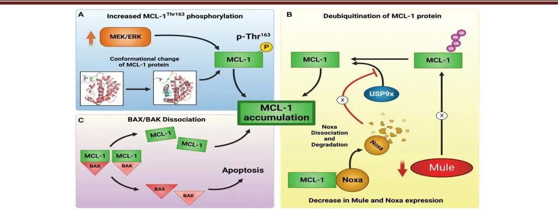


Fig 2:- Mechanisms of MCL-1 inhibitor-induced MCL-1 protein upregulation, stability, and induction of apoptosis


The increase of MCL-1 protein following treatment with MCL-1 inhibitors has been shown to occur via three main routes (A–C). (A) After interacting with MCL-1 inhibitors, the protein undergoes a conformational shift that increases MCL-1Thr163 phosphorylation by the upstream MEK/ERK signaling pathway, which helps to stabilize the MCL-1 protein. (B) Treatment with MCL-1 inhibitors increased DUB activity and caused Noxa to separate from MCL-1. [68]

followed by quick Noxa degradation, which strengthens the USP9x:MCL-1 association and results in MCL-1 stability. Furthermore, MCL-1 inhibitors cause improper ubiquitination of MCL-1 by lowering the amounts of the E3 ligase Mule. Increased stability of the MCL-1 protein is the overall result. (C) MCL-1 separation from the BAX/BAK pro-apoptotic protein complex is caused by MCL-1 inhibitor binding to MCL-1 protein, which facilitates their oligomerization and promotes apoptosis. Red arrows show an inhibitory impact, whereas black arrows show a potentiating effect. The typical course is disrupted, as indicated by X markings.Gene amplification and transcriptional activation of MCL1

Gene amplification of BCLX and MCL1 was found in a significant percentage of human malignancies, particularly lung and breast tumors, according to high resolution analyzes of somatic copy number alterations [83]. Furthermore, employing RNA interference to limit MCL1 expression has an impact on the proliferation of lung cancer cell lines with amplified MCL1 genes. [69] indicating that MCL1 plays a critical function in lung cancer. Results demonstrating that copy number variation of MCL1 predicts overall survival of patients with non-small cell lung cancer (NSCLC) also support this idea [70]. The MCL1 gene's transcription is increased by cytokines such as interleukin (IL)-3, IL-6, and others.growth factors such vascular endothelial growth factor and epidermal growth factor, as well as granulocyte-macrophage colony-stimulating factor10,11. Additionally, it has been demonstrated that the transcription factors activate the MCL-1 promoter.

STAT3 and NF-κ B activation are commonly seen in lung cancer, and their functions have been examined experimentally12–16. Furthermore, EGFR (epidermal growth factor receptor) activates STAT3 and ELK-113,14. Furthermore, a recent study revealed that lung cancer decreases the expression of a microRNA that directly inhibits MCL1 mRNA17, indicating that transcriptional and post-transcriptional regulation in lung cancer activates MCL1 mRNA expression. [71]

4.1       The MCL1 protein stabilization by inhibition of ubiquitin ligases

The ubiquitin proteasome system strongly regulates MCL1 protein production, according to mounting evidence10,18. Several ubiquitin ligases were discovered throughout these investigations. The BH3 domain of MULE (Mcl-1 ubiquitin ligase E3), a HECT domain ubiquitin ligase, enables MULE to preferentially bind with MCL119. It has been demonstrated that hepatocarcinogenesis is induced by suppression of MULE expression thru stabilization of MCL120; however, its role in the development of lung cancer has not been documented [72].

Furthermore, β-TrCP (β-transducin repeat-containing protein) phosphorylates MCL1 at serine 155, serine 159, and threonine 163 by GSK-3β (glycogen synthase kinase-3β)21β-TrCP is an F-box protein that belongs to the SCF (SKP1-cullin 1-F-box protein) family of ubiquitin ligases22 and serves as a substrate recognition component. Lung cancer patients' survival is adversely correlated with both EGFR expression and inactive phosphorylation of GSK-3β at serine 923. However, research is currently being done on the connection between GSK.[73]
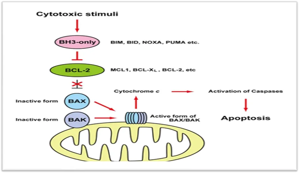


Figure 3: Mitochondrial apoptotic pathway


4.2       MCL1 protein stabilization by overexpression of deubiquitinase

After binding to MCL1, the deubiquitinate USP9X eliminates polyubiquitin chains that designate MCL1 for proteasomal degradation33. Furthermore, reduction of USP9X increases MCL1 turnover and elevated USP9X expression is correlated with increased MCL1 protein in human malignancies, indicating that USP9X stabilizes MCL1 and facilitates cancer cell survival. USP9X expression in NSCLC tissue has been found to be considerably greater in lung cancer.

compared to normal lung tissue, and that a worse prognosis is linked to an increased expression level of USP9X [74].

USP9X may be a p53 target gene, according to a worldwide map of p53 transcription factor-binding sites35. Radiation-induced USP9X makes cancer cells more resistant to treatment because elevated MCL1 protein levels inhibit apoptosis36. These results imply that USP9X stabilizes MCL1 in order to carry out its carcinogenic activity [75]. MCL1's function in lung cancer and its targeted treatment Cell cycle checkpoint activation, DNA replication stress, and/or numerous other stressors cause cells to express more pro-apoptotic proteins during the neoplastic transformation process. Nevertheless, cancer cells adapt to the effects of rising MCL1 and other anti-apoptotic BCL-2 family protein levels in order to survive. Despite the fact that some lung cancer cells do not 

rely on MCL1 for survival38; most lung cancer cells have increased MCL1 expression through a variety of methods. Numerous studies have demonstrated that MCL1 inhibition makes lung cancer cells more susceptible to anticancer medications. Given these findings, BH3 mimetics that selectively inhibit MCL1 and other anti-apoptotic BCL-2 proteins could be useful lung cancer treatments. [76-77]


TABLE 1: MCL-1 inhibitors in clinical trials: Comparative features.

Compound

Selectivity

and potency

Administration route

Response rates

Treatment duration

Reported clinical safety signals

Development status

S64315

High selectivity for MCL-1;

IC₅₀:1.81

nM

Intravenous

BOR

(treatment failure): 81.1%;

Progressive disease:

52.5%

Median treatment duration:

2            cycles

(42 days)

Troponin            I elevation; febrile neutropenia; alanine aminotransferase elevation

Phase   1            trial (acute   myeloid leukemia, myelodysplastic syndrome)

AZD5991

Potent and selective MCL-1

inhibitor;

IC₅₀:0.7

nM

Intravenous

Objective response: 3.8%

3–4 week cycles

Troponin elevation

Phase   1            trial (relapsed                       or refractory hematologic malignancies)

AMG 176

Selective

MCL-1

inhibitor;

IC₅₀:16

nM

Intravenous

No objective responses; transient blast reduction and decreased RBC

transfusion

burden

Median treatment duration:

3            cycles

(84 days

Neutropenia; anemia; hypertension; arrhythmias

(self-resolved)

Phase 1 trial (myelodysplastic syndromes)


5.         Working of Mcl-1 inhibitors

MCL-1 inhibitors are a major development in the field of targeted treatments for cancer. The protein known as myeloid cell leukemia 1 (Mcl-1) is a member of the B-cell lymphoma 2 (Bcl-2) family, which is mainly responsible for controlling apoptosis, the process of programd cell death. Mcl-1 is frequently overexpressed in a variety of malignancies, which helps cancer cells survive and proliferate. In order to take advantage of this vulnerability, researchers have shown a great deal of interest in the creation of Mcl-1 inhibitors.

Mcl-1 inhibitors function by focusing on the function of the Mcl-1 protein in the control of apoptosis. A crucial biological mechanism called apoptosis makes sure that damaged or undesirable cells are methodically broken down and eliminated. These medicinal substances upset the equilibrium of pro- and anti-apoptotic signals in the cell by blocking Mcl-1. In order to stop pro-apoptotic proteins like Bim, Bak, and Bax from causing cell death, Mcl-1 typically attaches to them. These pro-apoptotic proteins are released to start apoptosis when Mcl-1 inhibitors bind to the same locations in a competitive manner. When Mcl-1 is suppressed, cancer cells that are highly dependent on Mcl-1 for survival are particularly vulnerable to programd cell death [78].Safety and Toxicity of MCL-1 Inhibitors

i.    Cardiotoxicity

While the clinical conundrum of increased cardiomyocyte-specific troponin with AZD5991 indicates subclinical myocardial injury, MCL-1 maintains cardiomyocyte survival and hematopoietic stress levels, and its loss in the mouse heart results in decreased heart function independent of apoptosis. Consequently, methods to increase the danger of MCL-1 antagonists in the heart can guaranty suitable dosing techniques, like using cardiac biomarker management and intermittent dosing. In the phase 1 trial, 10.3% of patients receiving AZD5991 had asymptomatic increase of troponin I or T. [79].


II.        Hematopoietic Toxicity

Hematopoietic stem cells, granulocyte-monocyte progenitor cells, and common myeloid progenitor cells are lost when MCL-1 is deleted, which causes hematopoietic failure [80]. Adult mice that had one MCL-1 allele removed had less hematopoietic stem and progenitor cells (HSPCs), which caused them to die in two to three weeks. MCL-1 is necessary for neutrophil survival; mutant mice show 80%–90% decreases in quantities of mature neutrophils in the peritoneal exudates, spleen, and blood. Neutropenia and anemia were the most common grade ≥ 3 adverse events experienced by 62% of  quantities of mature neutrophils in the peritoneal exudates, spleen, and blood. Neutropenia and anemia were the most common grade ≥ 3 adverse events experienced by 62% of subjects in the AMG 176 study.. [81,82]

III.      Immune Effects

The diversity of lymphocyte populations may be reduced by long-term systemic suppression of MCL-1, which could hinder host immunity [83]. It is appropriate to use preclinical immunophenotyping and include solid tumors in clinical trials when the effects of MCL-One deletion manifest.

vii.       Off-Target Manifestations

Similar to cardiotoxicity, prior studies have shown that MCL-1 is an important anti-apoptotic agent for the liver, supporting hepatic homeostasis [84]. Toxicity in preclinical models must also be further assessed for level clarity. Drug transport and off-target mitochondrial disruptions are additional issues to take into account. Current MCL-1 inhibitors' high hydrophobicity may restrict tissue access, and systemic administration may cause undesired mitochondrial stress in areas that are not cancerous. To provide tailored delivery and reduce systemic toxicity, conjugates made especially for tumor targeting and nanoparticle-based delivery methods are being investigated. [85]

V.   Therapeutic Potential and Toxicity Profile

The safety and effectiveness of MCL-1 inhibitors in hematologic malignancies have been investigated in clinical trials, which have shed light on both their therapeutic potential and adverse effects. In a phase 1 study (n = 61) of patients with relapsed/refractory hematologic malignancies, the human MCL-1 inhibitor AZD5991 had poor clinical effect and was linked to a significant incidence of troponin increase [86]. Four patients died as a result of adverse events, which included nausea, vomiting, and diarrhea [87]. Three individuals showed an objective response, and five patients had dose-limiting toxicities. [88].

The safety and tolerability of the MCL-1 inhibitor tapotoclax (AMG 176) in patients with myelodysplastic syndromes (MDS) following hypomethylating agent (HMA) failure were evaluated in a phase I trial. According to the results of this investigation, the study population consisted of adult MDS patients who either showed no improvement after taking HMAs for at least four cycles or showed signs of progression or relapse at any time while using HMAs. The most frequent side effects of the treatment were palpitations, nausea, exhaustion, and diarrhea. In a high-risk group of patients with MDS following HMA failure, this trial found that tapotoclax was both safe and well tolerated; incidences of cardiac adverse events resolved on their own without the need to lower the dosage; and No patients had elevated troponin levels that were clinically significant. [89]

Strong, selective small-molecule inhibitors of the anti-apoptotic protein MCL-1 are S63845 and its clinical derivative S64315. It has been demonstrated that MCL-1 is overexpressed in many cancers, which enables cancer cells to survive and be effectively inhibited by these drugs, causing programd cell death (apoptosis) in tumors exposed to these drugs. [90]

The ability of these medications to cause on-target damage, such as hematopoietic suppression, in addition to their anticancer activity influences the choice to stick with intermittent dosage [91]. Overall, these findings show that MCL-1 inhibitors have therapeutic potential for a variety of hematologic malignancies and that the best dosage techniques are necessary to strike a balance between safety and efficacy.[92]

6.         Apoptosis and the Bcl-2 family of proteins

An organism's ability to maintain homeostasis and carry out regular physiological processes depends on the biological process of apoptosis. A wide range of intricate biological processes involving several proteins result in the cell's capacity to undergo apoptosis. The extrinsic pathway of apoptosis and the intrinsic/mitochondrial pathway of apoptosis are two different but connected processes that can cause apoptosis. In the extrinsic pathway, extracellular ligands like TNF or FasL activate cell surface death receptors (Fass, TNFR). When any of the death receptors are activated, caspase-8 is cleaved and activated, starting a signaling cascade that ends with the cell's demise. Numerous stress cues might trigger the intrinsic route, which includes. [93]The intricate connections of the Bcl-2 family of proteins are the main regulators of this mitochondrial pathway. The first protein in this family, Bcl-2, was identified during research on B-cell lymphoma. Bcl-2 homology (BH) domains allow the proteins in this family to have some sequence similarity. Each member of this family possesses at least one of the four BH domains. [94] One group of the family exhibits pro-apoptotic actions, while the other group exhibits anti-apoptotic effects. Two subgroups of the pro-apoptotic group are identified: one group comprises proteins like Bax and Bak, while the other group contains proteins

such as Bim, Bid, PUMA, and Noxa. Because the members of this subgroup only share sequence similarity with the remainder of the family through their BH3 domain, the latter group is frequently referred to as the BH3 only proteins. The proteins Bcl-2, Mcl-1, Bcl-XL, Bfl-1/A1, and Bcl-w are members of the anti-apoptotic group. An "indirect activation model" is the initial plan. The anti-apoptotic proteins in this scenario attach to Bax/Bak and prevent dimerization. By attaching to the anti-apoptotic proteins and displacing Bax and Bak, the BH3-only proteins carry out their pro-apoptotic functions. Now that Bax and Bak are free to form dimers, MOMP is produced. The second theory is a little more intricate "direct activation model." [95]

The BH3-only proteins in this model are separated into activator and sensitizer proteins. According to this concept, Bax/Bak oligomerization happens when BH3-only proteins attach to and activate Bax/Bak. The activator proteins must be bound by the anti-apoptotic proteins in order for them to bind and activate Bax/Bak. Consequently, the anti-apoptotic proteins are bound by the sensitizer BH3 only proteins, which stops them from engaging with the activator BH3 only proteins. [96]

Since apoptosis regulation is all about balance, it is likely that both of these models will occur. The ratio of pro-apoptotic to anti-apoptotic proteins determines whether a cell will survive or undergo apoptosis at a particular stage of its existence.97]

CONCLUSION

Many substances have been created, synthesized, or extracted from natural sources and tested as Mcl-1 ligands. It is difficult to find ligands that are particularly selective against Mcl1 since it shares structural characteristics with other members of the Bcl-2 family in its active site. The specific targeting of Mcl-1 was demonstrated by numerous reported inhibitors in this review. To do this, it appears that they must bind to the same area as the apoptotic proteins' BH3 helix. Although some are efficient inhibitors, it is true that some inhibitors raise Mcl-1 levels while others lower them, leading to distinct cell outcomes that cannot be anticipated by binding affinity measurement alone.. [98]

MCL-1 is an obvious target for cancer since it is essential to the normal variation of apoptosis and the cellular physiology of mitochondria. The viability of blocking MCL-1 to cause tumor cell death has been demonstrated by the creation of specific inhibitors, PROTACs, and reversible chemotypes. Despite worries about the effectiveness of such therapies in critical tissues, the combination of targeted, cytotoxic, and immune-mediated therapies offers the potential to improve patient-centric targeting therapies and increase persistence. This approach is currently receiving strong consideration. The ultimate goal is to reduce cancer fatalities by optimizing patient selection with MCL-1 affinities and the development of novel biomarkers. Selective MCL-1 inhibitors have been shown in preclinical and early clinical studies to boost apoptotic opportunity, improve chemosensitivity, and work in concert with BCL-2 inhibitors and other standard-of-care treatments—mechanisms that could all improve OS. [99]

REFERENCES

  1. M. Sancho, D. Leiva, E. Lucendo, and M. Orzáez, “Understanding MCL1: From Cellular Function and Regulation to Pharmacological Inhibition,” FEBS Journal 289, no. 20 (2022): 6209–6234.
  2. S. Escudero, E. Zaganjor, S. Lee, et al., “Dynamic Regulation of Long-Chain Fatty Acid Oxidation by a Noncanonical Interaction Between the MCL-1 BH3 Helix and VLCAD,” Molecular Cell 69, no. 5 (2018): 729–743.e7.
  3. K. J. Campbell, S. Dhayade, N. Ferrari, et al., “MCL-1 Is a Prognostic Indicator and Drug Target in Breast Cancer,” Cell Death & Disease 9, no. 2 (2018): 19.
  4. S. Dong and S. K. Alahari, “Small Molecule Mcl-1 Inhibitor for Triple Negative Breast Cancer Therapy,” Frontiers in Cell and Developmental Biology 12 (2024): 1408107.
  5. H. Wang, M. Guo, H. Wei, and Y. Chen, “Targeting MCL-1 in Cancer: Current Status and Perspectives,” Journal of Hematology & Oncology 14, no. 1 (2021): 67.
  6. S. I. Tantawy, N. Timofeeva, A. Sarkar, and V. Gandhi, “Targeting MCL-1 Protein to Treat Cancer: Opportunities and Challenges,” Frontiers in Oncology 13 (2023): 1226289.
  7. Q. Wen, Y. Zhan, H. Zheng, et al., “Elevated Expression of Mcl-1 Inhibits Apoptosis and Predicts Poor Prognosis in Patients With Surgically Resected Non-Small Cell Lung Cancer,” Diagnostic Pathology 14, no. 1 (2019): 108.
  8. O. S. Al-Odat, M. von Suskil, R. J. Chitren, et al., “Mcl-1 Inhibition: Managing Malignancy in Multiple Myeloma,” Frontiers in Pharmacology 12 (2021): 699629.
  9. H. E. Ramsey, M. A. Fischer, T. Lee, et al., “A Novel MCL1 Inhibitor Combined With Venetoclax Rescues Venetoclax-Resistant Acute Myelogenous Leukemia,” Cancer Discovery 8, no. 12 (2018): 1566–1581.
  10. A. Demelash, L. W. Pfannenstiel, L. Liu, and B. R. Gastman, “Mcl-1 Regulates Reactive Oxygen Species via NOX4 During Chemotherapy-Induced Senescence,” Oncotarget 8, no. 17 (2017): 28154–28168.
  11. T. Wright, M. E. Turnis, C. R. Grace, et al., “Anti-Apoptotic MCL-1 Promotes Long-Chain Fatty Acid Oxidation Through Interaction With ACSL1,” Molecular Cell 84, no. 7 (2024): 1338–1353.e8.
  12. H. Widden and W. J. Placzek, “The Multiple Mechanisms of MCL1 in the Regulation of Cell Fate,” Communications Biology 4, no. 1 (2021): 1029.
  13. M. L. Rasmussen, N. Taneja, A. C. Neininger, et al., “MCL-1 Inhibition by Selective BH3 Mimetics Disrupts Mitochondrial Dynamics Causing Loss of Viability and Functionality of Human Cardiomyocytes,” iScience 23, no. 4 (2020): 101015.
  14. X. Wang, M. Bathina, J. Lynch, et al., “Deletion of MCL-1 Causes Lethal Cardiac Failure and Mitochondrial Dysfunction,” Genes & Development 27, no. 12 (2013): 1351–1364.
  15. M. E. Turnis, E. Kaminska, K. H. Smith, et al., “Requirement for Antiapoptotic MCL-1 During Early Erythropoiesis,” Blood 137, no. 14 (2021): 1945–1958.
  16. S. Srivastava, G. Sekar, A. Ojoawo, et al., “Structural Basis of BAK Sequestration by MCL-1 in Apoptosis,” Molecular Cell 85, no. 8 (2025): 1606–1623.e10.
  17. 17. S. N. Willis, L. Chen, G. Dewson, et al., “Proapoptotic Bak Is Sequestered by Mcl-1 and Bcl-xL, but Not Bcl-2, Until Displaced by BH3-Only Proteins,” Genes & Development 19, no. 11 (2005): 1294–1305.
  18. Tong, P. Wang, S. Tan, et al., “Mcl-1 Degradation Is Required for Targeted Therapeutics to Eradicate Colon Cancer Cells,” Cancer Research 77, no. 9 (2017): 2512–2521.
  19. B. Mojsa, I. Lassot, and S. Desagher, “Mcl-1 Ubiquitination: Unique Regulation of an Essential Survival Protein,” Cells 3, no. 2 (2014): 418–437.
  20. S. I. Tantawy, A. Sarkar, S. Hubner, et al., “Mechanisms of MCL-1 Protein Stability Induced by MCL-1 Antagonists in B-Cell Malignancies,” Clinical Cancer Research 29, no. 2 (2023): 446–457.
  21. G. S. Choudhary, T. T. Tat, S. Misra, et al., “Correction: Cyclin E/ Cdk2-Dependent Phosphorylation of Mcl-1 Determines Its Stability and Cellular Sensitivity to BH3 Mimetics,” Oncotarget 15 (2024): 468–469.
  22. A. Slomp, L. M. Moesbergen, E. Eldering, M. J. Kersten, M. C. Minnema, and V. Peperzak, “Phosphatase PP2A Enhances MCL-1 Protein Half-Life in Multiple Myeloma Cells,” Cell Death & Disease 12, no. 3 (2021): 229.
  23. G. L. Q. Tang, J. X. H. Lai, and S. Pervaiz, “Ubiquitin-Proteasome Pathway-Mediated Regulation of the Bcl-2 Family: Effects and Therapeutic Approaches,” Haematologica 109, no. 1 (2024): 33–43
  24. H. Huang, K. Shah, N. A. Bradbury, C. Li, and C. White, “Mcl-1 Promotes Lung Cancer Cell Migration by Directly Interacting With VDAC to Increase Mitochondrial Ca2+ Uptake and Reactive Oxygen Species Generation,” Cell Death & Disease 5, no. 10 (2014): e1482.
  25. U. Anilkumar, M. Khacho, A. Cuillerier, et al., “MCL-1(Matrix) Maintains Neuronal Survival by Enhancing Mitochondrial Integrity and Bioenergetic Capacity Under Stress Conditions,” Cell Death & Disease 11, no. 5 (2020): 321.
  26. S. Jamil, C. Stoica, T. L. Hackett, and V. Duronio, “MCL-1 Localizes to Sites of DNA Damage and Regulates DNA Damage Response,” Cell Cycle 9, no. 14 (2010): 2915–2927.
  27. A. G. Moyzis, N. S. Lally, W. Liang, R. H. Najor, and Å. B. Gustafsson, “Mcl-1 Differentially Regulates Autophagy in Response to Changes in Energy Status and Mitochondrial Damage,” Cells 11, no. 9 (2022): 1469.
  28. J. P. Piret, E. Minet, J. P. Cosse, et al., “Hypoxia-Inducible Factor- 1-Dependent Overexpression of Myeloid Cell Factor-1 Protects Hypoxic Cells Against Tert-Butyl Hydroperoxide-Induced Apoptosis,” Journal of Biological Chemistry 280, no. 10 (2005): 9336–9344.
  29. I. Dzhagalov, A. St John, and Y. W. He, “The Antiapoptotic Protein Mcl-1 Is Essential for the Survival of Neutrophils but Not Macrophages,” Blood 109, no. 4 (2007): 1620–1626.
  30. A. H. Wei, A. W. Roberts, A. Spencer, et al., “Targeting MCL-1 in Hematologic Malignancies: Rationale and Progress,” Blood Reviews 44 (2020): 100672.
  31. M. Molgora and M. Colonna, “Turning Enemies Into Allies- Reprogramming Tumor-Associated Macrophages for Cancer Therapy,” Med 2, no. 6 (2021): 666–681.
  32. I. Truxova, D. Cibula, R. Spisek, and J. Fucikova, “Targeting Tumor- Associated Macrophages for Successful Immunotherapy of Ovarian Carcinoma,” Journal for Immunotherapy of Cancer 11, no. 2 (2023): e005968.
  33. P. Zhou, L. Qian, C. K. Bieszczad, et al., “Mcl-1 in Transgenic Mice Promotes Survival in a Spectrum of Hematopoietic Cell Types and Immortalization in the Myeloid Lineage,” Blood 92, no. 9 (1998): 3226–3239.
  34. V. Fleming, X. Hu, R. Weber, et al., “Targeting Myeloid-Derived Suppressor Cells to Bypass Tumor-Induced Immunosuppression,” Frontiers in Immunology 9 (2018): 398.
  35. Beroukhim R, Mermel CH, Porter D, et al. The landscape of somatic copy-number alteration across human cancers. Nature. 2010; 463: 899-905. doi:10.1038/nature08822
  36. Rosato R, Almenara J, Kolla S, Maggio S, Coe S, Gimnez M, et al. Mechanism and functional role of XIAP and mcl-1 down-regulation in flavopiridol/vorinostat antileukemic interactions. Molecular cancer therapeutics. 2007; 6(2):692. [PubMed: 17308065]
  37. Tan A, Yang X, Berman A, Zhai S, Sparreboom A, Parr A, et al. Phase I trial of the cyclindependent kinase inhibitor flavopiridol in combination with docetaxel in patients with metastatic breast cancer. Clinical cancer research. 2004; 10(15):5038. [PubMed: 15297405]
  38. Dai Y, Rahmani M, Grant S. Proteasome inhibitors potentiate leukemic cell apoptosis induced by the cyclin-dependent kinase inhibitor flavopiridol through a SAPK/JNK-and NF-kappaBdependent process. Oncogene. 2003; 22(46):7108. [PubMed: 14562039]
  39. Holkova B, Perkins EB, Ramakrishnan V, Tombes M, Shrader E, Talreja N, et al. Phase I trial of bortezomib (PS-341 NSC 681239) and alvocidib (flavopiridol NSC 649890) in patients with recurrent or refractory B-cell neoplasms. Clinical cancer research. 2011
  40. Chen R, Wierda W, Chubb S, Hawtin R, Fox J, Keating M, et al. Mechanism of action of SNS-032, a novel cyclin-dependent kinase inhibitor, in chronic lymphocytic leukemia. Blood. 2009; 113(19):4637. [PubMed: 19234140]
  41. Tong W, Chen R, Plunkett W, Siegel D, Sinha R, Harvey RD, et al. Phase I and pharmacologic study of SNS-032, a potent and selective Cdk2, 7, and 9 inhibitor, in patients with advanced chronic lymphocytic leukemia and multiple myeloma. Journal of clinical oncology. 2010; 28(18): 3015. [PubMed: 20479412] NIH-PA Author Manuscript NIH-PA Author Manuscript NIH-PA Author Manuscript
  42. Rahmani M, Davis E, Bauer C, Dent P, Grant S. Apoptosis induced by the kinase inhibitor BAY 43–9006 in human leukemia cells involves down-regulation of mcl-1 through inhibition of translation. J Biol Chem. 2005; 280(42):35217. [PubMed: 16109713]
  43. Yu C, Bruzek L, Meng X, Gores G, Carter C, Kaufmann S, et al. The role of mcl-1 downregulation in the proapoptotic activity of the multikinase inhibitor BAY 43–9006. Oncogene. 2005; 24(46): 6861. [PubMed: 16007148]
  44. Rahmani M, Davis E, Crabtree T, Habibi J, Nguyen T, Dent P, et al. The kinase inhibitor sorafenib induces cell death through a process involving induction of endoplasmic reticulum stress. Mol Cell Biol. 2007; 27(15):5499. [PubMed: 17548474]
  45. Schwickart M, Huang X, Lill J, Liu J, Ferrando R, French D, et al. Deubiquitinase USP9X stabilizes MCL1 and promotes tumour cell survival. Nature. 2010; 463(7277):103. [PubMed: 20023629]
  46. Sun H, Kapuria V, Peterson L, Fang D, Bornmann W, Bartholomeusz G, et al. Bcr-abl ubiquitination and Usp9x inhibition block kinase signaling and promote CML cell apoptosis. Blood. 2011; 117(11):3151. [PubMed: 21248063]
  47. Askari FK, McDonnell WM. Antisense-oligonucleotide therapy. N Engl J Med. 1996; 334(5):316. [PubMed: 8532029]
  48. Muchmore SW, Sattler M, Liang H, Meadows RP, Harlan JE, Yoon HS, et al. X-ray and NMR structure of human bcl-xL, an inhibitor of programmed cell death. Nature. 1996; 381(6580):335. [PubMed: 8692274]
  49. Oltersdorf T, Elmore S, Shoemaker A, Armstrong R, Augeri D, Belli B, et al. An inhibitor of bcl-2 family proteins induces regression of solid tumours. Nature. 2005; 435(7042):677. Paper introduces ABT-737, a compound that has become very important in the field of BH3 mimetics. [PubMed: 15902208]
  50. Chauhan D, Velankar M, Brahmandam M, Hideshima T, Podar K, Richardson P, et al. A novel bcl-2/Bcl-X(L)/Bcl-w inhibitor ABT-737 as therapy in multiple myeloma. Oncogene. 2007; 26(16):2374. [PubMed: 17016430]
  51. Tagscherer KE, Fassl A, Campos B, Farhadi M, Kraemer A, Bck BC, et al. Apoptosis-based treatment of glioblastomas with ABT-737, a novel small molecule inhibitor of bcl-2 family proteins. Oncogene. 2008; 27(52):6646. [PubMed: 18663354]
  52. Hann C, Daniel V, Sugar E, Dobromilskaya I, Murphy S, Cope L, et al. Therapeutic efficacy of ABT-737, a selective inhibitor of BCL-2, in small cell lung cancer. Cancer Res. 2008; 68(7):2321. [PubMed: 18381439]
  53. Mason K, Vandenberg C, Scott C, Wei A, Cory S, Huang DCS, et al. In vivo efficacy of the bcl-2 antagonist ABT-737 against aggressive myc-driven lymphomas. Proc Natl Acad Sci U S A. 2008; 105(46):17961. [PubMed: 19004807]
  54. Chonghaile TN, Letai A. Mimicking the BH3 domain to kill cancer cells. Oncogene. 2008; 27 (Suppl 1):S149. [PubMed: 19641500]
  55. van Delft M, Wei A, Mason K, Vandenberg C, Chen L, Czabotar P, et al. The BH3 mimetic ABT-737 targets selective bcl-2 proteins and efficiently induces apoptosis via Bak/Bax if mcl-1 is neutralized. Cancer cell. 2006; 10(5):389. [PubMed: 17097561]
  56. Okumura K, Huang S, Sinicrope F. Induction of noxa sensitizes human colorectal cancer cells expressing mcl-1 to the small-molecule bcl-2/Bcl-xL inhibitor, ABT-737. Clinical cancer research. 2008; 14(24):8132. [PubMed: 19088028]
  57. Keuling A, Felton KEA, Parker AAM, Akbari M, Andrew S, Tron V. RNA silencing of mcl-1 enhances ABT-737-mediated apoptosis in melanoma: Role for a caspase-8-dependent pathway. PloS one. 2009; 4(8):e6651. [PubMed: 19684859]
  58. Zall H, Weber A, Besch R, Zantl N, Hcker G. Chemotherapeutic drugs sensitize human renal cell carcinoma cells to ABT-737 by a mechanism involving the noxa-dependent inactivation of mcl-1 or A1. Molecular cancer. 2010; 9:164. [PubMed: 20576107]
  59. Chen S, Dai Y, Harada H, Dent P, Grant S. Mcl-1 down-regulation potentiates ABT-737 lethality by cooperatively inducing bak activation and bax translocation. Cancer Res. 2007; 67(2):782. [PubMed: 17234790] NIH-PA Author Manuscript NIH-PA Author Manuscript NIH-PA Author Manuscript
  60. An ongoing phase 1/2a study of ABT-263; pharmacokinetics (PK), safety and anti-tumor activity in patients (pts) with relapsed or refractory chronic lymphocytic leukemia (CLL) [Internet]. 2009. Available from:
  61. Gandhi L, Camidge DR, de Oliveira M, Bonomi P, Gandara D, Khaira D, et al. Phase I study of navitoclax (ABT-263), a novel bcl-2 family inhibitor, in patients with small-cell lung cancer and other solid tumors. Journal of clinical oncology. 2011; 29(7):909. [PubMed: 21282543]
  62. Konopleva M, Watt J, Contractor R, Tsao T, Harris D, Estrov Z, et al. Mechanisms of antileukemic activity of the novel bcl-2 homology domain-3 mimetic GX15-070 (obatoclax). Cancer Res. 2008; 68(9):3413. [PubMed: 18451169]
  63. Li J, Viallet J, Haura E. A small molecule pan-bcl-2 family inhibitor, GX15-070, induces apoptosis and enhances cisplatin-induced apoptosis in non-small cell lung cancer cells. Cancer Chemother Pharmacol. 2008; 61(3):525. [PubMed: 17505826]
  64. Li J, Viallet J, Haura E. A small molecule pan-bcl-2 family inhibitor, GX15-070, induces apoptosis and enhances cisplatin-induced apoptosis in non-small cell lung cancer cells. Cancer Chemother Pharmacol. 2008; 61(3):525. [PubMed: 17505826]
  65. Pan J, Cheng C, Verstovsek S, Chen Q, Jin Y, Cao Q. The BH3-mimetic GX15-070 induces autophagy, potentiates the cytotoxicity of carboplatin and 5-fluorouracil in esophageal carcinoma cells. Cancer Lett. 2010; 293(2):167. [PubMed: 20153924]
  66. Heidari N, Hicks MA, Harada H. GX15-070 (obatoclax) overcomes glucocorticoid resistance in acute lymphoblastic leukemia through induction of apoptosis and autophagy. Cell Death Disease. 2010; 1(9):e76. [PubMed: 21364679]
  67. Nguyen M, Marcellus R, Roulston A, Watson M, Serfass L, Murthy Madiraju SR, et al. Small molecule obatoclax (GX15-070) antagonizes MCL-1 and overcomes MCL-1mediated resistance to apoptosis. Proc Natl Acad Sci U S A. 2007; 104(49):19512. [PubMed: 18040043]
  68. Schimmer A, O’Brien S, Kantarjian H, Brandwein J, Cheson B, Minden M, et al. A phase I study of the pan bcl-2 family inhibitor obatoclax mesylate in patients with advanced hematologic malignancies. Clinical cancer research. 2008; 14(24):8295. [PubMed: 19088047]
  69. Paik P, Rudin C, Brown A, Rizvi N, Takebe N, Travis W, et al. A phase I study of obatoclax mesylate, a bcl-2 antagonist, plus topotecan in solid tumor malignancies. Cancer Chemother Pharmacol. 2010; 66(6):1079. [PubMed: 20165849]
  70. Hwang J, Kuruvilla J, Mendelson D, Pishvaian M, Deeken JF, Siu L, et al. Phase I dose finding studies of obatoclax (GX15-070), a small molecule pan-BCL-2 family antagonist, in patients with advanced solid tumors or lymphoma. Clinical cancer research. 2010; 16(15):4038. [PubMed: 20538761]
  71. Gilbert NE, O’Reilly JE, Chang CJ, Lin YC, Brueggemeier RW. Antiproliferative activity of gossypol and gossypolone on human breast cancer cells. Life Sci. 1995; 57(1):61. [PubMed: 7596222]
  72. Wei J, Kitada S, Rega M, Emdadi A, Yuan H, Cellitti J, et al. Apogossypol derivatives as antagonists of antiapoptotic bcl-2 family proteins. Molecular cancer therapeutics. 2009; 8(4):904. [PubMed: 19372563]
  73. Meng Y, Tang W, Dai Y, Wu X, Liu M, Ji Q, et al. Natural BH3 mimetic (−)-gossypol chemosensitizes human prostate cancer via bcl-xL inhibition accompanied by increase of puma and noxa. Molecular cancer therapeutics. 2008; 7(7):2192. [PubMed: 18645028]
  74. Voss V, Senft C, Lang V, Ronellenfitsch M, Steinbach J, Seifert V, et al. The pan-bcl-2 inhibitor (−)-gossypol triggers autophagic cell death in malignant glioma. Molecular cancer research. 2010; 8(7):1002. [PubMed: 20587533]
  75. Sung B, Ravindran J, Prasad S, Pandey M, Aggarwal B. Gossypol induces death receptor-5 through activation of the ROS-ERK-CHOP pathway and sensitizes colon cancer cells to TRAIL. J Biol Chem. 2010; 285(46):35418. [PubMed: 20837473]
  76. Pang X, Wu Y, Wu Y, Lu B, Chen J, Wang J, et al. (−)-Gossypol, a natural BH3 mimetic, suppresses the growth of human prostate cancer xenografts via modulating VEGF signalingmediated angiogenesis. Molecular cancer therapeutics. 2011
  77. Le Blanc M, Russo J, Kudelka A, Smith J. An in vitro study of inhibitory activity of gossypol, a cottonseed extract, in human carcinoma cell lines. Pharmacological research. 2002; 46(6):551.

[PubMed: 12457630] NIH-PA Author Manuscript NIH-PA Author Manuscript NIH-PA Author Manuscript

  1. Clinical trials with gossypol. provided by: US national institutes of health, US national library of medicine, US department of health and human services [Internet]. 2011. Available from: http://clinicaltrials.gov/ct2/results?term=gossypol
  2. Kitada S, Kress C, Krajewska M, Jia L, Pellecchia M, Reed J. Bcl-2 antagonist apogossypol (NSC736630) displays single-agent activity in bcl-2-transgenic mice and has superior efficacy with less toxicity compared with gossypol (NSC19048). Blood. 2008; 111(6):3211. [PubMed: 18202226]
  3. Wei J, Stebbins J, Kitada S, Dash R, Placzek W, Rega M, et al. BI-97C1, an optically pure apogossypol derivative as pan-active inhibitor of antiapoptotic B-cell lymphoma/leukemia-2 (bcl-2) family proteins. J Med Chem. 2010; 53(10):4166. Describes BI-97C1 for the first time, one of the newest and most promising BH3 mimetics. [PubMed: 20443627]
  4. Kazi A, Sun J, Doi K, Sung S, Takahashi Y, Yin H, et al. The BH3 alpha-helical mimic BH3-M6 disrupts bcl-X(L), bcl-2, and MCL-1 protein-protein interactions with bax, bak, bad, or bim and induces apoptosis in a bax-and bim-dependent manner. J Biol Chem. 2011; 286(11):9382–

92. [PubMed: 21148306]

  1. Lin X, Morgan-Lappe S, Huang X, et al. ‘Seed’ analysis of off-target siRNAs reveals an essential role of Mcl-1 in resistance to the smallmolecule Bcl-2/Bcl-XL inhibitor ABT737. Oncogene. 2007; 26: 39723979. doi:10.1038/sj.onc.1210166
  2. Yin J, Li Y, Zhao H, et al. Copy-number variation of MCL1 predicts overall survival of non-small-cell lung cancer in a Southern Chinese population. Cancer Med. 2016; 5: 2171-2179. doi:10.1002/cam4.774
  3. Zhang C, Cai T, Zhu H, Yang L, Jiang H, Dong X, et al. Synergistic antitumor activity of gemcitabine and ABT-737 in vitro and in vivo through disrupting the interaction of USP9X and mcl-1. Molecular cancer therapeutics. 2011
  4. Hikita H, Takehara T, Shimizu S, Kodama T, Shigekawa M, Iwase K, et al. The bcl-xL inhibitor, ABT-737, efficiently induces apoptosis and suppresses growth of hepatoma cells in combination with sorafenib. Hepatology. 2010; 52(4):1310. [PubMed: 20799354]
  5. Xu H, Krystal G. Actinomycin D decreases mcl-1 expression and acts synergistically with ABT-737 against small cell lung cancer cell lines. Clinical cancer research. 2010; 16(17):4392. [PubMed: 20732961]
  6. Pandit B, Gartel A. New potential anti-cancer agents synergize with bortezomib and ABT-737 against prostate cancer. Prostate. 2010; 70(8):825. [PubMed: 20058240]
  7. Dash R, Azab B, Quinn BA, Shen X, Wang X, Das SK, et al. An apogossypol derivative BI-97C1 (sabutoclax) targeting mcl-1 sensitizes prostate cancer cells to mda-7/IL-24mediated toxicity. Proc Natl Acad Sci U S A. 2011; 108(21):8785. [PubMed: 21555592]
  8. Jiang H, Lin JJ, Su ZZ, Goldstein NI, Fisher PB. Subtraction hybridization identifies a novel melanoma differentiation associated gene, mda-7, modulated during human melanoma differentiation, growth and progression. Oncogene. 1995; 11(12):2477. Discovery of mda-7/ IL-24, a promising protein that promotes cancer specific toxicity. [PubMed: 8545104]
  9. Dash R, Bhutia S, Azab B, Su Z, Quinn B, Kegelmen T, et al. Mda-7/IL-24: A unique member of the IL-10 gene family promoting cancer-targeted toxicity. Cytokine growth factor reviews. 2010; 21(5):381. [PubMed: 20926331]
  10. Emdad L, Lebedeva I, Su Z, Gupta P, Sauane M, Dash R, et al. Historical perspective and recent insights into our understanding of the molecular and biochemical basis of the antitumor properties of mda-7/IL-24. Cancer biology therapy. 2009; 8(5):391. [PubMed: 19276652]
  11. Wang X., Bathina M., Lynch J., et al., “Deletion of MCL-1 Causes Lethal Cardiac Failure and Mitochondrial Dysfunction,” Genes & Development 27, no. 12 (2013): 13511364.
  12. Widden H. and Placzek W. J., “The Multiple Mechanisms of MCL1 in the Regulation of Cell Fate,” Communications Biology 4, no. 1 (2021)
  13. Widden H. and Placzek W. J., “The Multiple Mechanisms of MCL1 in the Regulation of Cell Fate,” Communications Biology 4, no. 1 (2021)
  14. Molgora M. and Colonna M., “Turning Enemies Into Allies-Reprogramming TumorAssociated Macrophages for Cancer Therapy,” Med 2, no. 6 (2021)
  15. Bohler S., Afreen S., Fernandez-Orth J., et al., “Inhibition of the Anti-Apoptotic Protein MCL-1 Severely Suppresses Human Hematopoiesis,” Haematologica 106, no. 12 (2021)
  16. Zhang N., Hartig H., Dzhagalov I., Draper D., and He Y. W., “The Role of Apoptosis in the Development and Function of T Lymphocytes,” Cell Research 15, no. 10 (2005): 749–769.
  17. Vick B., Weber A., Urbanik T., et al., “Knockout of Myeloid Cell Leukemia-1 Induces Liver Damage and Increases Apoptosis Susceptibility of Murine Hepatocytes,” Hepatology 49, no. 2 (2009): 627–636. [ 98.
  18. Affects Intramedullary and Extramedullary Hematopoiesis,” Pharmaceutics 15, no. 4 (2023):

1085.

Reference

  1. M. Sancho, D. Leiva, E. Lucendo, and M. Orzáez, “Understanding MCL1: From Cellular Function and Regulation to Pharmacological Inhibition,” FEBS Journal 289, no. 20 (2022): 6209–6234.
  2. S. Escudero, E. Zaganjor, S. Lee, et al., “Dynamic Regulation of Long-Chain Fatty Acid Oxidation by a Noncanonical Interaction Between the MCL-1 BH3 Helix and VLCAD,” Molecular Cell 69, no. 5 (2018): 729–743.e7.
  3. K. J. Campbell, S. Dhayade, N. Ferrari, et al., “MCL-1 Is a Prognostic Indicator and Drug Target in Breast Cancer,” Cell Death & Disease 9, no. 2 (2018): 19.
  4. S. Dong and S. K. Alahari, “Small Molecule Mcl-1 Inhibitor for Triple Negative Breast Cancer Therapy,” Frontiers in Cell and Developmental Biology 12 (2024): 1408107.
  5. H. Wang, M. Guo, H. Wei, and Y. Chen, “Targeting MCL-1 in Cancer: Current Status and Perspectives,” Journal of Hematology & Oncology 14, no. 1 (2021): 67.
  6. S. I. Tantawy, N. Timofeeva, A. Sarkar, and V. Gandhi, “Targeting MCL-1 Protein to Treat Cancer: Opportunities and Challenges,” Frontiers in Oncology 13 (2023): 1226289.
  7. Q. Wen, Y. Zhan, H. Zheng, et al., “Elevated Expression of Mcl-1 Inhibits Apoptosis and Predicts Poor Prognosis in Patients With Surgically Resected Non-Small Cell Lung Cancer,” Diagnostic Pathology 14, no. 1 (2019): 108.
  8. O. S. Al-Odat, M. von Suskil, R. J. Chitren, et al., “Mcl-1 Inhibition: Managing Malignancy in Multiple Myeloma,” Frontiers in Pharmacology 12 (2021): 699629.
  9. H. E. Ramsey, M. A. Fischer, T. Lee, et al., “A Novel MCL1 Inhibitor Combined With Venetoclax Rescues Venetoclax-Resistant Acute Myelogenous Leukemia,” Cancer Discovery 8, no. 12 (2018): 1566–1581.
  10. A. Demelash, L. W. Pfannenstiel, L. Liu, and B. R. Gastman, “Mcl-1 Regulates Reactive Oxygen Species via NOX4 During Chemotherapy-Induced Senescence,” Oncotarget 8, no. 17 (2017): 28154–28168.
  11. T. Wright, M. E. Turnis, C. R. Grace, et al., “Anti-Apoptotic MCL-1 Promotes Long-Chain Fatty Acid Oxidation Through Interaction With ACSL1,” Molecular Cell 84, no. 7 (2024): 1338–1353.e8.
  12. H. Widden and W. J. Placzek, “The Multiple Mechanisms of MCL1 in the Regulation of Cell Fate,” Communications Biology 4, no. 1 (2021): 1029.
  13. M. L. Rasmussen, N. Taneja, A. C. Neininger, et al., “MCL-1 Inhibition by Selective BH3 Mimetics Disrupts Mitochondrial Dynamics Causing Loss of Viability and Functionality of Human Cardiomyocytes,” iScience 23, no. 4 (2020): 101015.
  14. X. Wang, M. Bathina, J. Lynch, et al., “Deletion of MCL-1 Causes Lethal Cardiac Failure and Mitochondrial Dysfunction,” Genes & Development 27, no. 12 (2013): 1351–1364.
  15. M. E. Turnis, E. Kaminska, K. H. Smith, et al., “Requirement for Antiapoptotic MCL-1 During Early Erythropoiesis,” Blood 137, no. 14 (2021): 1945–1958.
  16. S. Srivastava, G. Sekar, A. Ojoawo, et al., “Structural Basis of BAK Sequestration by MCL-1 in Apoptosis,” Molecular Cell 85, no. 8 (2025): 1606–1623.e10.
  17. 17. S. N. Willis, L. Chen, G. Dewson, et al., “Proapoptotic Bak Is Sequestered by Mcl-1 and Bcl-xL, but Not Bcl-2, Until Displaced by BH3-Only Proteins,” Genes & Development 19, no. 11 (2005): 1294–1305.
  18. Tong, P. Wang, S. Tan, et al., “Mcl-1 Degradation Is Required for Targeted Therapeutics to Eradicate Colon Cancer Cells,” Cancer Research 77, no. 9 (2017): 2512–2521.
  19. B. Mojsa, I. Lassot, and S. Desagher, “Mcl-1 Ubiquitination: Unique Regulation of an Essential Survival Protein,” Cells 3, no. 2 (2014): 418–437.
  20. S. I. Tantawy, A. Sarkar, S. Hubner, et al., “Mechanisms of MCL-1 Protein Stability Induced by MCL-1 Antagonists in B-Cell Malignancies,” Clinical Cancer Research 29, no. 2 (2023): 446–457.
  21. G. S. Choudhary, T. T. Tat, S. Misra, et al., “Correction: Cyclin E/ Cdk2-Dependent Phosphorylation of Mcl-1 Determines Its Stability and Cellular Sensitivity to BH3 Mimetics,” Oncotarget 15 (2024): 468–469.
  22. A. Slomp, L. M. Moesbergen, E. Eldering, M. J. Kersten, M. C. Minnema, and V. Peperzak, “Phosphatase PP2A Enhances MCL-1 Protein Half-Life in Multiple Myeloma Cells,” Cell Death & Disease 12, no. 3 (2021): 229.
  23. G. L. Q. Tang, J. X. H. Lai, and S. Pervaiz, “Ubiquitin-Proteasome Pathway-Mediated Regulation of the Bcl-2 Family: Effects and Therapeutic Approaches,” Haematologica 109, no. 1 (2024): 33–43
  24. H. Huang, K. Shah, N. A. Bradbury, C. Li, and C. White, “Mcl-1 Promotes Lung Cancer Cell Migration by Directly Interacting With VDAC to Increase Mitochondrial Ca2+ Uptake and Reactive Oxygen Species Generation,” Cell Death & Disease 5, no. 10 (2014): e1482.
  25. U. Anilkumar, M. Khacho, A. Cuillerier, et al., “MCL-1(Matrix) Maintains Neuronal Survival by Enhancing Mitochondrial Integrity and Bioenergetic Capacity Under Stress Conditions,” Cell Death & Disease 11, no. 5 (2020): 321.
  26. S. Jamil, C. Stoica, T. L. Hackett, and V. Duronio, “MCL-1 Localizes to Sites of DNA Damage and Regulates DNA Damage Response,” Cell Cycle 9, no. 14 (2010): 2915–2927.
  27. A. G. Moyzis, N. S. Lally, W. Liang, R. H. Najor, and Å. B. Gustafsson, “Mcl-1 Differentially Regulates Autophagy in Response to Changes in Energy Status and Mitochondrial Damage,” Cells 11, no. 9 (2022): 1469.
  28. J. P. Piret, E. Minet, J. P. Cosse, et al., “Hypoxia-Inducible Factor- 1-Dependent Overexpression of Myeloid Cell Factor-1 Protects Hypoxic Cells Against Tert-Butyl Hydroperoxide-Induced Apoptosis,” Journal of Biological Chemistry 280, no. 10 (2005): 9336–9344.
  29. I. Dzhagalov, A. St John, and Y. W. He, “The Antiapoptotic Protein Mcl-1 Is Essential for the Survival of Neutrophils but Not Macrophages,” Blood 109, no. 4 (2007): 1620–1626.
  30. A. H. Wei, A. W. Roberts, A. Spencer, et al., “Targeting MCL-1 in Hematologic Malignancies: Rationale and Progress,” Blood Reviews 44 (2020): 100672.
  31. M. Molgora and M. Colonna, “Turning Enemies Into Allies- Reprogramming Tumor-Associated Macrophages for Cancer Therapy,” Med 2, no. 6 (2021): 666–681.
  32. I. Truxova, D. Cibula, R. Spisek, and J. Fucikova, “Targeting Tumor- Associated Macrophages for Successful Immunotherapy of Ovarian Carcinoma,” Journal for Immunotherapy of Cancer 11, no. 2 (2023): e005968.
  33. P. Zhou, L. Qian, C. K. Bieszczad, et al., “Mcl-1 in Transgenic Mice Promotes Survival in a Spectrum of Hematopoietic Cell Types and Immortalization in the Myeloid Lineage,” Blood 92, no. 9 (1998): 3226–3239.
  34. V. Fleming, X. Hu, R. Weber, et al., “Targeting Myeloid-Derived Suppressor Cells to Bypass Tumor-Induced Immunosuppression,” Frontiers in Immunology 9 (2018): 398.
  35. Beroukhim R, Mermel CH, Porter D, et al. The landscape of somatic copy-number alteration across human cancers. Nature. 2010; 463: 899-905. doi:10.1038/nature08822
  36. Rosato R, Almenara J, Kolla S, Maggio S, Coe S, Gimnez M, et al. Mechanism and functional role of XIAP and mcl-1 down-regulation in flavopiridol/vorinostat antileukemic interactions. Molecular cancer therapeutics. 2007; 6(2):692. [PubMed: 17308065]
  37. Tan A, Yang X, Berman A, Zhai S, Sparreboom A, Parr A, et al. Phase I trial of the cyclindependent kinase inhibitor flavopiridol in combination with docetaxel in patients with metastatic breast cancer. Clinical cancer research. 2004; 10(15):5038. [PubMed: 15297405]
  38. Dai Y, Rahmani M, Grant S. Proteasome inhibitors potentiate leukemic cell apoptosis induced by the cyclin-dependent kinase inhibitor flavopiridol through a SAPK/JNK-and NF-kappaBdependent process. Oncogene. 2003; 22(46):7108. [PubMed: 14562039]
  39. Holkova B, Perkins EB, Ramakrishnan V, Tombes M, Shrader E, Talreja N, et al. Phase I trial of bortezomib (PS-341 NSC 681239) and alvocidib (flavopiridol NSC 649890) in patients with recurrent or refractory B-cell neoplasms. Clinical cancer research. 2011
  40. Chen R, Wierda W, Chubb S, Hawtin R, Fox J, Keating M, et al. Mechanism of action of SNS-032, a novel cyclin-dependent kinase inhibitor, in chronic lymphocytic leukemia. Blood. 2009; 113(19):4637. [PubMed: 19234140]
  41. Tong W, Chen R, Plunkett W, Siegel D, Sinha R, Harvey RD, et al. Phase I and pharmacologic study of SNS-032, a potent and selective Cdk2, 7, and 9 inhibitor, in patients with advanced chronic lymphocytic leukemia and multiple myeloma. Journal of clinical oncology. 2010; 28(18): 3015. [PubMed: 20479412] NIH-PA Author Manuscript NIH-PA Author Manuscript NIH-PA Author Manuscript
  42. Rahmani M, Davis E, Bauer C, Dent P, Grant S. Apoptosis induced by the kinase inhibitor BAY 43–9006 in human leukemia cells involves down-regulation of mcl-1 through inhibition of translation. J Biol Chem. 2005; 280(42):35217. [PubMed: 16109713]
  43. Yu C, Bruzek L, Meng X, Gores G, Carter C, Kaufmann S, et al. The role of mcl-1 downregulation in the proapoptotic activity of the multikinase inhibitor BAY 43–9006. Oncogene. 2005; 24(46): 6861. [PubMed: 16007148]
  44. Rahmani M, Davis E, Crabtree T, Habibi J, Nguyen T, Dent P, et al. The kinase inhibitor sorafenib induces cell death through a process involving induction of endoplasmic reticulum stress. Mol Cell Biol. 2007; 27(15):5499. [PubMed: 17548474]
  45. Schwickart M, Huang X, Lill J, Liu J, Ferrando R, French D, et al. Deubiquitinase USP9X stabilizes MCL1 and promotes tumour cell survival. Nature. 2010; 463(7277):103. [PubMed: 20023629]
  46. Sun H, Kapuria V, Peterson L, Fang D, Bornmann W, Bartholomeusz G, et al. Bcr-abl ubiquitination and Usp9x inhibition block kinase signaling and promote CML cell apoptosis. Blood. 2011; 117(11):3151. [PubMed: 21248063]
  47. Askari FK, McDonnell WM. Antisense-oligonucleotide therapy. N Engl J Med. 1996; 334(5):316. [PubMed: 8532029]
  48. Muchmore SW, Sattler M, Liang H, Meadows RP, Harlan JE, Yoon HS, et al. X-ray and NMR structure of human bcl-xL, an inhibitor of programmed cell death. Nature. 1996; 381(6580):335. [PubMed: 8692274]
  49. Oltersdorf T, Elmore S, Shoemaker A, Armstrong R, Augeri D, Belli B, et al. An inhibitor of bcl-2 family proteins induces regression of solid tumours. Nature. 2005; 435(7042):677. Paper introduces ABT-737, a compound that has become very important in the field of BH3 mimetics. [PubMed: 15902208]
  50. Chauhan D, Velankar M, Brahmandam M, Hideshima T, Podar K, Richardson P, et al. A novel bcl-2/Bcl-X(L)/Bcl-w inhibitor ABT-737 as therapy in multiple myeloma. Oncogene. 2007; 26(16):2374. [PubMed: 17016430]
  51. Tagscherer KE, Fassl A, Campos B, Farhadi M, Kraemer A, Bck BC, et al. Apoptosis-based treatment of glioblastomas with ABT-737, a novel small molecule inhibitor of bcl-2 family proteins. Oncogene. 2008; 27(52):6646. [PubMed: 18663354]
  52. Hann C, Daniel V, Sugar E, Dobromilskaya I, Murphy S, Cope L, et al. Therapeutic efficacy of ABT-737, a selective inhibitor of BCL-2, in small cell lung cancer. Cancer Res. 2008; 68(7):2321. [PubMed: 18381439]
  53. Mason K, Vandenberg C, Scott C, Wei A, Cory S, Huang DCS, et al. In vivo efficacy of the bcl-2 antagonist ABT-737 against aggressive myc-driven lymphomas. Proc Natl Acad Sci U S A. 2008; 105(46):17961. [PubMed: 19004807]
  54. Chonghaile TN, Letai A. Mimicking the BH3 domain to kill cancer cells. Oncogene. 2008; 27 (Suppl 1):S149. [PubMed: 19641500]
  55. van Delft M, Wei A, Mason K, Vandenberg C, Chen L, Czabotar P, et al. The BH3 mimetic ABT-737 targets selective bcl-2 proteins and efficiently induces apoptosis via Bak/Bax if mcl-1 is neutralized. Cancer cell. 2006; 10(5):389. [PubMed: 17097561]
  56. Okumura K, Huang S, Sinicrope F. Induction of noxa sensitizes human colorectal cancer cells expressing mcl-1 to the small-molecule bcl-2/Bcl-xL inhibitor, ABT-737. Clinical cancer research. 2008; 14(24):8132. [PubMed: 19088028]
  57. Keuling A, Felton KEA, Parker AAM, Akbari M, Andrew S, Tron V. RNA silencing of mcl-1 enhances ABT-737-mediated apoptosis in melanoma: Role for a caspase-8-dependent pathway. PloS one. 2009; 4(8):e6651. [PubMed: 19684859]
  58. Zall H, Weber A, Besch R, Zantl N, Hcker G. Chemotherapeutic drugs sensitize human renal cell carcinoma cells to ABT-737 by a mechanism involving the noxa-dependent inactivation of mcl-1 or A1. Molecular cancer. 2010; 9:164. [PubMed: 20576107]
  59. Chen S, Dai Y, Harada H, Dent P, Grant S. Mcl-1 down-regulation potentiates ABT-737 lethality by cooperatively inducing bak activation and bax translocation. Cancer Res. 2007; 67(2):782. [PubMed: 17234790] NIH-PA Author Manuscript NIH-PA Author Manuscript NIH-PA Author Manuscript
  60. An ongoing phase 1/2a study of ABT-263; pharmacokinetics (PK), safety and anti-tumor activity in patients (pts) with relapsed or refractory chronic lymphocytic leukemia (CLL) [Internet]. 2009. Available from:
  61. Gandhi L, Camidge DR, de Oliveira M, Bonomi P, Gandara D, Khaira D, et al. Phase I study of navitoclax (ABT-263), a novel bcl-2 family inhibitor, in patients with small-cell lung cancer and other solid tumors. Journal of clinical oncology. 2011; 29(7):909. [PubMed: 21282543]
  62. Konopleva M, Watt J, Contractor R, Tsao T, Harris D, Estrov Z, et al. Mechanisms of antileukemic activity of the novel bcl-2 homology domain-3 mimetic GX15-070 (obatoclax). Cancer Res. 2008; 68(9):3413. [PubMed: 18451169]
  63. Li J, Viallet J, Haura E. A small molecule pan-bcl-2 family inhibitor, GX15-070, induces apoptosis and enhances cisplatin-induced apoptosis in non-small cell lung cancer cells. Cancer Chemother Pharmacol. 2008; 61(3):525. [PubMed: 17505826]
  64. Li J, Viallet J, Haura E. A small molecule pan-bcl-2 family inhibitor, GX15-070, induces apoptosis and enhances cisplatin-induced apoptosis in non-small cell lung cancer cells. Cancer Chemother Pharmacol. 2008; 61(3):525. [PubMed: 17505826]
  65. Pan J, Cheng C, Verstovsek S, Chen Q, Jin Y, Cao Q. The BH3-mimetic GX15-070 induces autophagy, potentiates the cytotoxicity of carboplatin and 5-fluorouracil in esophageal carcinoma cells. Cancer Lett. 2010; 293(2):167. [PubMed: 20153924]
  66. Heidari N, Hicks MA, Harada H. GX15-070 (obatoclax) overcomes glucocorticoid resistance in acute lymphoblastic leukemia through induction of apoptosis and autophagy. Cell Death Disease. 2010; 1(9):e76. [PubMed: 21364679]
  67. Nguyen M, Marcellus R, Roulston A, Watson M, Serfass L, Murthy Madiraju SR, et al. Small molecule obatoclax (GX15-070) antagonizes MCL-1 and overcomes MCL-1mediated resistance to apoptosis. Proc Natl Acad Sci U S A. 2007; 104(49):19512. [PubMed: 18040043]
  68. Schimmer A, O’Brien S, Kantarjian H, Brandwein J, Cheson B, Minden M, et al. A phase I study of the pan bcl-2 family inhibitor obatoclax mesylate in patients with advanced hematologic malignancies. Clinical cancer research. 2008; 14(24):8295. [PubMed: 19088047]
  69. Paik P, Rudin C, Brown A, Rizvi N, Takebe N, Travis W, et al. A phase I study of obatoclax mesylate, a bcl-2 antagonist, plus topotecan in solid tumor malignancies. Cancer Chemother Pharmacol. 2010; 66(6):1079. [PubMed: 20165849]
  70. Hwang J, Kuruvilla J, Mendelson D, Pishvaian M, Deeken JF, Siu L, et al. Phase I dose finding studies of obatoclax (GX15-070), a small molecule pan-BCL-2 family antagonist, in patients with advanced solid tumors or lymphoma. Clinical cancer research. 2010; 16(15):4038. [PubMed: 20538761]
  71. Gilbert NE, O’Reilly JE, Chang CJ, Lin YC, Brueggemeier RW. Antiproliferative activity of gossypol and gossypolone on human breast cancer cells. Life Sci. 1995; 57(1):61. [PubMed: 7596222]
  72. Wei J, Kitada S, Rega M, Emdadi A, Yuan H, Cellitti J, et al. Apogossypol derivatives as antagonists of antiapoptotic bcl-2 family proteins. Molecular cancer therapeutics. 2009; 8(4):904. [PubMed: 19372563]
  73. Meng Y, Tang W, Dai Y, Wu X, Liu M, Ji Q, et al. Natural BH3 mimetic (−)-gossypol chemosensitizes human prostate cancer via bcl-xL inhibition accompanied by increase of puma and noxa. Molecular cancer therapeutics. 2008; 7(7):2192. [PubMed: 18645028]
  74. Voss V, Senft C, Lang V, Ronellenfitsch M, Steinbach J, Seifert V, et al. The pan-bcl-2 inhibitor (−)-gossypol triggers autophagic cell death in malignant glioma. Molecular cancer research. 2010; 8(7):1002. [PubMed: 20587533]
  75. Sung B, Ravindran J, Prasad S, Pandey M, Aggarwal B. Gossypol induces death receptor-5 through activation of the ROS-ERK-CHOP pathway and sensitizes colon cancer cells to TRAIL. J Biol Chem. 2010; 285(46):35418. [PubMed: 20837473]
  76. Pang X, Wu Y, Wu Y, Lu B, Chen J, Wang J, et al. (−)-Gossypol, a natural BH3 mimetic, suppresses the growth of human prostate cancer xenografts via modulating VEGF signalingmediated angiogenesis. Molecular cancer therapeutics. 2011
  77. Le Blanc M, Russo J, Kudelka A, Smith J. An in vitro study of inhibitory activity of gossypol, a cottonseed extract, in human carcinoma cell lines. Pharmacological research. 2002; 46(6):551.

[PubMed: 12457630] NIH-PA Author Manuscript NIH-PA Author Manuscript NIH-PA Author Manuscript

  1. Clinical trials with gossypol. provided by: US national institutes of health, US national library of medicine, US department of health and human services [Internet]. 2011. Available from: http://clinicaltrials.gov/ct2/results?term=gossypol
  2. Kitada S, Kress C, Krajewska M, Jia L, Pellecchia M, Reed J. Bcl-2 antagonist apogossypol (NSC736630) displays single-agent activity in bcl-2-transgenic mice and has superior efficacy with less toxicity compared with gossypol (NSC19048). Blood. 2008; 111(6):3211. [PubMed: 18202226]
  3. Wei J, Stebbins J, Kitada S, Dash R, Placzek W, Rega M, et al. BI-97C1, an optically pure apogossypol derivative as pan-active inhibitor of antiapoptotic B-cell lymphoma/leukemia-2 (bcl-2) family proteins. J Med Chem. 2010; 53(10):4166. Describes BI-97C1 for the first time, one of the newest and most promising BH3 mimetics. [PubMed: 20443627]
  4. Kazi A, Sun J, Doi K, Sung S, Takahashi Y, Yin H, et al. The BH3 alpha-helical mimic BH3-M6 disrupts bcl-X(L), bcl-2, and MCL-1 protein-protein interactions with bax, bak, bad, or bim and induces apoptosis in a bax-and bim-dependent manner. J Biol Chem. 2011; 286(11):9382–

92. [PubMed: 21148306]

  1. Lin X, Morgan-Lappe S, Huang X, et al. ‘Seed’ analysis of off-target siRNAs reveals an essential role of Mcl-1 in resistance to the smallmolecule Bcl-2/Bcl-XL inhibitor ABT737. Oncogene. 2007; 26: 39723979. doi:10.1038/sj.onc.1210166
  2. Yin J, Li Y, Zhao H, et al. Copy-number variation of MCL1 predicts overall survival of non-small-cell lung cancer in a Southern Chinese population. Cancer Med. 2016; 5: 2171-2179. doi:10.1002/cam4.774
  3. Zhang C, Cai T, Zhu H, Yang L, Jiang H, Dong X, et al. Synergistic antitumor activity of gemcitabine and ABT-737 in vitro and in vivo through disrupting the interaction of USP9X and mcl-1. Molecular cancer therapeutics. 2011
  4. Hikita H, Takehara T, Shimizu S, Kodama T, Shigekawa M, Iwase K, et al. The bcl-xL inhibitor, ABT-737, efficiently induces apoptosis and suppresses growth of hepatoma cells in combination with sorafenib. Hepatology. 2010; 52(4):1310. [PubMed: 20799354]
  5. Xu H, Krystal G. Actinomycin D decreases mcl-1 expression and acts synergistically with ABT-737 against small cell lung cancer cell lines. Clinical cancer research. 2010; 16(17):4392. [PubMed: 20732961]
  6. Pandit B, Gartel A. New potential anti-cancer agents synergize with bortezomib and ABT-737 against prostate cancer. Prostate. 2010; 70(8):825. [PubMed: 20058240]
  7. Dash R, Azab B, Quinn BA, Shen X, Wang X, Das SK, et al. An apogossypol derivative BI-97C1 (sabutoclax) targeting mcl-1 sensitizes prostate cancer cells to mda-7/IL-24mediated toxicity. Proc Natl Acad Sci U S A. 2011; 108(21):8785. [PubMed: 21555592]
  8. Jiang H, Lin JJ, Su ZZ, Goldstein NI, Fisher PB. Subtraction hybridization identifies a novel melanoma differentiation associated gene, mda-7, modulated during human melanoma differentiation, growth and progression. Oncogene. 1995; 11(12):2477. Discovery of mda-7/ IL-24, a promising protein that promotes cancer specific toxicity. [PubMed: 8545104]
  9. Dash R, Bhutia S, Azab B, Su Z, Quinn B, Kegelmen T, et al. Mda-7/IL-24: A unique member of the IL-10 gene family promoting cancer-targeted toxicity. Cytokine growth factor reviews. 2010; 21(5):381. [PubMed: 20926331]
  10. Emdad L, Lebedeva I, Su Z, Gupta P, Sauane M, Dash R, et al. Historical perspective and recent insights into our understanding of the molecular and biochemical basis of the antitumor properties of mda-7/IL-24. Cancer biology therapy. 2009; 8(5):391. [PubMed: 19276652]
  11. Wang X., Bathina M., Lynch J., et al., “Deletion of MCL-1 Causes Lethal Cardiac Failure and Mitochondrial Dysfunction,” Genes & Development 27, no. 12 (2013): 13511364.
  12. Widden H. and Placzek W. J., “The Multiple Mechanisms of MCL1 in the Regulation of Cell Fate,” Communications Biology 4, no. 1 (2021)
  13. Widden H. and Placzek W. J., “The Multiple Mechanisms of MCL1 in the Regulation of Cell Fate,” Communications Biology 4, no. 1 (2021)
  14. Molgora M. and Colonna M., “Turning Enemies Into Allies-Reprogramming TumorAssociated Macrophages for Cancer Therapy,” Med 2, no. 6 (2021)
  15. Bohler S., Afreen S., Fernandez-Orth J., et al., “Inhibition of the Anti-Apoptotic Protein MCL-1 Severely Suppresses Human Hematopoiesis,” Haematologica 106, no. 12 (2021)
  16. Zhang N., Hartig H., Dzhagalov I., Draper D., and He Y. W., “The Role of Apoptosis in the Development and Function of T Lymphocytes,” Cell Research 15, no. 10 (2005): 749–769.
  17. Vick B., Weber A., Urbanik T., et al., “Knockout of Myeloid Cell Leukemia-1 Induces Liver Damage and Increases Apoptosis Susceptibility of Murine Hepatocytes,” Hepatology 49, no. 2 (2009): 627–636. [ 98.
  18. Affects Intramedullary and Extramedullary Hematopoiesis,” Pharmaceutics 15, no. 4 (2023):1085.

Photo
Sudhir Kumar Sah
Corresponding author

B.Pharm Final Year Student, A and E College of Pharmacy, Samastipur, India

Photo
Sana Nusrat Praween
Co-author

A and E College of Pharmacy, Samastipur, India

Sudhir Kumar Sah, Sana Nusrat Praween, A Review on Targeted Cancer Therapy Using MCL -1 Inhibitor, Int. J. of Pharm. Sci., 2026, Vol 4, Issue 10, 1993-2012, https://doi.org/10.5281/zenodo.23357289

More related articles
Design, Formulation, and Evaluation of Norethindro...
Sawase Kiran, Nishant Mali, Avinash Hosmani, Rutuja Jedhe, Yogesh...
Development and Characterization of Diclofenac Sod...
Ritish Sankpal, Vasant Lokhande, Kailas Karande, Smita Borkar...
Evaluation of Ethanolic Extract of Moringa oleifer...
Himani Bharadwaj, Rajesh Asija, Richa Agarwal...
Related Articles
Nose-to-Brain Delivery of Levodopa: A Nasal Route Strategy for Parkinson's Disea...
Janvi kakde, Tushar Shelke, Sushma Nakhate, Sakshi Kakade, Ashish Kalambate...
Genomic Drivers of Cancer Progression and Their Implications for Targeted Therap...
Zakiya Sultana, Mohammad Zulekha, Arindom Patar, Janani Ramachandran...
Design, Formulation, and Evaluation of Norethindrone Acetate-Loaded Cubosomal Hy...
Sawase Kiran, Nishant Mali, Avinash Hosmani, Rutuja Jedhe, Yogesh Patil...
More related articles
Design, Formulation, and Evaluation of Norethindrone Acetate-Loaded Cubosomal Hy...
Sawase Kiran, Nishant Mali, Avinash Hosmani, Rutuja Jedhe, Yogesh Patil...
Development and Characterization of Diclofenac Sodium-Loaded Thermoresponsive Hy...
Ritish Sankpal, Vasant Lokhande, Kailas Karande, Smita Borkar...
Design, Formulation, and Evaluation of Norethindrone Acetate-Loaded Cubosomal Hy...
Sawase Kiran, Nishant Mali, Avinash Hosmani, Rutuja Jedhe, Yogesh Patil...
Development and Characterization of Diclofenac Sodium-Loaded Thermoresponsive Hy...
Ritish Sankpal, Vasant Lokhande, Kailas Karande, Smita Borkar...
