

We use cookies to ensure our website works properly and to personalise your experience. Cookies policy
Department of Pharmaceutics, K. M. Kundnani College of Pharmacy
Stimuli-responsive drug delivery systems have emerged as transformative platforms in precision medicine, offering site-specific, temporally controlled, and minimally invasive therapeutic options. Among various external triggers, ultraviolet (UV) light has garnered considerable attention due to its ability to induce rapid, localized drug release with precise spatiotemporal control. This review comprehensively explores recent advances in UV-responsive nanocarriers developed for controlled drug delivery, including polymeric nanoparticles, liposomes, nanogels, micelles, dendrimers, and metal-based systems. Mechanistic insights into photo-cleavage, photoisomerization, and photothermal effects that drive UV-mediated drug release are critically discussed. The application of these nanocarriers in dermatology, oncology, ophthalmology, antimicrobial therapy, and gene delivery is elaborated, highlighting their potential to revolutionize targeted therapy. Despite promising progress, challenges such as limited tissue penetration of UV light, phototoxicity, and formulation scalability remain significant barriers to clinical translation. Emerging strategies such as dual-stimuli responsiveness, photostable materials, and integration with wearable photonic devices offer potential solutions. This review provides an in-depth analysis of the current landscape, limitations, and future prospects of UV-triggered nanocarriers, with a focus on advancing safer, more effective, and personalized drug delivery systems.
Over recent decades, drug delivery has shifted from conventional methods to controlled drug delivery systems (CDDS) that allow more precise and sustained drug release, ultimately improving patient compliance. Building on this progress, stimuli-responsive (smart) systems have emerged, which can release drugs in response to internal triggers such as pH, enzymes, and redox conditions, or external stimuli like heat, magnetic fields, and light. Among these, UV light stands out due to its ability to provide precise spatial and temporal control, enabling non-invasive and targeted drug release. Various nanocarriers, including liposomes, micelles, hydrogels, and metallic nanoparticles, have been developed to respond to UV exposure through mechanisms like bond cleavage or structural changes. These UV-responsive systems hold strong potential in treating skin conditions such as psoriasis, superficial cancers, and ocular diseases, as well as in antimicrobial and gene delivery applications, offering controlled and on-demand therapeutic effects.
2. UV Light as an External Trigger:
2.1 Types of UV Radiation: Ultraviolet (UV) radiation is a form of electromagnetic energy that lies just beyond the violet region of the visible light spectrum, with wavelengths between 100 and 400 nanometers (nm). Based on their wavelength and energy level, UV rays are categorized into three types: UVA (315–400 nm), UVB (280–315 nm), and UVC (100–280 nm). These variants differ significantly in their tissue penetration depth, biological impact, and suitability for use in UV-responsive nanocarrier systems [1].
UVA radiation, the longest and least energetic, can penetrate into the dermis — the deeper layer of skin. Though less damaging than other forms, UVA contributes to long-term oxidative stress and skin aging by generating reactive oxygen species (ROS). Its deeper tissue reach and lower phototoxicity make it the preferred trigger in many nanocarrier-based drug delivery systems [2,3].
UVB, with its shorter wavelength and higher energy, is largely absorbed in the epidermis. It plays a major role in causing DNA damage, erythema (sunburn), and skin cancer. While UVB can activate drug release from nanocarriers, its cytotoxicity and shallow penetration limit its practical application [4].
UVC radiation has the highest energy but does not naturally reach living tissue, as it is completely filtered by the Earth’s ozone layer. Due to its extreme mutagenic potential and lack of penetration, UVC is not considered suitable for biomedical use, though it is effective for external sterilization processes [6].
Thus, among the three UV types, UVA is most widely employed in drug delivery systems due to its favorable balance between penetration depth and safety. UVB is less favored due to its higher toxicity, and UVC is generally avoided in biomedical applications [5,7].
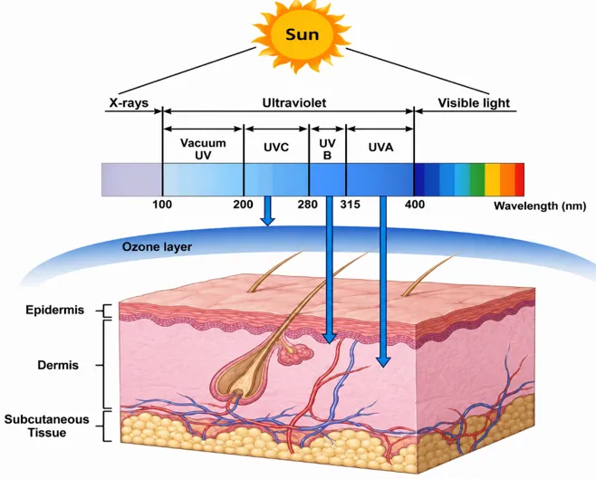
Fig. 2.1.1: UV radiation types and their skin penetration depth and biological effects [7]
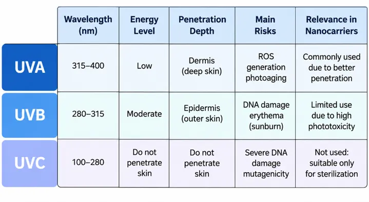
Fig 2.1.2: Overview of UV types and their relevance in nanocarrier drug delivery [1].
2.2 Mechanism:
2.2.1. Photoisomerization: Recent advancements in nanomedicine have enabled the design of light-responsive liposomal nanocarriers that incorporate azobenzene groups within the lipid bilayer structure. Azobenzene molecules exhibit reversible trans–cis isomerization when exposed to UV-A or NIR irradiation, producing conformational alterations that disturb lipid packing and increase membrane permeability. Such structural changes allow controlled, on-demand drug release with improved spatial and temporal accuracy. To address limitations associated with UV light, including limited tissue penetration and possible phototoxic effects, upconversion nanoparticles (UCNPs) are co-encapsulated inside the liposomal carriers. These UCNPs convert deeply penetrating NIR radiation into localized UV emission, which subsequently activates azobenzene isomerization even in subdermal or tumor regions. This hybrid platform merges NIR light penetration with azobenzene photo-switching, creating a stable, biocompatible, externally controlled system for precise therapeutic delivery in advanced drug therapy. [11,12,13].
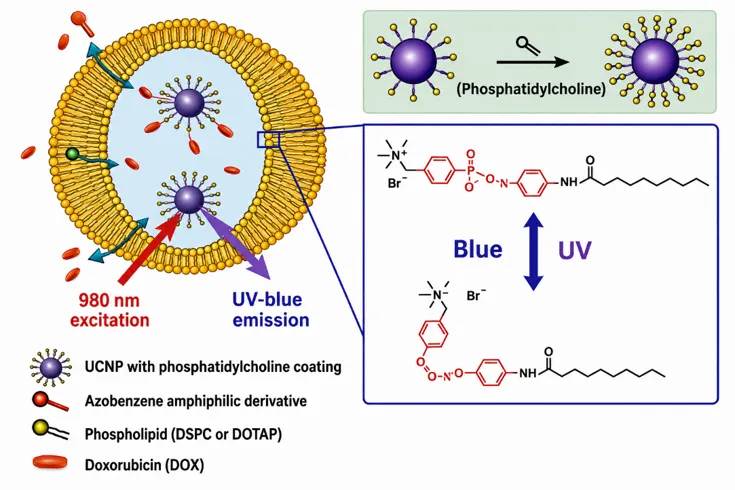
Fig. 2.2.1: UV-triggered photoisomerization of azobenzene for light-controlled drug release [14 ]
2.2.2. Photocleavage: One of the most commonly used strategies for UV-responsive nanocarriers involves photocleavable o-nitrobenzyl (o-NB) derivatives that function as molecular switches for controlled drug release. These groups are covalently attached to therapeutic molecules or polymer backbones and remain stable under normal physiological conditions. However, when exposed to UV-A light (~365 nm), they undergo rapid and irreversible bond cleavage. This photolytic reaction leads to the release of the active drug or an intermediate product with high spatial precision. Importantly, o-NB linkers demonstrate efficient cleavage kinetics, with more than 80% bond dissociation occurring within minutes at relatively low light intensities (≈3.5 mW/cm²), which supports mild and noninvasive activation. In addition, the localized nature of UV irradiation allows site-specific drug release at targeted tissues such as tumors or inflamed regions. Structural modification of o-NB derivatives also enables adjustment of photoreactivity and stability, offering flexibility in nanocarrier design. [17,18].
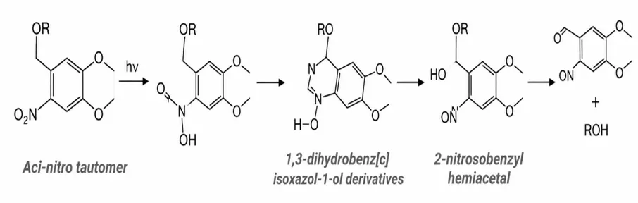
Fig 2.2.2: UV-triggered photocleavage of o-nitrobenzyl for controlled drug release [19].
2.2.3. Photothermal Effects: Another emerging approach for light-controlled drug delivery leverages the photothermal effect, wherein nanomaterials absorb light—typically in the near-infrared (NIR) region—and convert it into localized heat. This heat induces thermoresponsive phase transitions in carrier materials such as poly(N-isopropylacrylamide) (pNIPAAm), which has a lower critical solution temperature (LCST) around 39–42 °C. Below this threshold, the polymer remains hydrated and swollen, but upon NIR-induced heating above the LCST, it collapses and expels the encapsulated drug. To facilitate this response, photothermal agents like gold nanorods, carbon nanotubes, and indocyanine green (ICG) are integrated into the carrier matrix to efficiently generate heat upon irradiation. This strategy enables deep-tissue activation, reversible control, and non-invasive, repeatable release without requiring chemical bond cleavage. Consequently, photothermal-responsive systems show great promise for injectable or implantable nanogels and micelles, offering precise, on-demand therapeutic delivery for cancer treatment and localized inflammation management. [22,23].
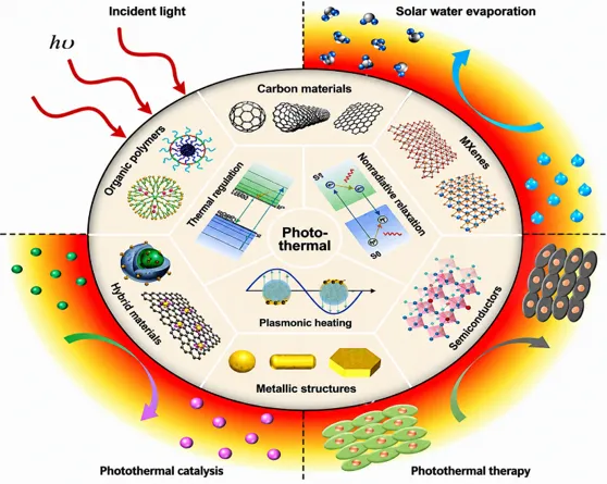
Fig 2.2.3: NIR-triggered photothermal release via heat-activated liposomes [24].
Table 2.2: Comparative Summary of UV-Responsive Mechanisms in Drug Delivery[14,19,24]
|
Sr No. |
Mechanism |
Example |
Trigger Light |
UV Responsive Behaviour |
Type of Release |
|
1. |
Photoisomerization |
Azobenzene- containing liposomes |
UV- A (365nm) or NIR (via UCNPs) |
Reversible Cis–trans Isomerization |
Reversible, on–off control |
|
2. |
Photocleavage |
O-nitrobenzyl- based nano carriers |
UV-A (365nm) |
UV-induced bond cleavage |
Irreversible payload release |
|
3. |
Photothermal Effects |
Thermo responsive polymers + gold/ICG agents |
NIR or Visible |
Localized heating above LCST |
Thermal- Triggered burst |
2.3 Safety considerations and Dose Dependence: Efficient use of light-activated nanocarriers for drug delivery hinges critically on balancing activation efficacy with safety. It showcases how different light-responsive systems—including UV-triggered ones—need precise dose control to avoid undesirable side effects. It emphasizes that too-high exposure levels can exaggerate oxidative stress, damage DNA, or elicit inflammatory responses depending on the light type used [2,6]. When designing such systems, researchers often operate within a "safe window" of light dose: sufficient to trigger photochemical changes (e.g., isomerization or cleavage), yet low enough to minimize collateral cellular damage. In UV-based nanocarriers, this targeted activation is frequently achieved using brief, localized exposure, avoiding full-body irradiation. Technologies like upconversion nanoparticles (UCNPs) further enhance safety by enabling deep-tissue NIR activation that converts into the UV stimulus only at the nanocarrier site, reducing phototoxic risk [25,26].
Table 2.3: Safety factors and strategies for UV-triggered nanocarrier drug delivery [27].
|
Safety Factor |
Recommended Strategy |
|
Optimal dose window |
Defined light intensity and duration |
|
Phototoxicity |
Use of short pulses and local targeting |
|
Excessive heat / oxidation |
Incorporation of stabilizers or filters |
|
Deeper tissue reach |
NIR activation via UCNPs |
|
Controlled released timing |
On/off mechanisms like isomerization switching |
3. Types of UV-Responsive Nanocarriers:
3.1. Liposomes – Liposomes are biocompatible vesicular nanocarriers formed from phospholipid bilayers that are capable of encapsulating both hydrophilic and hydrophobic drugs, making them effective carriers for psoriasis therapy. In UV-responsive liposomes, photosensitive groups such as azobenzene or o-nitrobenzyl are incorporated into the structure. These groups undergo photocleavage or membrane destabilization when exposed to UV light (200–400 nm), which results in controlled drug release specifically at psoriatic lesions during phototherapy. Drugs are commonly incorporated using thin-film hydration or gradient-based active loading techniques. These approaches maintain formulation stability under dark conditions while allowing rapid drug release following light exposure. Such targeted systems enhance skin penetration, concentrate drug activity at diseased sites, and minimize systemic side effects. For instance, docetaxel-loaded liposome-in-gel formulations have demonstrated improved skin retention and reduced inflammatory markers in psoriatic models.[28,29]
Table 3.1.1: Key Features of UV-Responsive Liposomes for Controlled Drug Delivery[30]
|
Feature |
Details |
|
Composition |
Phospholipid bilayers + UV‑sensitive moieties (e.g., azobenzene, nitrobenzyl) |
|
Drug Loading |
Thin-film hydration; gradient-based methods (optional) |
|
Stimulus trigger |
UV light (200–400 nm) |
|
Release behavior |
On-demand drug release via increased membrane permeability |
|
Interactions |
Cancer therapy (proof of concept); potential use in psoriasis treatment |
Table 3.1.2: Examples of Liposomal Formulations for Psoriasis Treatment and Their Therapeutic Effects[30]
|
Nanocarrier |
Drug (API) |
Loading method |
Effects (in vitro/in vivo) |
|
Liposome-in-gel |
Docetaxel |
Thin-film reformulated into gel |
↓IL‑6, HIF‑1α, VEGF; improved skin retention |
|
Standard liposomes |
Tacrolimus, Methotrexate |
Hydration & ultrasonication |
Enhanced penetration, reduced PASI scores |
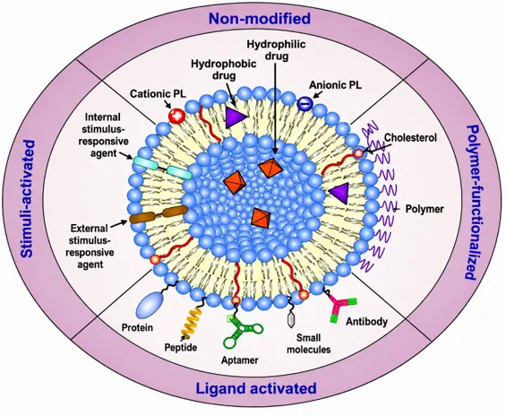
Fig3.1: Surface modification strategies of liposomes with their classification.[28]
1. Polymeric Nanoparticles – Polymeric nanoparticles (PNPs) are nanosized drug carriers (10–1000 nm) composed of biodegradable polymers that allow precise and controlled drug delivery in psoriasis treatment. Designed with photo-cleavable linkers such as o-nitrobenzyl or azobenzene, UV-responsive PNPs enable selective drug release when exposed to light. Following topical application, they penetrate the skin through intercellular or follicular pathways, providing sustained or stimuli-triggered release depending on polymer composition and environmental factors like UV, pH, or temperature. Formulations based on PLGA, PEG, or chitosan integrated with photo-responsive groups have shown UV-induced drug release, improved skin permeation, and effective localized therapeutic action.[31,32]
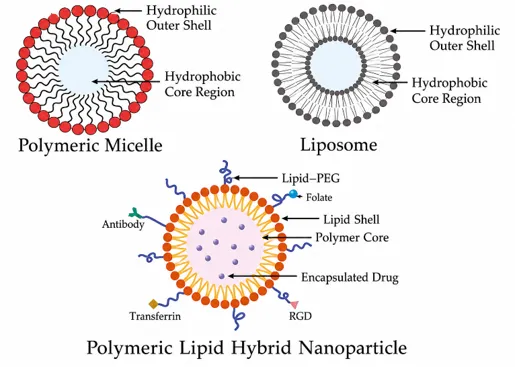
Fig 3.2: Polymeric Micelle, Liposome & Polymeric Hybrid Nanoparticle.[33]
3. Dendrimers – Dendrimers are highly branched, monodisperse polymers with a central core and multiple surface groups that allow both drug encapsulation and conjugation, making them versatile carriers for psoriasis therapy. They are synthesized using divergent or convergent methods, and low-generation dendrimers can penetrate the skin via intercellular and follicular pathways, with positive surface charges further enhancing transdermal delivery. The incorporation of photo-labile groups such as azobenzene or o-nitrobenzyl provides UV-responsiveness, enabling light-triggered bond cleavage and site-specific drug release. Drugs may be loaded within internal cavities or covalently attached to the surface, ensuring controlled and predictable release profiles. [34,35]
Table 3.3.1: Drug Loading Modes and UV-Triggered Release Mechanisms in Dendrimers[36]
|
Loading mode |
Site |
Release trigger |
|
Encapsulation |
Within core cavities |
Diffusion+stimuli |
|
Surface conjugation |
Surface groups |
UV-sensitive bond cleavage |
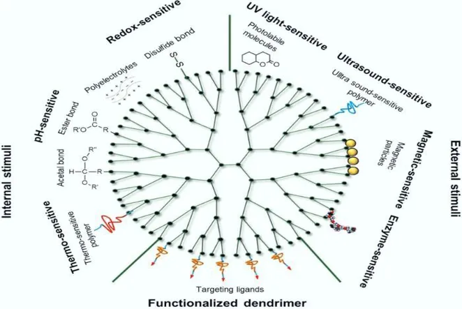
Fig 3.3: Schematic Representation of a Functionalized Dendrimer [34]
4. Micelles – Micelles are nanosized colloidal carriers (10–100 nm) formed through the self-assembly of amphiphilic copolymers, making them efficient systems for targeted drug delivery in psoriasis. UV-responsive micelles incorporate photoactive groups such as spiropyran, azobenzene, or o-nitrobenzyl, which undergo structural changes when exposed to UV light (254–365 nm), altering their polarity and initiating drug release. This photoisomerization destabilizes the micelle core, resulting in controlled or burst drug release depending on the formulation. Drug loading and encapsulation efficiency depend on polymer composition, micelle size, and drug–polymer interactions, with release generally occurring rapidly (within 1–2 hours) under UV exposure while remaining stable in dark conditions. In psoriasis therapy, UV-triggered micelles improve skin penetration, localize drug action at inflammatory lesions, and reduce systemic toxicity, particularly when combined with phototherapy for precise, light-controlled treatment.[37,38]
Table 3.4.1: Composition and Benefits of UV-Responsive Polymeric Micelles [39]
|
Aspect |
Details |
|
Core Material
|
Amphiphilic block copolymers (e.g., PEG-PLA, PEG-PLGA |
|
Responsive moiety |
Spiropyran, Azobenzene, o-Nitrobenzyl |
|
Drug examples |
Curcumin, Tacrolimus |
|
Skin benefits |
Enhanced penetration, localized release, reduced side effects |
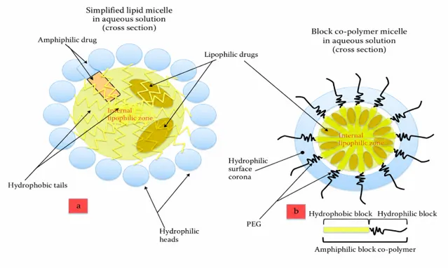
Fig 3.4: Illustration of Polymeric Micelles Demonstrating Drug Encapsulation Mechanisms [37]
5. Hydrogels – UV-responsive hydrogels are smart polymeric networks that undergo structural or chemical changes—such as bond cleavage, isomerization, or swelling—when exposed to UV light, allowing precise and controlled drug release for targeted skin therapy. Their mechanisms involve photocleavage of bonds (e.g., o-nitrobenzyl, azobenzene), photoisomerization (trans–cis conversion that alters pore size), and photothermal effects using embedded agents like gold nanorods or TiO₂, which generate localized heat to trigger drug release. Drugs may be encapsulated within the gel matrix or attached through photosensitive linkages, providing high loading efficiency and adjustable release depending on light intensity and exposure duration. For example, SiO₂/PVP hydrogels have shown up to 50% faster drug release under UV irradiation. Their applications go beyond psoriasis, extending to wound healing (with antibacterial and photothermal hydrogels promoting tissue regeneration) and photoaging treatment (using antioxidant or microalgae-based systems to protect against UV-induced damage).[40,41]
Table 3.5.1: Light-Responsive Drug Delivery Systems and Their Kinetic Profiles [42]
|
System |
Trigger |
Kinetic profile |
Notes |
|
CPT‑nanoparticle hybrid |
UV on/off |
Burst on cycles |
Controlled dosing (Lidsen, 2024) |
|
AuNP‑capped TiO₂ nanotubes |
Visible light |
Hydrophobic cap opens |
Antibiotic release (arXiv, 2020) |
|
PTX‑PLGA NPs in photopolymerizable gel |
UV polymerization |
Initial burst, sustained layered |
Survival benefit in GBM model (Lidsen, 2024) |
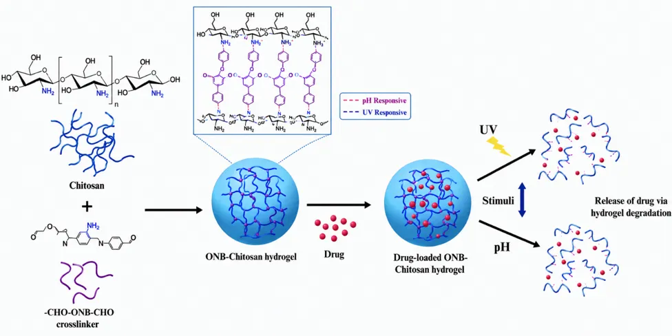
Fig 3.5: Schematic Representation of UV-Responsive Hydrogel Polymer. [40]
6. Metallic Nanoparticles – Metallic nanoparticles (MNPs) are nanoscale carriers composed of metals such as gold, silver, zinc oxide, or titanium dioxide, often surface-modified for biomedical applications. When designed to be UV-responsive, these MNPs allow precise, light-triggered drug release for targeted skin therapy. Their mechanisms include photocatalytic bond cleavage (e.g., o-nitrophenyl, aminoacrylate linkers), photothermal heating generated by metallic cores (e.g., Au, TiO₂), structural shrinkage of core–shell systems that improves penetration, and reactive oxygen species (ROS) generation for combined photodynamic and therapeutic effects. Drugs are typically encapsulated within polymer or silica shells, sealed using UV-cleavable linkers, or coated with photosensitive polymers that degrade upon irradiation. [43,44]
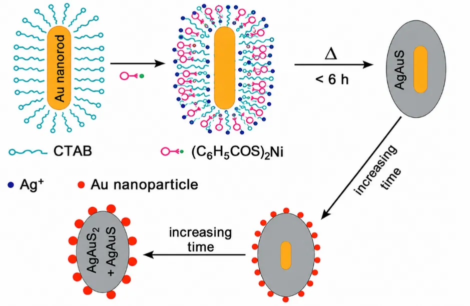
Fig 3.6: Illustration of Metallic Nanoparticles Demonstrating UV Activated Drug Delivery Mechanism[45]
4. Applications of UV-Responsive Nanocarriers:
4.1. Dermatological Applications –
4.1.1 Psoriasis – Liposomal gels have demonstrated strong potential in psoriasis treatment. Methotrexate-loaded liposomes combined with HA-microneedles improved drug delivery in psoriatic mouse models, reducing PASI scores, epidermal thickness, and inflammatory markers (IL-17A, IL-23, TNF-α, Ki67). Ginsenoside Rg3 liposomes incorporated into microneedles provided sustained drug release and reduced skin inflammation. Cationic liposomes containing peptides such as omiganan enhanced solubility, improved skin penetration, and significantly reduced pro-inflammatory cytokines.[51]
4.1.2 Vitiligo – Ultradeformable liposomes co-loaded with psoralen and resveratrol (100–150 nm, +46 mV) have shown effective skin penetration, enhancing melanogenesis and tyrosinase activity in vitro. Resveratrol also offered antioxidant protection, making this dual-loaded system a promising approach for vitiligo management.[46]
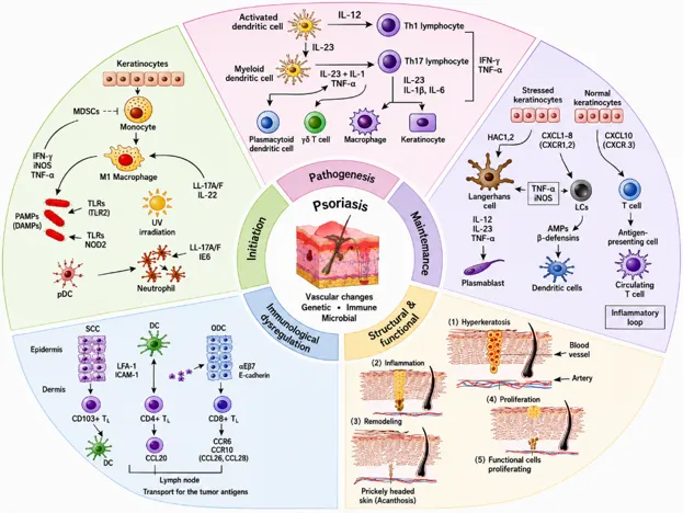
Fig 4.1: Dermatological applications and therapeutic strategies for skin disorders. [46]
4.2. Oncology –Photocleavable nanoparticles are innovative UV-responsive carriers that enable precise, site-specific delivery of chemotherapeutic drugs like methotrexate (MTX) with minimal systemic toxicity. These nanocarriers remain stable in circulation and release their payload only upon targeted UV irradiation, where light-sensitive linkers within the matrix cleave, causing rapid disassembly and localized drug release at the tumor site. This light-triggered mechanism offers exceptional spatial and temporal control, minimizing off-target effects and protecting healthy tissues.[50]
Experimental studies show that these systems enhance MTX uptake, promote ROS generation, and induce apoptosis in tumor cells, demonstrating superior anticancer efficacy. Their controllable, repeatable release under light exposure allows for personalized and non-invasive therapy.[52]
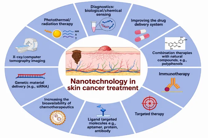
Fig 4.2: Oncological applications for targeted chemotherapeutic interventions [52]
4.3. Ophthalmic Delivery –Delivering drugs to the posterior segment of the eye poses significant hurdles due to anatomical barriers like the blood-retinal barrier and rapid intraocular clearance. Conventional treatments for retinal diseases such as age-related macular degeneration (AMD) and diabetic retinopathy often rely on repeated intravitreal injections, which can be invasive, and carry risks such as infection or retinal detachment.
A promising approach involves the use of UV-A light (365 nm) to trigger in situ crosslinking of injectable hydrogels directly inside the eye. In this method, a preloaded solution containing bevacizumab, a VEGF-inhibiting monoclonal antibody, is injected into the vitreous and then exposed to UV-A light. This exposure initiates rapid hydrogel formation, creating a stable matrix that allows for sustained release of bevacizumab over four months.[47]
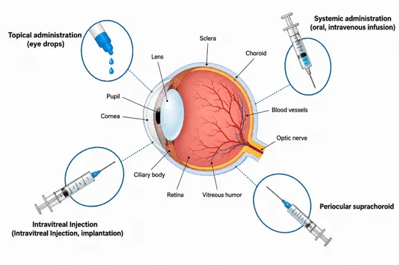
Fig 4.3: Ophthalmic applications for sustained posterior-segment drug release [47]
4.4. Antimicrobial Therapy –To combat antibiotic resistance and improve targeted antibacterial therapy, light-responsive nanocarrier systems have been developed using berberine hydrochloride (BH) loaded into mesoporous titanium nanoparticles (MTNs) coated with polyethyleneimine (PEI) for enhanced stability and drug loading. Upon UV exposure, these MTNs exhibit a dual antibacterial action - controlled release of berberine and photocatalytic generation of reactive oxygen species (ROS). The released berberine disrupts bacterial membranes and metabolism, while ROS cause oxidative damage, together achieving potent, localized bacterial eradication. This synergistic, UV-triggered approach offers a promising strategy for treating persistent infections, especially in wounds or implant-associated sites, with precise control, minimal side effects, and reduced risk of resistance.[48]
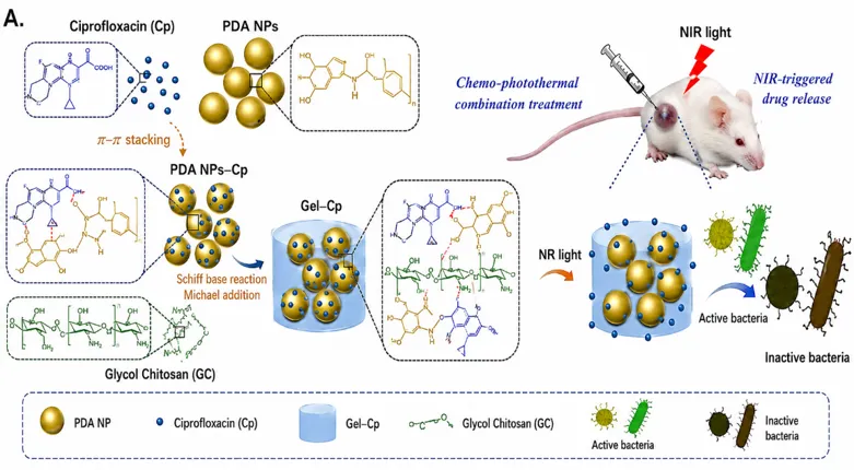
Fig 4.4: Antimicrobial applications showing dual antibacterial action via berberine release [48]
4.5. Gene Delivery –A cutting-edge approach to gene silencing uses azobenzene-functionalized DNA nanostructures that enable precise, UV-controlled release of small interfering RNA (siRNA) for targeted cancer therapy. These nanostructures remain stable under physiological conditions but undergo a conformational change when exposed to 365 nm UV light, as azobenzene groups switch from trans to cis form, disrupting DNA base pairing and triggering siRNA release exactly at the desired site. This light-responsive system offers exceptional spatial and temporal control, reducing systemic exposure and minimizing off-target effects. Moreover, the reversible nature of azobenzene isomerization allows for potential on-demand reactivation, making it a promising, selective, and safe platform for next-generation gene-based therapies.[49]
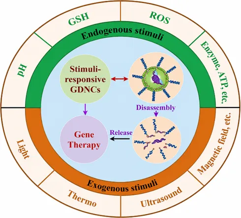
Fig 4.5: Gene delivery applications for controlled siRNA release. [49]
5. Formulation Challenges:
5.1. Limited Tissue Penetration of UV Light –Ultraviolet (UV) light in the wavelength range of 200–400 nm possesses sufficient photon energy to initiate photochemical reactions. This characteristic makes UV light an attractive external stimulus for triggering drug release from photosensitive nanocarriers, particularly through mechanisms like bond cleavage (e.g., o-nitrobenzyl linkers) or structural isomerization (e.g., azobenzene moieties). However, a major challenge that limits the clinical application of UV-responsive systems is the inherently low tissue penetration capability of UV radiation.Biological tissues present multiple barriers to UV light transmission. UV light is readily scattered and absorbed by components such as water and especially melanin, which is abundant in the skin’s epidermal layer. This leads to substantial attenuation of UV energy before it can reach subdermal or deeper tissues.[7,53] Prolonged or repeated exposure to UV light raises safety concerns due to potential DNA damage, oxidative stress, and inflammatory responses. [2,54]
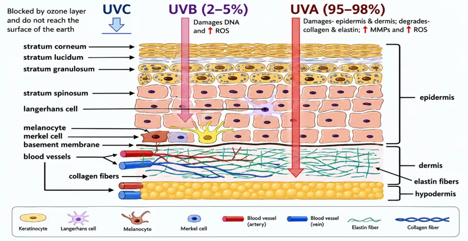
Fig 5.1: UV penetration depth limits for drug delivery to skin layers.[55]
5.2. UV‑Induced Drug / Photo‑Degradation –Ultraviolet (UV) radiation has a dual role in drug delivery and treatment. While its ability to break specific chemical bonds makes it a powerful tool for controlled drug release. It also presents a significant challenge in terms of drug stability. Many drugs, particularly those containing photo-labile groups like nitrobenzyl esters or aromatic chromophores, are vulnerable to degradation upon exposure to UV light. [27]
It restricts the range of active pharmaceutical ingredients (APIs) that can be safely used in UV-based drug delivery systems. Therefore, designing such formulations requires a strategic selection of molecules that either resist photodegradation or are activated only under specific UV conditions.[25] Additionally, shorter wavelengths like UVC and UVB are more energetic and can cause deeper molecular damage, while UVA, though less energetic, can still induce degradation over longer exposure durations.[7]
Example:
Methoxsalen Degradation in Psoriasis Treatment: Methoxsalen is a photosensitizing agent used in PUVA (Psoralen + UVA) therapy for the treatment of psoriasis and other skin conditions. When activated by UVA radiation, Methoxsalen binds to DNA in skin cells and slows their abnormal growth, providing therapeutic benefit. However, excessive exposure to UVA can lead to photodegradation of Methoxsalen, producing harmful by-products or rendering the drug inactive. To overcome this, researchers are exploring UV-responsive nanocarrier systems that can protect Methoxsalen from premature degradation while still allowing for controlled UVA-triggered activation at the target site. These nanocarriers act as both protectants and regulators, improving drug stability, minimizing side effects, and enhancing patient outcomes in phototherapy-based treatments.[56]
5.3. Cytotoxicity and Phototoxicity –Ultraviolet (UV) radiation is known to induce the formation of reactive oxygen species (ROS) and cause direct DNA damage, which can trigger harmful effects on living cells. These effects are particularly concerning when they impact non-target tissues, where unintended damage can occur. Phototoxic reactions such as - inflammation, cell membrane disruption are often reported in laboratory cell models, especially when cells are repeatedly exposed to high doses of UV light. Such exposure may result in mitochondrial dysfunction and apoptosis, contributing to tissue injury. [2,26] To address these risks, UV-responsive nanocarriers must be carefully engineered optimizing their physicochemical properties, such as surface charge, size, and photo reactivity to improve safety. [25,57]
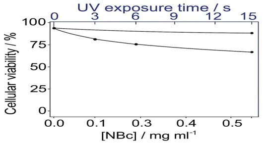
Fig 5.3: UV exposure lowers cell viability, showing phototoxic risk.[58]
5.4. Scale-Up Challenges and Reproducibility –The development of UV-responsive nanocarriers for drug delivery presents significant challenges when transitioning from laboratory-scale experiments to large-scale manufacturing. One major hurdle is the complexity of formulation, which often involves delicate chemical linkages, such as photo-cleavable bonds or photosensitive moieties, that require precise control during synthesis. These systems also demand highly specific and sensitive fabrication protocols, making them susceptible to inconsistencies when produced in bulk. Furthermore, batch-to-batch reproducibility becomes difficult to achieve due to the sensitivity of these carriers to small variations in formulation conditions, such as pH, light intensity, and temperature. These inconsistencies can impact the functional performance of the nanocarrier, including drug release profiles and targeting capabilities. [31,59, 60]
5.5. Regulatory and Safety Concerns –Despite their advanced functionality, UV-triggered nanocarriers raise serious safety and regulatory concerns, particularly due to the phototoxicity and mutagenic potential of UV radiation. UV light, especially in the UVB and UVC ranges, can induce DNA damage, oxidative stress, and inflammation in biological tissues. Regulatory bodies like the FDA (Food and Drug Administration) and EMA (European Medicines Agency) have therefore established strict limits on UV exposure for medical applications. This makes approval pathways for UV-activated systems highly complex. In addition, the long-term systemic effects of UV-triggered drug release are not yet fully understood, which introduces further uncertainty in clinical adoption. Most notably, nanocarriers that rely on external UV activation may face challenges in terms of justifying safety and efficacy data, particularly in deep tissue applications where UV penetration is limited. [61,62, 63]
6. Recent Challenges:
|
Sr. No. |
Challenge |
Cause |
Impact |
|
1 |
Stratum Corneum Resistance[64] |
Hyperkeratosis thickens stratum corneum |
Acts as strong barrier, limiting nanogel penetration |
|
2 |
Limited Drug Loading Capacity[65] |
Restricted internal space for hydrophobic drugs |
Causes instability, viscosity changes, and poor performance |
|
3 |
Environmental Instability[66] |
Sensitive to light, heat, humidity |
Leads to drug leakage, degradation, and reduced shelf life |
|
4 |
Photocatalyst Toxicity[67] |
TiO₂/ZnO generate ROS under UV |
Induces oxidative stress and skin irritation |
|
5 |
Inconsistent Light Exposure[68] |
Variation in UV intensity/time |
Causes uneven drug release and variable results |
|
6 |
Poor Adhesion on Oily/Scalp Areas[69] |
Presence of sebum and hair |
Reduces contact time and treatment efficiency |
|
7 |
Patient Application Issues[70] |
Manual application inconsistency |
Leads to uneven absorption and reduced efficacy |
|
8 |
Lack of Approved Products[71] |
Mostly preclinical stage
|
No FDA-approved nanogels; limits clinical adoption |
7. Future Directions and Research Gaps:
|
Sr. No. |
Focus Area |
Approaches |
Impact |
|
1 |
Dual-Responsive Nanogels[72] |
Respond to both light & heat. |
Precise, site-specific drug release. |
|
2 |
AI-Based Formulation[73] |
Use AI/ML for optimization |
Predicts behavior, saves time, enables personalization |
|
3 |
Gene-Targeted Nanogels[74] |
Deliver siRNA/CRISPR tools |
Silences psoriasis-related genes (IL-17, TNF-α) |
|
4 |
Antioxidant Co-Loading[75] |
Add resveratrol or curcumin |
Reduces UV-induced ROS damage |
|
5 |
Long-Acting Depot Gels[76] |
Form skin drug depots |
Sustained release for 3–7 days |
|
6 |
Personalized Nanogels[77] |
Tailor to genetic/cytokine profile |
Improves efficacy, reduces side effects |
|
7 |
Microneedle Delivery[78] |
Use dissolvable microneedles |
Enhances skin penetration |
|
8 |
Spray/Roll-On Formats[79] |
Develop easy-apply systems |
Improves convenience and compliance |
CONCLUSION
UV-responsive nanocarriers represent a cutting-edge approach in smart drug delivery, enabling precise spatial and temporal control of therapeutic release through UV light activation. These systems enhance localized drug action, reduce systemic toxicity, and are particularly beneficial in skin disorders like psoriasis. Upon UV exposure, they undergo structural or chemical transformations—such as bond cleavage or polymer degradation—facilitating controlled release. Diverse materials, including photo-cleavable hydrogels, micelles, liposomes, metallic nanoparticles, and dendrimers, have shown high potential due to their adaptability and efficiency.
However, clinical translation remains limited by challenges such as UV-induced phototoxicity, DNA damage risks, synthesis complexity, and scalability issues. Future advancements should prioritize safer stimuli (e.g., UV-A or NIR), biodegradable polymers, and green synthesis methods. Integrating multi-stimuli responsiveness (e.g., UV + pH or redox) may further enhance precision and therapeutic outcomes. In summary, despite existing barriers, UV-responsive nanocarriers hold immense promise for next-generation, patient-centric drug delivery—offering targeted, on-demand, and minimally invasive treatment solutions.
REFERENCES

Dr. Monal Jain, Auston Rodrigues, Pratik Chavan, Abhijit Das, Rushi Busamwar, Advances in UV-Responsive Nanocarriers for Controlled Drug Delivery: A Comprehensive Review, Int. J. of Pharm. Sci., 2026, Vol 4, Issue 9, 568-588. https://doi.org/10.5281/zenodo.22309088
 10.5281/zenodo.22309088