

We use cookies to ensure our website works properly and to personalise your experience. Cookies policy
Department of Pharmaceutics, KK College of Pharmacy, The Tamil Nadu Dr. M.G.R. Medical University, Chennai India.
Topical drug delivery offers a major improvement over traditional systemic routes. It provides localized therapeutic effects, avoids first-pass metabolism, and reduces gastrointestinal irritation. However, getting drugs to effectively penetrate the skin remains a significant challenge due to the tough barrier of the stratum corneum. Common topical formulations like creams, ointments, and gels often struggle with poor drug absorption, instability, skin irritation, and low patient adherence. Vesicular drug delivery systems have emerged as new nanocarriers to address these issues. This review gives an insightful look into the skin's structure, as well as the main routes for drug entry: transepidermal and transappendageal. It also discusses several innovative vesicular systems, such as liposomes, niosomes, transfersomes, ethosomes, transethosomes, spanlastics, and novasomes. Each system is examined for its unique structure, how it helps drugs penetrate the skin, its advantages, and its drawbacks. Liposomes are biocompatible and allow for sustained release. Niosomes offer better chemical stability. Transfersomes have excellent deformability for deeper penetration. Ethosomes use ethanol to boost penetration. Spanlastics combine elasticity with a surfactant structure. Novasomes provide high encapsulation efficiency in multilamellar cores. The review also describes general preparation methods showing their importance in creating scalable and reproducible formulations. In conclusion, vesicular systems represent a significant step forward in topical drug delivery. They allow controlled release, better bioavailability, fewer side effects, and enhanced therapeutic effectiveness. Continued innovation in vesicle design and production has great potential to expand the use of topical treatments in dermatology, cosmetics, and transdermal medicine.
Topical drug delivery offers a new alternative to traditional drug delivery methods. It avoids passing through the gastrointestinal tract, the liver's first pass effect, and gastrointestinal irritation. These products are meant for external use and are applied directly to the skin. They act locally by penetrating one or more layers of the skin or reaching the systemic circulation (1,2,3). The skin is the largest organ in the human body. It protects against the external environment and has three layers: the epidermis, dermis, and hypodermis. The skin acts as a barrier against the absorption of various chemicals and biological agents. The stratum corneum (SC) is the outermost layer of the epidermis. It serves as the main barrier that limits drug transport through the skin (2).
Traditional topical products deliver drugs into different skin layers, such as creams, lotions, ointments, and gels. Often, the area of application is not covered, leading to potential loss or evaporation of the drug into the atmosphere or into the skin. Clothing can also remove some products. A major drawback of topical drug delivery is localized skin irritation. Some medications have low permeability, making it difficult for them to pass through the skin, which leads to low drug concentration availability. Larger particles are also not effective (4,5,7). Certain enzymes in the skin can metabolize some drugs before they are absorbed. Applying these products can take time, and the regimen might be complicated, messy, or uncomfortable. New drug delivery systems aim to tackle these challenges while improving the safety and effectiveness of topical treatments. These methods provide significant benefits over traditional forms, such as better drug penetration, less skin irritation, and improved stability (6).
Topical vesicular systems are used to enhance drug delivery to the skin. They improve patient compliance by maintaining controlled and steady drug levels, reducing the risk of over- or under-dosing, and decreasing how often the drug needs to be applied. Vesicular carriers include liposomes, niosomes, ethosomes, deformable vesicles, and other specialized novel carriers. Liposomes contain phospholipids as the lipophilic phase and an aqueous core, giving them their dual nature. Niosomes consists of non-ionic surfactants within lipid vesicles. Ethosomes are made from ethanol, which acts as a permeation enhancer. Transfersomes are phospholipid-based vesicles with an edge activator. Transethosomes add a permeation enhancer to ethosomes. Spanlastics are ultra-deformable niosomal vesicles made with span and an edge activator. Novasomes are multi-bilayer vesicular drug-delivery systems made from non-ionic surfactants, free fatty acids, and cholesterol (8,9,22).
2. STRUCTURE OF THE SKIN
The skin is one of the largest organs in the human body. In a person weighing 70 kg, the skin accounts for over 5 kg of body weight and covers about 2 m². It serves as the body’s primary line of defense (5,12). The skin temperature varies from 30 to 40°C based on environmental conditions. It is a complex barrier made up of three layers: epidermis, dermis and hypodermis (16). Its main functions are to act as a barrier against environmental threats like UV radiation, chemicals, physical damage, and microbes. It also prevents water loss, regulates temperature, and allows for self repair (10).
2.1. Epidermis:
The epidermis, which is the skin’s outer layer, varies in thickness from 0.05 to 1 mm depending on the specific area of the body (10). The epidermis is mainly made up of three types of cells: keratinocytes, melanocytes, and Langerhans cells.
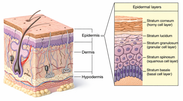
Figure 1: Structure of the skin and the epidermal layers
Keratinocytes account for about 95% of the cells in the epidermis. Typically, the epidermis has four layers: stratum basale, stratum spinosum, stratum granulosum, and stratum corneum (SC) (13).
The SC is the outermost layer of the epidermis. It acts as a barrier, controlling what substances enter and exit the body. This model describes the SC as a brick-and-mortar system. In this system, terminally differentiated keratinocytes (corneocytes) act as bricks, surrounded by lipid membranes. The lipid membranes of the SC mainly consist of free fatty acids, ceramides, and sterols. The barrier properties of the stratum corneum mainly depend on its makeup, which is about 5–10% lipids, 5–10% water, and 75–80% protein when dry. When fully hydrated, it can expand several times its thickness to around 10 µm while remaining somewhat waterproof but flexible (5). The dermal-epidermal junction (DEJ) is a special membrane zone that connects the epidermis and dermis. It links the two layers together and helps resist external shearing forces (10,16,17).
2.2. Dermis and Hypodermis:
The dermis lies beneath the epidermis and is linked to it by a network of proteins and glycoproteins. Collagen, comprising approximately 80–85% of dermal dry weight, provides tensile strength, while elastin and associated microfibrils contribute to skin elasticity and resilience. The dermis also contains non-collagenous glycoproteins, including fibronectins, fibulins, and integrins, which facilitate cell adhesion and migration. Most blood vessels are located in the dermis and hypodermis, though they may extend into the epidermis during abnormal growth. This layer is critical for drug distribution, as drugs reaching the dermis can enter systemic circulation or undergo metabolism. Beneath it, the hypodermis consists mainly of adipose tissue and serves as a reservoir for lipophilic drugs, affecting their retention and release. Therefore, topical drug delivery systems should ensure controlled skin penetration while limiting systemic exposure (12,14).
2.3 Routes of Drug Penetration via Skin
To reach systemic circulation, drugs must first penetrate the skin’s molecular barrier. Drug absorption through the stratum corneum (SC) generally occurs via two main pathways: transepidermal and transappendageal.
2.3.1. Transepidermal Route: The transepidermal route is the primary pathway for skin absorption, allowing drugs to diffuse across the SC either through cells (transcellular) or between cells (intercellular). In the transcellular pathway, drugs pass through SC cells and must cross lipid bilayer membranes, making this route more favorable for hydrophobic drugs due to the lipid-rich nature of cell membranes. The intercellular pathway involves diffusion through interlamellar regions composed of ordered lipids and hydrophobic chains, forming non-planar channels between cells. Hydrophilic molecules mainly diffuse laterally through aqueous regions or less densely packed lipid domains (18,19).
2.3.2. Transappendageal Route: The transappendageal route involves drug penetration through skin appendages such as hair follicles and sebaceous glands. This pathway is particularly useful for polar, ionizable, and larger drug molecules that may have difficulty crossing the dense SC. These appendageal structures provide alternative penetration channels, expanding the range of compounds capable of effective dermal absorption (14).
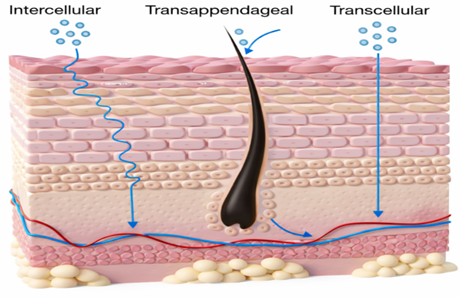
Figure 2: Routes of drug permeation across the skin
3. VESICULAR DRUG DELIVERY SYSTEM
Over recent decades, considerable attention has been directed toward the development of novel drug delivery systems (NDDS). Ideally, these systems should release drugs at rates that match physiological needs and target the active agent to the intended site of action. Targeted delivery can also be achieved by positioning controlled-release systems near or within diseased tissues or by using carriers or chemical modifications to direct drugs to specific cell types (Biju et al., 2006). Vesicular systems are organized structures composed of one or more concentric lipid bilayers formed when amphiphilic molecules interact with water. First reported in 1965 by Bangham and termed Bingham bodies, these vesicles can be prepared from various amphiphilic components (Bangham et al., 1965). Conventional drug delivery is often ineffective against intracellular infections due to poor cellular uptake; vesicular drug delivery systems offer a solution by enhancing intracellular drug delivery (20).
Drug targeting involves directing therapeutic agents to specific receptors, organs, or tissues where they are required. The concept was pioneered by Paul Ehrlich in 1909, leading to the development of multiple drug carriers such as immunoglobulins, serum proteins, synthetic polymers, microspheres, liposomes, niosomes, and erythrocytes. Among these, vesicular drug delivery systems are widely recognized for their effectiveness (21). These systems provide several benefits, including prolonged systemic circulation, reduced toxicity through site-specific delivery, improved bioavailability of poorly soluble drugs, compatibility with both hydrophilic and lipophilic drugs, sustained drug release, enhanced stability, reduced treatment costs, and improved cellular permeation (20,21). However, limitations such as oxidative and hydrolytic degradation, drug leakage, vesicle fusion, stability issues, high production costs, and short half-lives remain challenges (22).
3.1. Liposomes
Liposomes were discovered in the mid-1960s by Alec Bangham and were initially termed Banghosomes, serving as membrane models. They form when lipids self-assemble into vesicles capable of encapsulating drugs, making them effective carriers due to their ease of transport and minimal biological barriers (23). The term liposome derives from the Greek words ‘lipos’ (fat) and ‘soma’ (body) (24). Liposomes are lipid-based vesicles composed of single (unilamellar) or multiple (multilamellar) concentric phospholipid bilayers surrounding an aqueous core. Their size ranges from approximately 30 nm to several micrometers, with a bilayer thickness of 4–5 nm (25).
Liposomes have been extensively investigated for delivering small-molecule drugs, proteins, nucleic acids, and imaging agents via multiple administration routes, including parenteral, pulmonary, oral, transdermal, ophthalmic, and nasal pathways, to enhance therapeutic efficacy and patient compliance. They are also applied in food and cosmetic industries. As drug carriers, liposomes protect encapsulated agents from degradation, prolong drug half-life, enable controlled release, and offer excellent biocompatibility and safety. Moreover, they facilitate passive or active targeted delivery, reducing systemic toxicity, increasing tolerated doses, and improving therapeutic outcomes (25).
3.1.1. Classification of Liposomes
Liposomes are generally classified by their structure (lamellarity and particle size), composition (phospholipids and cholesterol), application (conventional, charged, stealth stable, actively targeted, stimulatory, and bubble liposomes), and preparation methods (classical, mechanical, drying-based, and advanced). Depending on compartment structure and lamellarity, liposomes are categorized as unilamellar vesicles (ULVs), oligolamellar vesicles (OLVs, 100-1000 nm), multilamellar vesicles (MLVs, > 400 nm), and multivesicular liposomes (MVLs). By particle size, they can be divided into small unilamellar vesicles (SUVs, 25–100 nm), large unilamellar vesicles (LUVs, 100–400 nm), and giant unilamellar vesicles (GUVs, >1000 nm) (26).
3.1.2. Structure and Composition of Liposomes
The main components of liposomes are phospholipids, cholesterol, steric stabilizers, and load generators.
Phospholipids: Phospholipids are lipid molecules composed of a hydrophilic head and a hydrophobic tail. The tail consists of two hydrocarbon chains—one saturated and one unsaturated—while the polar head contains glycerol, phosphate, and choline groups, giving the molecule both polar and non-polar characteristics. Various phospholipids, including phosphatidic acid, phosphatidylinositol, phosphatidylserine, phosphatidylcholine (lecithin), phosphatidylethanolamine, phosphatidylglycerol, cardiolipin, dioleoyl phosphatidylcholine, distearoyl phosphatidylcholine, and dioleoyl phosphatidylethanolamine, are commonly used in liposome formulation (23,24).
Cholesterol: Cholesterol can be incorporated into phospholipid bilayers at molar ratios of up to 1:1 or 2:1 relative to phosphatidylcholine. As an amphipathic molecule, it orients its hydroxyl group toward the aqueous phase while its hydrophobic chain aligns with the acyl chains within the bilayer core. This arrangement increases spacing between choline head groups and reduces electrostatic and hydrogen bonding interactions, thereby enhancing membrane stability (24).
Steric stabilizers and charge inducers: Sterilamine and diacetyl phosphate are commonly used to impart positive or negative surface charges to liposomes. These agents improve vesicle stability, prevent aggregation, and enhance cellular uptake and absorption (23).
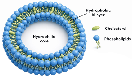
Figure 3: Structure of Liposomes
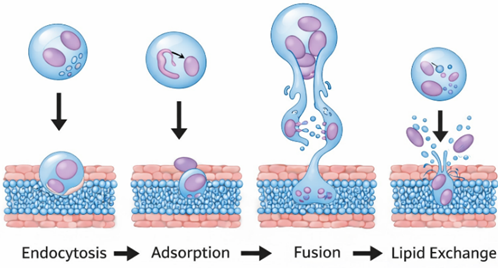
Figure 4: Mechanism of drug permeation via liposomes
3.1.3. Mechanism of Skin permeation
A liposome consists of an aqueous core enclosed within a hydrophobic lipid bilayer. Hydrophobic drugs can integrate into the lipid membrane, while hydrophilic drugs are retained in the aqueous interior, enabling liposomes to carry both types of molecules. Drug localization depends on its physicochemical properties and lipid composition. At the target site, liposomes can fuse with cellular membranes to release their contents (40,41).
Liposomes exert their effects through four primary mechanisms (24,40):
3.1.4. Advantages (24,27,28,29)
3.1.5. Limitations (24,27,28,29)
3.2. Niosomes
The first niosome formulations were patented in 1975 by L’Oréal (France) for cosmetic applications. Unlike liposomes, which are composed of phospholipid bilayers, niosomes are vesicles formed from non-ionic surfactants, with or without cholesterol. In aqueous media, these surfactants self-assemble into concentric bilayer vesicles capable of encapsulating both hydrophilic and lipophilic drugs (30). Compared with lipid-based nanocarriers such as liposomes, solid lipid nanoparticles (SLNs), and nanostructured lipid carriers (NLCs), niosomes demonstrate superior chemical stability because non-ionic surfactants are less susceptible to oxidation and hydrolysis. This enhanced stability reduces the need for specialized manufacturing conditions, such as inert gas protection, and lowers production costs due to the cost-effectiveness and availability of surfactants.
Niosomes have been extensively investigated for drug delivery in fungal infections, wound healing, rheumatoid arthritis, psoriasis, and other inflammatory conditions, as well as in transdermal gene delivery. Studies on pergolide indicate that skin penetration is influenced by formulation pH, highlighting the importance of niosomal pH in transdermal delivery. Due to their favorable physicochemical properties, niosomes can encapsulate diverse drugs and improve bioavailability by overcoming biological barriers, including the skin and gastrointestinal tract, making them promising carriers for topical and transdermal drug delivery (31).
3.2.1. Classification of Niosomes (31,32,33)
Niosomes are spherical vesicular systems composed of one or more concentric bilayers. Depending on their size, they can be divided into four categories:
Several types of niosomes exists, including proniosomes, ethosomes, aspasomes, discosomes, polyhedral niosomes, and PEGylated niosomes.
3.2.2. Structure and Composition of Niosomes
Niosomes are vesicular systems composed of non-ionic surfactants, cholesterol, charge-inducing agents, and a hydration medium, forming a stable bilayer structure. This bilayer creates two distinct regions—an aqueous core and a lipid domain—allowing encapsulation of both hydrophilic drugs in the core and hydrophobic drugs within the lipid bilayer (33).
Non-ionic surfactants: Non-ionic surfactants are the primary components of niosomes and possess amphiphilic properties with a polar head and a non-polar tail. They are preferred due to their stability, biocompatibility, low toxicity, and ease of handling and storage. Key surfactant properties influencing niosome formation include the hydrophilic–lipophilic balance (HLB), critical packing parameter, chemical structure, and phase transition temperature (30).
Cholesterol: Cholesterol interacts with surfactant head groups via hydrogen bonding, enhancing membrane rigidity and stability. Its concentration significantly affects vesicle entrapment efficiency, drug release behavior, permeability, long-term stability, and resistance to plasma- and serum-induced destabilization. Cholesterol also reduces membrane permeability, minimizing drug leakage (32).
Charge inducing molecules: Charge-inducing agents stabilize niosomes by imparting positive or negative surface charges, thereby preventing aggregation through electrostatic repulsion. Commonly used agents include negatively charged dicetyl phosphate (DCP) and positively charged stearylpyridinium chloride or stearylamine (STR). These compounds are typically incorporated at 2.5–5 mol%, as excessive amounts may disrupt bilayer integrity and reduce vesicle stability (31).
Hydration medium: The hydration medium, often a buffer, is essential for vesicle formation and is selected based on the solubility and stability of the encapsulated drug. For example, phosphate buffer (pH 5.5) has been used for ketoconazole-loaded niosomes. A thorough understanding of formulation physicochemical properties is crucial for designing stable nanovesicles capable of effective drug or biomolecule delivery (33).
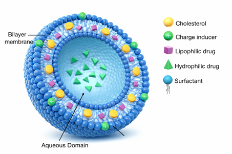
Figure 5: Structure of Niosomes
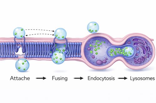
Figure 6: Mechanism of drug permeation via niosomes
3.2.3. Mechanism of Skin permeation (34,42,43)
The exact mechanism by which niosomes enhance skin permeation is not fully understood, but several contributing factors have been proposed. Niosomes may temporarily disrupt the lipid organization of the stratum corneum, altering its barrier function. They can also reduce transepidermal water loss, increasing skin hydration and loosening the tightly packed corneocyte structure. Additionally, adsorption or fusion of niosomes on the skin surface creates a high thermodynamic activity gradient at the interface, which promotes drug permeation, as demonstrated by freeze-fracture electron microscopy and small-angle X-ray scattering studies.
Niosomes may adhere to the cell surface with minimal internalization of lipid or aqueous components, enabling direct drug transfer through physical interactions or receptor–ligand binding. Alternatively, they can fuse with cell membranes, releasing their contents into the cytoplasm, or be internalized via endocytosis. In the latter case, lysosomal enzymes degrade the vesicle membrane, releasing the encapsulated drug into surrounding tissues.
3.2.4. Advantages (32,34)
3.2.5. Limitations (32,34)
3.3. Transfersomes
The high flexibility of ultra-flexible lipid nanoparticles (LNPs) enhances transdermal drug delivery by improving drug entrapment efficiency and stability. These properties have been further optimized in transfersomes, ultra-deformable vesicles developed in the early 1990s by Cevc et al., designed to enhance skin permeability. The term transfersome originates from Latin and Greek, meaning “to carry the body.” By generating an osmotic gradient, transfersomes penetrate the stratum corneum via intercellular or transcellular pathways and can encapsulate both hydrophilic and hydrophobic drugs (35). They are composed of phospholipids and an edge activator (EA)—such as Tween 80, Span 80, or sodium cholate—which imparts exceptional membrane flexibility. This self-optimizing deformability enables transfersomes to adjust their shape and pass through narrow skin pores with minimal resistance.
Several transfersome-based formulations are currently undergoing clinical evaluation. A Phase III trial of ketoprofen-loaded transfersomes (Diractin®) demonstrated superior pain relief in knee osteoarthritis with fewer side effects compared to placebo. Insulin-loaded transfersomes (Transfersulin®) are under Phase I clinical investigation for topical glucose control and have shown promising hypoglycemic effects in diabetic rabbit models. Additionally, randomized controlled trials of triamcinolone acetonide-loaded transfersomes reported improved therapeutic efficacy and safety compared with conventional topical formulations. These findings highlight transfersomes as one of the most advanced and promising carriers for transdermal drug delivery (36).
3.3.1. Structure and Composition of Transfersomes
Transfersomes are vesicular carrier systems consisting of an inner aqueous core enclosed by a lipid bilayer and a single-chain surfactant acting as an edge activator. This structure produces ultra-deformable vesicles with self-optimizing and self-regulating properties. Unlike conventional liposomes formulated from natural phospholipids such as egg phosphatidylcholine (EPC) and soybean phosphatidylcholine (SPC), or synthetic phospholipids including dimyristoyl phosphatidylcholine (DMPC), dipalmitoyl phosphatidylcholine (DPPC), and dipalmitoyl phosphatidyl glycerol (DPPG), transfersomes depend primarily on edge activators, which typically constitute 10–25% of the formulation (35). These agents soften the bilayer, enhancing vesicle flexibility and skin permeation.
Common edge activators include stearylamine, dodecyltrimethyl ammonium bromide, cetylpyridinium chloride monohydrate, sodium cholate, sodium deoxycholate, sodium lauryl sulfate, Tweens, Spans, and Brij 30. Oils such as oleic acid, eucalyptus oil, limonene, and castor oil may also serve as edge activators. The choice and concentration of edge activator influence vesicle deformability, size, entrapment efficiency, and zeta potential; increasing surfactant content generally reduces vesicle size but may promote drug leakage due to increased membrane fluidity and pore formation. Positively charged vesicles often exhibit superior skin permeation due to electrostatic repulsion from negatively charged skin surfaces. When formulated at optimal lipid–surfactant ratios, transfersomes become highly deformable and can traverse pores smaller than their own diameter without fragmentation. Additional formulation components commonly include cholesterol as a stabilizer, alcohol (3–10%), and a suitable hydration medium (36,37).
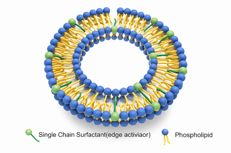
Figure 7: Structure of Transfersomes
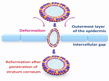
Figure 8: Mechanism of drug permeation via transfersomes
3.3.2. Mechanism of Skin permeation
The transport of transfersomes across the skin is primarily driven by an osmotic gradient generated when water evaporates from the lipid suspension after topical application. This creates a transepidermal hydration gradient that acts as the main driving force for vesicle movement. Owing to their high elasticity and deformability, transferosomes can squeeze through intercellular pores of the stratum corneum that are smaller than their own diameter. Under non-occlusive conditions, the vesicles migrate toward the more hydrated deeper skin layers to maintain water balance. During penetration, the bilayer undergoes reversible deformation and partial dehydration while preserving vesicle integrity. Upon reaching water-rich regions, the vesicles rehydrate and regain their original structure. Because transferosomes are too large for passive diffusion, they effectively create and navigate their own pathways through the skin barrier. Drug delivery may further involve lipid interaction or membrane fusion processes. Overall, transferosome permeation is governed by a combination of vesicle elasticity, hydration/osmotic forces, and elasto-mechanical behavior (37,38).
3.3.3. Advantages (36,37,39)
3.3.4. Limitations (37,38,39)
3.4. Ethosomes
Touitou et al. (2000) developed ethosomes, a novel lipid-based vesicular carrier designed to enhance skin delivery. Unlike conventional liposomes, ethosomes contain a high concentration of ethanol, which increases vesicle flexibility and significantly improves skin penetration. The elevated ethanol content enhances intracellular drug delivery within the stratum corneum by disrupting its lipid structure, enabling deeper tissue penetration and targeted drug release. Ethosomes were developed to overcome the poor skin permeation associated with earlier transdermal delivery systems, which struggled to transport active compounds across the stratum corneum. Following the development of transfersomes and niosomes, ethosomes represented a major advancement in nano-vesicular drug delivery. As a result, ethosomes are considered a highly effective and promising carrier for topical and transdermal drug delivery (44,45).
3.4.1. Classification of Ethosomes
Ethosomes are classified into classical ethosomes, binary ethosomes, transethosomes, composite phospholipid ethosomes and actively targeted (functionalized) ethosomes, each designed to enhance transdermal drug delivery.
Classical Ethosomes: Classical ethosomes are lipid-based nanocarriers composed of phospholipids, ethanol (20–45%), and water. They exhibit high skin permeability, good stability, and strong skin compatibility, and are simpler and more cost-effective to formulate than other ethosomal variants. Unlike binary ethosomes, which require additional penetration enhancers, or transethosomes, which may cause irritation due to surfactants, classical ethosomes offer a balanced combination of safety, efficacy, and manufacturability (44,45).
Binary Ethosomes: Binary ethosomes are an advanced form of classical ethosomes that incorporate a second alcohol, such as propylene glycol or isopropyl alcohol, alongside ethanol and phospholipids. The additional alcohol enhances stratum corneum fluidization, improving skin permeability, drug retention, and vesicle stability. Binary ethosomes demonstrate improved skin compatibility compared to transethosomes and are suitable for delivering both hydrophilic and lipophilic drugs in dermatological and systemic applications (44,45).
Transethosomes: Transethosomes are highly deformable vesicles containing edge activators and penetration enhancers, enabling them to pass through pores 5–10 times smaller than their diameter. Edge activators—amphiphilic molecules with charged head groups—destabilize lipid bilayers, increasing flexibility and deformability. Penetration enhancers such as pyrrolidones, alkanols, sulfoxides, glycols, azones, and peptide chains disrupt stratum corneum lipid layers to reduce skin barrier resistance. Compared to classical ethosomes, transethosomes exhibit higher encapsulation efficiency, greater formulation variability, and reduced dependence on drug molecular weight, charge, or polarity.
An advanced form, multiethosomes, incorporating ethanol, propylene glycol, Tween 80, and cinnamaldehyde, has demonstrated improved skin delivery of terbinafine hydrochloride with reduced irritation and allergic reactions. Safety studies, including Draize and ocular irritation tests in New Zealand white rabbits, confirmed that transethosomes are biocompatible and safe for ocular use (46).
Composite phospholipid ethosomes: Composite phospholipid ethosomes are designed to prevent oxidation of unsaturated phospholipids and have shown improved drug entrapment and enhanced topical delivery compared with classical ethosomes.
Actively Targeted (Functionalized) ethosomes: Functionalized ethosomes are engineered for site-specific drug delivery, such as targeting acne and skin infections. They offer enhanced stability, prolonged drug release, improved transdermal performance, and targeted therapeutic action. These systems are increasingly applied in the treatment of various dermatological conditions (45).
3.4.2. Structure and Composition of Ethosomes (44,47)
Ethosomes have three main components:
Phospholipids: Phospholipids form the vesicle bilayer and provide biocompatibility. e.g. Soy phosphatidylcholine, Egg phosphatidylcholine, Dipalmityl phosphatidylcholine, Distearyl phosphatidylcholine.
Ethanol: Ethanol (20-45%) improves vesicle flexibility and disrupts skin lipids to help drug penetration.
Water: Water acts as the hydration medium and stabilizing phase for ethosomal vesicles.
This composition lets ethosomes carry a wide range of drugs, including hydrophilic, lipophilic, and amphiphilic molecules. They are versatile carriers for both dermatological and transdermal applications. This unique composition enables ethosomes to encapsulate diverse drug molecules and makes them highly suitable for topical and transdermal delivery.
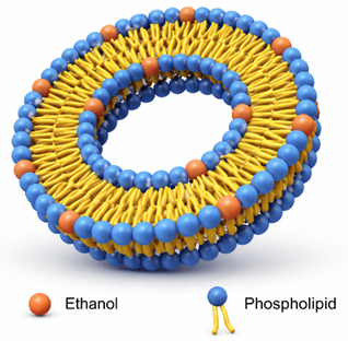
Figure 9: Structure of Ethosomes
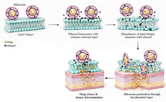
Figure 10: Mechanism of drug permeation via ethosomes
3.4.3. Mechanism of Skin permeation
The mechanism of drug delivery from ethosomes is based on the interaction between ethanol, lipid bilayers, and vesicles. It occurs in two main stages: the ethanol effect and the ethosome effect (47).
In the ethanol effect, ethanol interacts with phospholipid head groups, reducing the lipid transition temperature, increasing membrane fluidity, and lowering lipid packing density. This enhances drug solubility and encapsulation for both hydrophilic and hydrophobic drugs. Ethanol also disrupts the lipid organization of the stratum corneum, weakens the skin barrier, and increases lipid fluidity, enabling deeper penetration. Additionally, ethanol improves vesicle flexibility, allowing ethosomes to deform and pass through narrow intercellular spaces.
The ethosome effect involves vesicle malleability and fusion with skin lipids, promoting efficient drug release. Ethosomes can fuse with stratum corneum and penetrate deeper skin layers, delivering drugs directly into the tissue. They may penetrate into the dermis or systemic circulation in transdermal applications and act as a drug reservoir, enabling controlled and sustained release to maintain stable therapeutic levels (45).
3.4.4. Advantages of Ethosomes (44,47,48)
3.4.5. Limitations (44,47,48)
3.5. Spanlastics
Spanlastics are elastic, surfactant-based nanovesicular drug delivery systems primarily composed of nonionic sorbitan surfactants (Span) and an edge activator that provides high flexibility and deformability. The term Spanlastic (Span + Elastic) was introduced by Kakkar and Kaur in 2011 to describe these highly elastic vesicles (49). Drugs are encapsulated within a bilayer vesicular core, forming an amphiphilic carrier capable of entrapping both hydrophilic and lipophilic molecules. Similar to transfersomes, spanlastics exhibit remarkable elasticity, enhancing permeability and drug penetration compared with conventional formulations. Designed to overcome liposomal limitations such as oxidative instability and phospholipid purity issues, spanlastics offer nanoscale size, high deformability, and site-targeting capability, making them suitable for ocular, oral, topical, nasal, and translingual drug delivery (50).
3.5.1. Classification of spanlastics (50,53)
Based on lamellarity and size, spanlastics are classified as:
3.5.2. Structure and Composition of Spanlastics
Spanlastics are spheroidal nanovesicles composed of amphiphilic molecules that form an efficient matrix for drug bioencapsulation. Structurally, they consist of concentric lipid bilayers, similar to liposomes, and may exist as either unilamellar or multilamellar vesicles (53).
Components of Nanospanlastics (51):
Non-ionic surfactants (e.g., Span 60, Span 80, Tween 60, Tween 80): Form the vesicular bilayer, provide structural stability, regulate hydrophilic–lipophilic balance, and influence drug entrapment efficiency.
Edge activators (e.g., sodium cholate, sodium deoxycholate, Tween 80, Span 20): Enhance membrane flexibility and elasticity, enabling vesicle deformation and passage through narrow intercellular spaces.
Drug payload: Encapsulates hydrophilic drugs in the aqueous core, lipophilic drugs within the lipid bilayer, and amphiphilic drugs across both regions.
Optional additives:
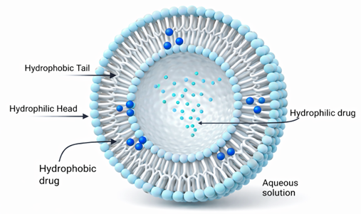
Figure 11: Structure of Spanlastics
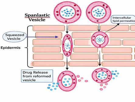
Figure 12: Mechanism of drug permeation via spanlastics
3.5.3. Mechanism of Skin permeation
Spanlastic (SP) vesicles penetrate the skin through two complementary mechanisms. First, they act as drug reservoirs and carriers, enabling rapid transport of drug molecules across the stratum corneum by passing through skin pores. Second, SP vesicles function as penetration enhancers by disrupting the intercellular lipid lamellae, thereby reducing the skin barrier and promoting deeper drug permeation (52). Drug transport occurs either through vesicle-mediated delivery, where intact elastic vesicles traverse intercellular spaces, or through lipid fluidization, where vesicle components alter epithelial membrane structure to facilitate diffusion. The dominant mechanism depends on the drug’s physicochemical nature (hydrophilic or lipophilic) and vesicle composition.
Efficient penetration is supported by (53):
3.5.4. Advantages (49,52,53)
3.5.5. Limitations (49,53)
3.6. Novasomes
Novasomes are advanced vesicular drug delivery systems developed to address the limitations of conventional carriers such as liposomes and niosomes (54). Initially created by Novavax and later licensed to IGI Laboratories, novasomes are enhanced liposomal/niosomal structures with superior drug-loading capacity and performance. They are spherical paucilamellar vesicles (0.1–1.0 µm) composed of polyoxyethylene fatty acid monoesters, cholesterol, and free fatty acids, typically in a 74:22:4 ratio. Structurally, novasomes contain two to seven bilayer membranes surrounding a large central core capable of encapsulating high amounts of both hydrophilic and hydrophobic drugs. Owing to their high encapsulation efficiency, stability, and versatility, novasomes have been widely investigated in pharmaceutical applications, including use as vaccine adjuvants and carriers for drugs such as zolmitriptan, terconazole, and fenticonazole nitrate, showing improved performance over niosomes and strong antifungal and therapeutic potential (55).
3.6.1. Characteristics of Novasomes (56,57)
3.6.2. Structure and Composition of Novasomes (58,59)
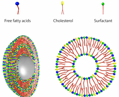
Figure 13: Structure of Novasomes
3.6.3. Mechanism of Action (56,57,59)
3.6.4. Advantages (55,56,58,59)
3.6.5. Limitations
Although novasomes represent an advanced vesicular drug delivery system, they face certain limitations, such as potential storage instability and challenges in large-scale manufacturing. Ongoing research focuses on improving their stability, scalability, and quality through optimized formulation techniques and stringent quality control. With continued development, novasomes hold significant promise for enhancing drug delivery efficiency and improving therapeutic outcomes (59).
4. GENERAL METHODS OF PREPARATION
4.1. Thin Film Hydration Method (Hand-Shaking Technique) (26,43,51,56,59)
The thin film hydration method is a widely used laboratory technique for preparing vesicular drug delivery systems, especially multilamellar vesicles (MLVs). In this process, vesicle-forming components such as phospholipids, surfactants, cholesterol, and lipophilic drugs are dissolved in a volatile organic solvent and evaporated under reduced pressure using a rotary evaporator to form a thin lipid film on the flask wall. The dried film is then hydrated with an aqueous phase at a temperature above the lipid phase transition point, leading to spontaneous vesicle formation upon gentle agitation. Hydrophilic drugs are incorporated into the aqueous phase, while lipophilic drugs are added to the lipid solution before evaporation.
Vesicle characteristics such as size, lamellarity, and encapsulation efficiency depend on formulation and process variables including hydration temperature, lipid composition, and agitation rate. Post-processing techniques like extrusion, sonication, and freeze–thaw cycling can improve vesicle uniformity, although excessive sonication may cause vesicle damage.
Despite its simplicity and versatility, the method has limitations such as size variability, moderate drug entrapment, solvent residue risks, sterilization challenges, and poor scalability. Nevertheless, it remains a commonly used technique for preparing vesicles for small molecules, proteins, nucleic acids, and other therapeutic agents.
4.2. Reverse phase evaporation method (26,33,39,57)
The reverse phase evaporation (REV) method is a common technique for preparing vesicular systems. It is especially effective for encapsulating hydrophilic drugs and large biomolecules. In this method, lipids or surfactants dissolve in an organic solvent and mix with an aqueous drug phase to create a water-in-oil system. This generally happens through sonication or mechanical agitation. Gradually removing the organic solvent under reduced pressure changes the emulsion into a gel-like phase, which then collapses to form vesicles.
The characteristics of vesicles, including size, stability, and encapsulation efficiency, depend on several formulation parameters. These include the lipid-to-aqueous phase ratio, solvent type, temperature, and sonication intensity. Under optimized conditions, entrapment efficiencies of up to 65% have been reported.
While this method works well for encapsulating nucleic acids, hydrophilic drugs, and larger molecules, it has limitations. These include exposure to organic solvents, potential drug degradation, high solvent use, sterilization challenges, and difficulties in scaling up. Nonetheless, REV remains a promising approach for developing controlled-release nanocarriers, such as topical and transdermal delivery systems.
4.3. Ethanol injection method (26,32,52,58)
The ethanol injection method is a simple and widely used technique for preparing vesicular nanocarriers, in which lipids, surfactants, and drugs are dissolved in ethanol and rapidly injected into a heated aqueous phase under stirring, homogenization, or sonication. Rapid dilution of ethanol in the aqueous medium causes lipid precipitation and self-assembly into planar bilayer fragments, which subsequently fuse to form closed unilamellar vesicles. This method can produce small, relatively uniform particles (approximately 100 nm) and has been applied in the formulation of transferosomes, liposomes, and other lipid-based carriers for pulmonary, transdermal, and systemic drug delivery, including therapies for rheumatoid arthritis, cancer, and hormone-related conditions.
Vesicle size and encapsulation efficiency depend on factors such as lipid composition, injection rate, temperature, and mixing intensity. However, the technique has notable limitations, including difficulty in removing residual ethanol due to azeotrope formation, limited lipid solubility in ethanol, potential degradation of sensitive drugs or macromolecules, and reduced encapsulation efficiency for hydrophilic drugs. Despite these challenges, the ethanol injection method remains a valuable platform for nanocarrier development, and newer adaptations—such as microfluidic, transverse injection, and membrane contactor systems—have been introduced to improve scalability, reproducibility, and formulation control.
4.4. Ether injection method (26,59,61)
The ether injection method is a vesicle preparation technique in which lipids or surfactants dissolved in diethyl ether are slowly injected into a preheated aqueous drug solution maintained above the boiling point of ether. As ether rapidly evaporates upon contact with the warm aqueous phase, the dispersed lipids self-assemble to form unilamellar vesicles. Compared with ethanol injection, this method allows more efficient solvent removal, enabling prolonged processing and producing vesicles with relatively high encapsulation capacity. Vesicle size is influenced by formulation variables such as cholesterol concentration, surfactant-to-drug ratio, injection rate, needle gauge, and temperature, with reported particle sizes ranging from approximately 50 to 1000 nm. However, rapid injection rates may cool the aqueous phase due to ether evaporation, leading to nozzle blockage or formation of multilamellar vesicles. Additionally, the use of ether poses safety concerns and limits compatibility with thermolabile compounds, although fluorinated hydrocarbons have been explored as lower-temperature alternatives. Despite these limitations, the ether injection method remains a useful approach for producing lipid-based vesicles with high drug-loading potential.
4.5. Sonication method (26,44,50,57)
The sonication method is a widely used technique for preparing and size-reducing vesicular drug delivery systems by applying ultrasonic energy to lipid or surfactant dispersions. In this approach, a drug solution in buffer is mixed with a surfactant–cholesterol combination and subjected to probe or bath sonication, typically at elevated temperatures, to promote vesicle formation. Sonication is commonly employed to convert multilamellar vesicles (MLVs) into smaller unilamellar vesicles (SUVs) with more uniform particle size. Vesicle characteristics such as size distribution and zeta potential are influenced by factors including ultrasound power, duration, probe depth, and processing temperature. While probe sonication delivers high energy and enables efficient size reduction, it may cause temperature rise, lipid degradation, metal contamination, and loss of encapsulated drug if not carefully controlled. Bath sonication offers gentler and more sterile processing but generally results in lower encapsulation efficiency and smaller internal volume. Despite these limitations, sonication remains a popular and effective post-processing technique for controlling vesicle size in liposomal and niosomal formulations.
4.6. Microfluidization (23,49,51,56)
The microfluidization (submerged-jet) method is an advanced high-pressure homogenization technique used to produce uniform nanosized elastic vesicles such as spanlastics, niosomes, novasomes, and nanoliposomes. In this approach, surfactants, drugs, and edge activators are pumped through a microfluidizer, where two high-velocity liquid streams collide within precisely engineered microchannels under extreme pressure. This collision generates intense shear forces that break down large droplets and promote the formation of small, homogeneous unilamellar vesicles with narrow size distributions. The submerged-jet principle ensures that energy is concentrated within the formulation zone, improving particle uniformity, size control, and batch-to-batch reproducibility. Additionally, this method can produce nanocarriers without the use of toxic organic solvents, making it suitable for sensitive pharmaceutical applications. However, the high pressures involved may cause temperature elevation, requiring cooling systems to preserve formulation stability. Despite these considerations, microfluidization remains a highly efficient and scalable technique for producing reproducible, nanosized vesicular drug delivery systems.
4.7. Multiple membrane extrusion method (56,57,59)
The multiple membrane extrusion method is an effective technique for producing vesicles with controlled and uniform particle size. In this approach, a mixture of surfactant, cholesterol, and diacetyl phosphate dissolved in chloroform is evaporated to form a thin lipid film. The dried film is then hydrated with an aqueous drug solution to generate a vesicular suspension. This suspension is subsequently passed multiple times through polycarbonate membrane filters arranged in a series of channels, enabling progressive size reduction and improved uniformity. Repeated extrusion through membranes with defined pore sizes allows precise control over vesicle diameter and enhances reproducibility. Owing to its simplicity and efficiency in size regulation, this method is widely used for producing well-defined niosomal and liposomal formulations.
4.8. Cold method (44,45,47)
The cold method is one of the simplest and most widely used techniques for preparing ethosomal formulations. In this approach, phospholipids, drugs, and other lipid components are dissolved in ethanol at room temperature under vigorous stirring. Polyols such as propylene glycol are then gradually added, and the mixture is gently heated to approximately 30 °C. In a separate vessel, water is preheated to the same temperature and slowly incorporated into the ethanolic lipid phase with continuous stirring, initiating vesicle formation within a few minutes. The resulting ethosomal dispersion can be further processed by sonication or extrusion to reduce vesicle size and improve uniformity. To preserve stability, the final formulation is typically stored under refrigerated conditions. Due to its simplicity, reproducibility, and mild processing conditions, the cold method remains a preferred technique for ethosome preparation in topical and transdermal drug delivery applications.
4.9. Hot method (44,45,47)
The hot method is an efficient technique for preparing ethosomes, particularly suitable for thermally stable drugs. In this method, phospholipids are dispersed in water and heated (typically around 40–60 °C) until a colloidal aqueous phase is formed. In parallel, ethanol and propylene glycol are mixed and heated to the same temperature to form the organic phase. Once both phases reach thermal equilibrium, the organic phase is gradually added to the aqueous phase under continuous stirring, promoting ethosome formation. Depending on the drug’s solubility, it is dissolved in either the aqueous or ethanolic phase prior to mixing. Vesicle size and uniformity can be further optimized using probe sonication or extrusion. While this method offers good vesicle formation and high encapsulation efficiency, it is generally unsuitable for heat-sensitive drugs due to the elevated processing temperature.
CONCLUSION
In summary, vesicular drug delivery systems represent a transformative advancement in topical and transdermal therapy, effectively overcoming the inherent limitations of conventional formulations and the skin's formidable barrier function. By utilizing innovative carriers such as liposomes, niosomes, transfersomes, ethosomes, spanlastics, and novasomes, these systems enable enhanced drug penetration, improved bioavailability, targeted delivery, and sustained release—all while minimizing systemic side effects and improving patient compliance. Each vesicular platform offers unique mechanistic advantages, from the bilayer flexibility of transfersomes to the ethanol-enhanced permeation of ethosomes, allowing for tailored therapeutic approaches across a wide range of dermatological and systemic conditions. Although challenges related to stability, scalability, and regulatory approval remain, ongoing research into hybrid systems, smart carriers, and optimized preparation methods continues to drive the field forward. Ultimately, vesicular drug delivery systems stand at the forefront of modern pharmacotherapy, offering a versatile, efficient, and patient-friendly paradigm for the future of topical medicine.
CONFLICT OF INTEREST
The authors declare that there is no conflict of interest.
REFERENCES

Sam Roshan, Surya, Ashik Rahuman, Vani, An Insight into Vesicular Drug Delivery Systems for Topical Skin Delivery: A Review, Int. J. of Pharm. Sci., 2026, Vol 4, Issue 4, 557-581. https://doi.org/10.5281/zenodo.19410458
 10.5281/zenodo.19410458