

We use cookies to ensure our website works properly and to personalise your experience. Cookies policy
1 Shri Shankaracharya College of Pharmaceutical Sciences Junwani, Bhilai Chhattisgarh
2 J K Institute of Pharmaceutical Education and Research, Bilaspur Chhattisgarh
3 Raigarh College of Pharmacy, Raigarh Chhattisgarh
4 Pt. Ravishankar Shukla University, Raipur Chhattisgarh.
Silver nanotechnology is an advanced and fascinating field of nanoparticles among all metal nanoparticles used in biomedicine. Silver nanoparticles play an important role in nanotechnology and nanoscience. Green synthesis is an eco-friendly method and an alternative synthesis process to the physical and chemical techniques to reduce the use of toxic chemicals. The main purpose of the review is to discuss the synthesis process of the silver nanotechnology, their characterisation process, and advanced applications of the silver nanoparticles. Biological sources for green technology are plant extracts, algae, fungi, yeast, enzymes, and biomolecules. Phytochemicals present in these sources reduce Ag? ions into metallic Ag? and act as stabilising agents. Visual characterisation of nanotechnology is by using the UV-Visible. FTIR, XRD, and SEMTEM are used to analyse the formation structure and morphology of the silver nanotechnology. More importantly, we discuss the bio application of the silver nanotechnology, for example, as antibacterial, antifungal, antiviral, anti-inflammatory, anti-angiogenic, and anti-cancer agents. However, challenges such as toxicity, large-scale production and reproducibility remain. Finally, we conclude the prospects of the silver nanoparticles.
A significant area of contemporary research is nanotechnology, which deals with the creation, synthesis, and manipulation of particle shapes with sizes ranging from around 1 to 100 nm. Nanotechnology is an essential topic for contemporary researchers since nanoparticles (NPs) offer a wide range of applications in fields including cosmetics, environmental wellness, healthcare, biomedical science, chemical and food industries, etc. Recently, there has been significant interest in nanotechnology due to the unique properties of nanoparticles (1–100 nm) [1,2]. Since these characteristics are derived from size, morphology, and shape, numerous studies have been carried out to alter the size and form of nanoparticles during their synthesis for a range of purposes. The numerous uses of silver nanoparticles in wastewater treatment, solar energy conversion, medicine, and catalysis are driving research on their manufacture. Research on bio-inspired nanoparticles as innovative delivery methods for the treatment of eye conditions has been reported [3,5]. Researchers have also paid close attention to the application of nanomaterials in the battle against cancer. Each of these efforts contributes significantly to the goals of sustainable development. Silver nanoparticles made from plant extracts have been employed as antibacterial agents. Additional applications of silver nanoparticles in textiles, biomedical services, wound care, and keyboards have been demonstrated. Silver nanoparticles are widely employed in many scientific fields because silver is easily accessible, moderately inexpensive, and possesses well-known antifungal, antibacterial, and anticancer properties. These procedures hurt the ecology because certain dangerous substances may adsorb on the outer layer of chemically produced nanoparticles. In this review paper, we highlight the green strategy for the production of silver nanoparticles. Chemical treatments are detrimental to the environment, according to Karthick et al. Green synthesis offers a one-step, sustainable, and safe method of creating nanoparticles. According to earlier studies, green synthesis does not require high energy, pressure, or temperature. Microbes may be used to produce eco-friendly nanoparticles as the new generation of nano compounds for a range of Tran's field of study applications, according to some recent research. Plant-based extracts were selected and employed in this experiment because it has been demonstrated that they decrease metallic ions in sustainable synthesis more quickly than microorganisms [4].The most common phytochemicals found in plant extracts include flavonoids, terpenoids, ketones, amides, aldehydes, and carboxylic acids. These phytochemicals participate in the reduction of metal ions to create the required nanoparticles in addition to serving as capping agents. In a prior study, we synthesised silver nanoparticles utilising Euphorbia species confinalis and Scelocarya birrea and evaluated their antibacterial effectiveness [7]. In other studies, a number of other plant extracts have been employed to create silver nanoparticles in a way that is safe for the environment [6,11]. In this experiment, silver nanofragments are made using a range of plant extracts.[8,9,10]
2.SynthesisTechnique:
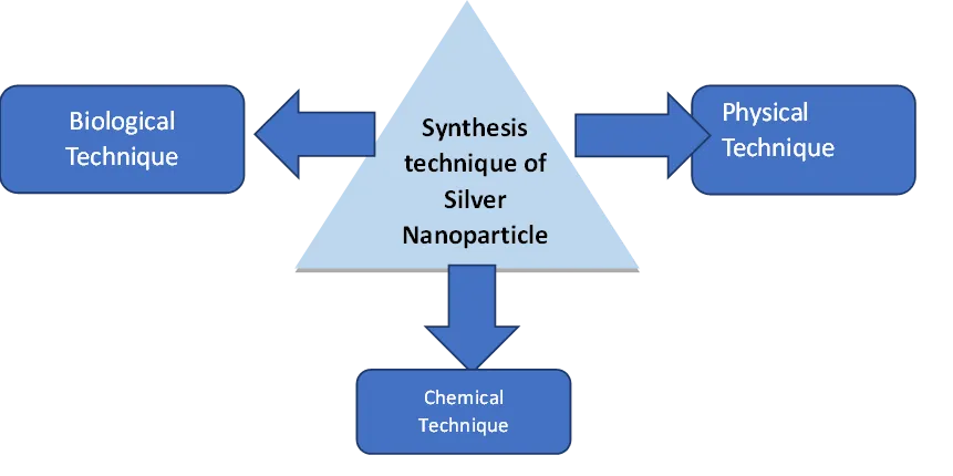
The creation of nanoparticles can be done in three different ways. These techniques are enumerated.
1.PhysicalTechniques
2.ChemicalTechniques
3.BiologicalApproaches
A review of chemical, physical, and green synthesis approaches for creating silver nanoparticles. is offered in this review article.
2.1 Physical Techniques:
a) Mechanical Approach
b) Ablation using Pulse Laser
c)Method of Pulsed Wire Discharge
d).Deposition of Chemical Vapour
e).Pyrolysis using a laser
f). Deposition of Ionised Cluster Beams
The two most significant physical methods for creating silver nanoparticles are evaporation-condensation and laser annihilation.[12] Two advantages of physical production methods over chemical ones are the similar distribution of NPs and the lack of solvent interference in the resulting thin films. It is quite difficult to physically produce silver nanoparticles in a tube furnace at atmospheric pressure. For example, the tube boiler requires a lot of power to raise the ambient temperature, requires a lot of space, and takes a long time to achieve thermal stability. Additionally, standard tube combustion requires more than a few kilowatts of electricity and must reheat itself for several tens of minutes in order to reach a constant operating temperature. It has been demonstrated that silver nanoparticles (NPs) can form in a small ceramic furnace with a restricted heating area. The small ceramic heater was used to evaporate the source materials. The temperature gradient near the heater surface is far steeper than in a tube boiler, allowing the evaporated vapour to cool at a suitable rate.[13,14] This physical technique can be used to calibrate nanoparticle measurement apparatus and produce nanoparticles for long-term inhalation toxicity investigations.[14].
Laser ablation of metal-based bulk materials in solution 19 can yield silver nanoparticles (NPs) depending on the laser's wavelength range and pulse lengths that come into contact with the metal-based target. (in the femto-, pico-, and nanosecond regime) control both the ablation efficiency and the characteristics of the generated nano-silver particles. One of the main advantages of laser ablation over other techniques for creating metal particles is the lack of chemical-based reagents in solutions. Therefore, this technique can be applied to create pure, contaminant-free metal colloids for later applications. [15].
Advantages:
Disadvantages-
2.2 Chemical Techniques
Chemical Reduction Technique
Chemical reduction with either organic or inorganic reducing agents is the most popular technique for creating silver nanoparticles. Silver ions (Ag+) can be reduced in water-based or non-aqueous remedies using a range of reducing agents, such as sodium citrate, ascorbate, sodium borohydride (NaBH4), elemental hydrogen, polyol process, Tollens reagent, N, N-dimethyl formamide (DMF), poly (ethylene glycol)-block copolymers, etc. Ag+ ions are transformed by these reducing agents into metallic silver (Ag0), which subsequently forms oligomeric clusters[16,17]. Eventually, metallic colloidal silver particles are produced by these clusters. Polymeric substances, including poly (vinyl alcohol), poly (vinyl pyrrolidone), poly (ethylene glycol), poly (methacrylic acid), and poly (methyl methacrylate), are the best protective agents for stabilising NPs[18]. Kim and colleagues reported the synthesis of spherical silver NPs with a regulated size and high monodispersity utilising the polyol process and a modified precursor injection technique, whereas Oliveira and colleagues generated dodecanethiol-capped silver NPs. For the precursor injection method to produce evenly sized silver nanoparticles, the injection velocity and reaction temperature were essential. Listed a few chemical processes that are utilised to produce silver nanoparticles [19].
a) Synthesis via Sono chemistry
b) Method of Co-precipitation
c) The Method of Inert Gas Condensation
d) Synthesis via Hydrothermal
Sono chemical fusion with copper salt in the presence of palladium and water has effectively formed Pd-CuO nano hybrids. When palladium and water exist, ultrasonic frequencies can transform metal salts into their oxides.[17]
Co-precipitation Technique
This method is sometimes referred to as the wet chemical procedure or the solvent shifting technique. Polymer solvents include ethanol, acetone, hexane, and non-solvent-based polymers; the polymeric phase might be synthetic or organic. By mixing the polymer solution, the polymer-solvent rapidly diffuses into a non-solvent polymer phase, creating nanoparticles. (18).
This process is commonly used to manufacture metal micro particles. A typical approach for manufacturing fine nano particles is the unused gas compression technique, which makes nano particles by dis appearing to be a metallic source in an inactive gas. At a temperature that can be reached, metals evaporate at an acceptable pace. The technique of generating copper metal nanoparticles requires vaporising the metal in a chamber filled with argon, helium, or neon. Liquid nitrogen cools the gases, forming nanoparticles that range in size from 2 to 100 nm[19]
It is one of the most used approaches for producing nanoparticles. It is largely a technique based on chemical reactions. In order to make nanoparticles, hydrothermal production uses a wide range of temperatures, from the ambient temperature to very high temperatures. The chemical technique has some advantages over physical and biological procedures (20,21).
2.3 Biological methods
Biologically generated silver nano particles (AgNPs) have attracted a lot of interest as environmentally acceptable and sustainable substitutes for traditional physical and chemical processes[22]. These techniques convert silver ions (Ag⁺) into metallic silver nanoparticles (Ag₄) using naturally occurring reducing and stabilising agents such as bacteria, fungi, algae, yeast, and plant-based extracts. Proteins, enzymes, phytochemicals, and other biomolecules present in these biological systems enhance the production of nanoparticles under mild reaction conditions. Numerous benefits of biological synthesis include low environmental impact, cost-effectiveness, biocompatibility, and little toxicity. Additionally, the produced AgNPs are suitable for a range of applications in drug transport, antimicrobial therapy, biosensing, and nanomedicine due to their unique size, shape, and stability [45.46]. Thus, in recent years, biological synthesis has provided a viable and eco-friendly way to produce silver nanoparticles on a large scale. There are two primary ways that bacteria can produce nanoparticles: extracellular biosynthesis and intracellular biosynthesis. Certain bacteria have the ability to separate metal ions. Heavy metals can be stored and detoxified by microorganisms thanks to a variety of reductase enzymes.[23,24]
Ag-NP Synthesis Utilising Fungus: Because of their ability to bio accumulate metals, as well as their tolerance, high binding capacity, and intracellular absorption, fungi have the potential to synthesise metallic nanoparticles (NPs) that are easier to handle in a research facility than bacteria (Sastry et al. 2003). According to Mandal et al. (2006), fungi can be used in a variety of ways to synthesize nano particles (NPs) by secreting large amounts of enzymes that are used to decrease the AgNO3 solution. Ag-NPs produced extracellularly by et al. (2007). According to Vigneshwaran et al. (2007), the fungus Aspergillus flavus can be used to create monodisperse Ag-NPs, with an average size of 8.92 ± 1.61 as determined by Transmission Electron Microscopy (TEM). Balaji et al. discovered the extracellular synthesis of Ag-NPs utilising the fungus Cladosporium cladosporioides, and TEM revealed that the size of the NPs was between 10 and 100 nm (Balaji et al. 2009).[24,25]
Synthesis Using Yeast: Yeast-mediated synthesis of silver nanoparticles (AgNPs) is an eco-friendly and cost-effective biological approach that utilises yeast cells as natural reducing and stabilising agents. Yeast species such as Saccharomyces cerevisiae are capable of reducing silver ions (Ag⁺) into metallic silver nanoparticles (Ag⁰) through enzymatic and metabolic processes. The synthesis may occur intracellularly or extracellularly, resulting in stable and uniformly sized nanoparticles. Bio molecules such as proteins, polysaccharides, and enzymes play a critical role in nanoparticle production and capping. This green synthesis method operates under mild conditions, avoids toxic chemicals, and produces biocompatible AgNPs. Yeast-synthesised silver nanoparticles exhibit significant antimicrobial activity and hold potential applications in medicine, biotechnology, and nanotechnology[26,27].
Ag-NP Synthesis Employing Bacteria: Bacteria can generate inorganic substances extracellularly or intracellularly. Because of this, they could be employed as bio factories to generate noble metal nano particles (NPs) like gold and silver. Although Ag-NPs are known to be biologically compatible, a few bacteria are known to be resistant to Ag. Because of their ability to aggregate silver on their cell walls, these bacteria are recommended for use in the commercial recovery of silver from mining materials (Pooley 1982). Initially, it was reported by Klaus et al. (1999) that Pseudomonas stutzeri AG259, an Ag-resistant bacterial strain, was used to create Ag-NPs. Ag-NPs are accumulated in these cells in significant quantities up to 200 nm. Shivaji et al. (2011) used the culture supernatants of psychrophilic bacteria to create Ag-NPs. The production of Ag-NPs by Bacillus licheniformis was demonstrated by Kalimuthu et al. (2008).
Ag-NP Synthesis Employing Plants and Plant Extracts: Plant extracts are the primary source of nanoparticle formation. This approach is also known as "green synthesis" or "green nanoparticle manufacturing". The current processes for making NPs are costly, dangerous, and environmentally harmful. Researchers have discovered the exact green routes—that is, naturally occurring sources and their products that can be used to synthesise NPs—to overcome these obstacles. Green synthesis can be classified into two categories: (a) employing microorganisms such as bacteria, fungi, yeasts (eukaryotes), and actinomycetes; and (b) using plants and plant-based extracts. The following sections discuss green synthesis with bacteria, fungi, plants, and plant extracts. The first method of using plants for the production of metallic NPs using lucerne sprouts was described by Gardea-Torresdey et al. (2003), 64, 65, 66. Silver from agar media may be absorbed by lucerne roots and transferred to plant shoots in the same oxidation state. The arrangement of these Ag atoms produced Ag-NPs in shoots. Ahmad and Sharma (2012) state that Ag-NPs are made by adding Argemone mexicana leaf extract as a capping and reducing agent to the aqueous solution of AgNO3. The produced NPs were examined using High Resolution Transmission Electron Microscopy (HRTEM), UV-Vis spectrophotometer, and Energy Dispersive X-ray Spectroscopy (EDX). The characteristics of NPs are examined using UV-Vis spectrometers, X-ray diffractometers (XRD), scanning electron microscopy (SEM), and Fourier Transmission Infrared (FTIR) spectrophotometers67, 68. According to Velmurugan et al. (2015), Ag-NPs are made from peanut shell extract and their properties and antifungal activity are compared with those of commercial Ag-NPs. Ag-NPs were made by reducing an aqueous AgNO3 solution using an extract of neem and triphala, and their properties were examined using EDX, nanoparticle tracking analysis (NTA), and TEM69. Roy et al. (2014) also used fruit extract from Malus domestica as a capping agent to create spherical Ag-NPs with an average diameter of 20 nm. The synthesis of NPs is analysed using UV-Vis spectral analysis, different phases and morphology are confirmed using XRD and TEM, and the biomolecules responsible for NP reduction and stability are identified using FTIR. FTIR and absorption spectroscopy are utilised to investigate the papaya fruit extract 71, 72. According to Rout et al. (2012), spherical-shaped Ag-NPs were created using Ocimum sanctum leaf extract as a stabilising agent, and the particles' properties were examined using an XRD, SEM, and UV-Vis spectrometer. Bar et al. (2009) reported that Ag-NPs were produced by reducing an aqueous AgNO3 solution using Jatropha curcas latex as a capping agent73, 74, while Awwad et al. (2013) investigated the synthesis of spherical Ag-NPs using carbo leaf extract. Ag-NPs were also produced by employing Cassia auriculata leaf extract as a reducing and capping agent, as demonstrated by Udayasoorian et al. (2011). Ag-NPs were created by Kasthuri et al. (2009) using leaf extract of Apiin as a capping and reducing agent to reduce aqueous Ag ions. TEM measurements revealed the average particle diameter 75, 76. Shankar et al. (2003) demonstrated the extracellular synthesis of Ag-NPs using Geranium leaf extract; Nakkala et al. (2014) investigated the use of Acorus calamus extract as a capping agent for the synthesis of Ag-NPs; Kumar et al. (2014) reported that Ag-NPs are synthesised using leaf extracts of Boerhaavia diffusa and Acalypha indica (Euphorbiaceae), which produced silver NPs (20–30 nm) in 30 minutes. These NPs showed remarkable antibacterial effectiveness against water-borne pathogens like E. coli and V. cholera with a minimum inhibition concentration (MIC) of 10μg/ml.
3. CHARACTERIZATION OF NANOPARTICLES
The characterization of nanoparticles is important to determine the size, shape, structure, surface chemistry, optical properties, and stability. Various analytical techniques for the characterisation of nanoparticles include XRD, FTIR, SEM, TEM, UV-Vis Spectrometry, DLS, and LEIS.(32,33)
3.1. UV VISIBLE
This technique is commonly used to monitor the creation and stability of metal-based nanoparticles in order to characterise them. When a metallic nanoparticle is formed from its specific salt, it produces a characteristic peak with notable absorptions in the visible range. The absorption band is most useful for identifying particles in the size range of 2–100 nm because silver nanoparticles are quite near to one another. A surface Plasmon resonance absorption band is produced by the free mobility of electrons in these bands. The dielectric medium, particle size, and chemical environment all affect how much light the silver nanoparticles absorb. Using UV-Vis spectro photometry, a surface Plasmon resonance peak at the same wavelength was discovered. (32,35)
3.2 X-RAY DIFFRACTION
By deeply permeating the material with X-rays, X-ray diffraction analysis (XRD) is a commonly used analytical technique to view the structure of crystalline metallic nanoparticles. The formation of crystalline nanoparticles is confirmed by the subsequent diffraction pattern. The Debye-Scherrer equation is used to determine the particle size from the XRD data by calculating the width of the Bragg reflection law using the following formula: d = Kλ/β cos θ, where d is the particle size (nm), K is the Scherrer constant, λ is the X-ray wavelength, β is the full width half maximum, and θ is the diffraction angle. XRD is an effective method for researching nanomaterials and can be used to evaluate the structural properties of a wide range of materials, including proteins, polymers, glasses, and superconductors. (36,32)
3.3 FOURIER TRANSFORM INFRARED SPECTROSCOPY
Fourier transform infrared spectroscopy (FTIR) can be used to investigate the surface chemistry of generated metal nanoparticles and the role of biomolecules in nanoparticle production. In FTIR, the sample is exposed to infrared radiation; part of the rays is absorbed by the sample, while the rest passes through. The obtained spectra show the usual absorption and transmission of the sample material. (37,32)
3.4 DYNAMIC LIGHT SCATTERING
DLS is a popular method for figuring out a molecule's size and size distribution. It has been used to measure the size of nanoparticles and is commonly used to characterise them. Furthermore, DLS has been widely utilised to size magnetised nanoparticles in the liquid phase and has been demonstrated to be helpful in characterising a variety of nanoparticle kinds. Because of the impact of Brownian motion, the size of the nanoparticle as estimated by DLS is usually bigger than that of TEM. This technique can be used to determine the average size of nanoparticles in liquids. (38,32)
3.5 TRANSMISSION ELECTRON SPECTROSCOPY
The size and shape of nanoparticles can be determined using TEM, a very helpful tool. TEM's resolution is 1,000 times greater than SEM's, and its images offer more accurate information on the nanoparticles' size, shape, and crystallisation. (39,32)
3.6 SCANNING ELECTRON MICROSCOPY
SEM is used to measure the size of individual nanoparticles at the micro- (10−6) and nano- (10−9) scales, as well as to analyse the topography and morphology of nanoparticles. The sample nanoparticles' surface is exposed to a high-energy electron beam generated by SEM, and the backscattered electrons that arise are what give the sample its distinctive properties. The morphological alterations of the cell before and after treatment with nanoparticles are examined using electron microscope analysis. As markers of the antibacterial qualities of nanoparticles, numerous investigations have used visible alterations in cell shape and nanoparticle perforations in the cell wall. Making use of SEM. (39,32)
3.7 LOW ENERGY ION SCATTERING
One widely used method of surface analysis that is renowned for its excellent surface
Synthesis of silver nanoparticles and their biological uses: 825 sensitivity. A sample's elemental makeup and structure can be ascertained using this technique. Furthermore, a relevant surface analytical technique for evaluating SAM-functionalized nano components has high sensitivity. (34)
3.8 ENERGY DISPERSIVE X-RAY SPECTROSCOPY
EDX is a crucial method for figuring out a sample's elemental makeup, and its use in the field of nanotechnology has been documented. Each element has a distinct set of peaks in the X-ray spectrum due to its various atomic structures, which can be used to identify the elemental makeup of any nanoparticle. (34,32)
4. APPLICATION OF SILVER NANOPARTICLE
Uses of silver nanoparticles in biomedical sciences
Antiviral Activity:
To cure viral diseases and prevent their spread, nanoparticles can be employed instead of pharmaceuticals. Strong antiviral medications that prevent viruses from operating may be produced by the biogenesis of silver nanoparticles. Bio-silver nanoparticles with strong anti-HIV properties at an early stage of the reversible transcription process were studied by Suriyakalaa et al. In order to regulate the activity of the virus, bio-produced metallic nanoparticles have several attachment sites for the gp120 on the viral membrane. But according to a different study, bio-based nanoparticles are very powerful virucidal agents against cell-associated viruses or free HIV. Silver nanoparticles have been shown to exhibit antiviral properties against HIV-1 at non-cytotoxic concentrations. These silver nanoparticles were analysed using a variety of in vitro tests to see how they exhibited antiviral activity against HIV. Silver nanoparticles with or without a polysaccharide covering had antiviral activity against the monkeypox virus, according to another study. This study demonstrates that silver nanoparticles considerably reduce monkeypox virus infection in vitro. Before infection, tacaribe to silver nanoparticles facilitated the virus's absorption into the host cells. On the other hand, the virus treated with silver demonstrated a notable decrease in viral RNA synthesis, suggesting that silver nanoparticles can stop viral infection in vitro.[40,41]. Ag30-MHCs had the highest efficiency for viral inactivation out of the three types of silver nanoparticle-MHCs evaluated, according to another study.
Antifungal Activity:
To cure viral illnesses and prevent their spread, nanoparticles can take the role of medications.
The biogenesis of silver nanoparticles may result in the production of potent antiviral drugs that block the activity of viruses. Bio-silver nanoparticles that showed strong anti-HIV effects early in the reverse transcription process were studied by Suriyakalaa et al. The viral membrane's gp120 has several attachment sites on bio-generated metallic nanoparticles to regulate the activity of the virus. Bio-based nanoparticles, however, are potent virucidal agents against free HIV or cell-associated viruses, according to a different investigation. At non-cytotoxic doses, silver nanoparticles have been demonstrated to have antiviral action against HIV-1 . To elucidate these silver nanoparticles' method of antiviral action against HIV-1, a variety of novel in vitro approaches were employed. Silver nanoparticles with or without a polysaccharide covering had antiviral activity against the monkeypox virus, according to another study. Silver nanoparticles dramatically lower monkeypox virus infection in vitro, according to this study. Before infection, the virus was exposed to silver nanoparticles, which allowed it to enter the host cells. However, the virus treated with silver demonstrated a notable decrease in viral RNA production, suggesting that silver nanoparticles can prevent our virus infection in vitro.[42,43]. Another study found that among the three types of silver nanoparticle-MHCs examined, Ag30-MHCs showed the highest efficiency for viral inactivation (the biosynthesised silver nanoparticle 92, 93). Silver nanoparticles were generated in three hours by applying Trichodermaharzianum cell filtrate, and TEM analysis showed spherical and ellipsoid particles with an average size of 34.77 nm and a size range of 19–63 nm. Jalal et al. concluded that adding silver nanoparticles to Candida cells resulted in significant cell deformation, utilising TEM analysis. Additionally, the cell contraction was enhanced by the nanoparticles' contact with the fungal cell wall and membrane. It disrupted the structure of the cell membrane and stopped the normal budding process due to the breakdown and loss of membrane integrity.[44,45]
Anti-Parasitic Activity:
It has been discovered that silver nanoparticles exhibit larvacidal properties against the dengue vectors Culexquinquefasciatus and Aedes aegypti. Research by Allahverdiyev et al. assessed how silver nanoparticles affected Leishmaniatropicala biological parameters. Because silver nanoparticles can impede promastigotes' ability to proliferate, our study verified that they had anti-leishmanial properties. Additionally, it was discovered that silver nanoparticles prevented amastigotes from surviving in host cells, and that this effect was amplified when UV radiation was present. After synthesising copper and silver nanoparticles and evaluating their antiparasitic properties, Saad and associates discovered that silver nanoparticles dramatically decreased Cryptosporidium parvum oocyst survival. These results imply that silver nanoparticles were safe and very efficient against Entamoeba histolytica and Cryptosporidium parvum parasite infections (46,47).
Antibacterial Activity:
The antimicrobial properties of silver nanoparticles are significant. Furthermore, it has been shown that silver nanoparticles have a strong capacity to stop bacteria and other microbes from growing. Because they don't induce infections, silver nanoparticle-based devices are frequently employed in cardiovascular and dental implants. Silver nanoparticles have been shown to have a potent antibacterial impact on both Gram-positive and Gram-negative bacteria 98, 99. Research has shown that Gram-negative bacteria are more susceptible to silver nanoparticles than Gram-positive bacteria; other studies have yielded conflicting results. They proposed that the two bacterial species' different sensitivity might be due to the size and shape of the silver nanoparticles, as well as structural differences. showed that the bactericidal qualities of numerous antibiotic classes were improved by the presence of silver nanoparticles. Nanda and Saravanan looked into the antibacterial properties of silver nanoparticles against several serious illnesses. The most potent antibacterial action was shown by methicillin-resistant Staphylococcus aureus . Both alone and in combination with other medications, the antibacterial and anti-biofilm properties of silver nanoparticles against a variety of human pathogenic microorganisms were investigated. The study's findings demonstrated significant antibacterial and anti-biofilm properties at the lowest concentration of silver nanoparticles that were biosynthesised using an Allophylus cobbe plant extract when combined with antibiotics 102. Silver nanoparticles' bactericidal qualities are strongly influenced by their size. Qasim and associates investigated the antibacterial properties of silver nanoparticles encapsulated in polymeric nanoparticles based on poly-N-isopropylacrylamide, according to Morones et al. The study found that the polymeric nanoparticle's size and AgNO3 concentration affected its bacteriostatic properties. The mechanism of silver nanoparticles' antibacterial action has been studied in a groundbreaking study.[47]
Antifouling Activity:
It is often known that one of the most significant problems affecting the water sector and public health is biofouling. Rhizopus oryzae is a fungus that has been used to test silver nanoparticles on tainted water. Silver nanoparticles derived from Lactobacillus fermentum cells have been demonstrated to exhibit antifouling qualities and regulate the formation of biofilms. Additionally, silver nanoparticles are employed to address a number of environmental problems, including surface, water, and air disinfection. A recent study found that simply spraying ecologically friendly surfaces with silver nanoparticles can effectively control biofouling.
Antibiotic Film Activity
The food business and communities throughout the world face issues related to microbial biofilms. Johani and associates carried out research to assess the presence of biofilm and any lingering bacterial contamination in decontaminated endoscope channels. They showed that 47% of channels were culture positive, and the most often isolated species were coliforms from colonoscopies and α-haemolytic streptococci from gastroscopes. However, biofilm was found in all 39 of the channels that were analysed. Furthermore, it was shown that whereas strong pathogens were present in every sample, ambient bacteria were the main constituents of this biofilm. Since antibiotics have a limited impact on bacterial biofilm and antimicrobial resistance is developing quickly, other approaches like green silver nanotechnology are becoming more popular because of the distinct size, shape, and structure of the nanoparticles that are created using this technique. Although the precise mechanism of silver nanoparticles' inhibitory activity is unclear, individuals have recently begun employing them to prevent the formation of biofilms. Chen et al. divided anti bio film tactics into two categories: (i) therapies that directly prevent the production of bio films, and (ii) avoiding bio films and using biomaterials that have been changed to make biomedical equipment resistant to bio film formation The innovative methods for altering the surface of biomedical equipment to stop microbial attachment, adhesion, and growth were validated by earlier studies. Silver nanoparticles successfully inhibited the development of biofilms in a study of their anti-biofilm efficacy against isolates of Gram-negative bacteria that were resistant to several drugs. Martinez-Gutierrez et al. deduced from their research that silver nanoparticles inhibited the development of biofilms and that bacteria in known biofilms were eliminated. Research by Palanisamy et al. examined how silver nanoparticles affected the development of biofilm. They showed that silver nanoparticles prevented biofilms from forming in resistant strains. To assess the interactions between silver nanoparticles and Pseudomonas putida biofilms, another recent investigation was conducted. Silver nanoparticle therapy was demonstrated to inhibit the biofilms. Silver nanoparticles' anti-biofilm activity against Pseudomonas aeruginosa and Staphylococcus epidermidis biofilms was examined by Kalishwarala et al..
Pharmacological uses of silver Nanoparticles
Wound Healing
Cell division, reorganisation, inflammatory response, and extracellular matrix material synthesis are stages of the molecular mechanisms underlying wound healing []. Silver nanoparticles are widely utilised to promote infection-free wound healing, either by themselves or in conjunction with antibacterial medications. Human partial thickness burns and an in vitro fibroblast cell culture have both been treated with silver nanoparticle-based dressings. According to a study, dressings based on silver nanoparticles did not affect the proliferation of keratinocytes and fibroblasts, which leads to the restoration of normal skin. Additionally, Silver nanoparticles and antibiotics like tetracycline work better together to lower bacterial loads and increase macroscopic wound contraction than either silver nanoparticles or tetracycline therapy alone. These results also imply that infected skin wounds may be treated with a mix of antibacterial drugs and silver nanoparticles.
Rujitanaroj et al. used gelatin fibre mats and silver nanoparticles to create a wound-dressing pad. The effectiveness of two antibacterial treatments, such as cadexomer iodine and nanocrystalline silver, was investigated in another study. Community nursing clients with bacterially challenged leg ulcers were selected for the randomised controlled experiment in this study. Iodine or silver was used to treat their wounds. The findings demonstrated that the use of silver compounds during therapy increased the rate of recovery.108
The use of silver nanoparticles in the food industry
Silver nanoparticles are powerful antimicrobials against bacteria and viruses, although being safe for humans in small amounts. As a result, they can be used to disinfect food.
Silver nanoparticles are included in widely used fresh food bags, like Sunriver Industrial Co.'s nano silver food bags.
Other Therapeutic Uses
Anti-Tumour Activity
One of the many complex diseases that result in cancer is a shift in cell signalling pathways. It has been demonstrated that natural products or the active components of medicinal plants can help prevent cancer by killing cancer cells. In this regard, it has been discovered that silver nanoparticles play a crucial role in inhibiting cancer cells, thereby preventing the illness from developing and spreading. Gold and silver nanoparticles have been demonstrated to be essential in stopping the growth of cancer cells. Silver nanoparticles' potential as an in vitro and in vivo anticancer treatment was investigated utilising lymphoma cell lines. The study found that silver nanoparticles initiated apoptosis and killed lymphoma cells in vitro in a dose-dependent manner. Additionally, it was found that nanoparticles significantly extended the survival duration in the tumour mouse model and reduced the amount of ascetic fluid in tumour-bearing animals. The effects of silver nanoparticles on gene expression in a human lung epithelial cell line were examined. According to the study, exposure to silver nanoparticles disrupted the cell cycle and resulted in a G2/M phase arrest. According to a recent study, silver nanoparticles stimulated the PtdIns3K signalling pathway, which in turn encouraged autophagy in cancer cells. Furthermore, green-produced silver nanoparticles showed a dose-dependent response in the human lung cancer investigation, and wortmannin, an autophagy inhibitor, greatly increased the anticancer effect of silver nanoparticles in a melanoma cell model. Human cancer cell lines were used to study the cytotoxic and oxidative effects of silver nanoparticles derived from Panax ginseng leaves. According to the study, the nano formulation showed an anticancer effect. Khateef and associates investigated the cytotoxicity of various silver nanoparticle concentrations. It was discovered that the suppression of cell development increased along with concentrations[48,49].
Drug-Delivery Systems
The delivery of pharmaceutical or natural chemicals to produce a desired potential therapeutic effect is referred to as drug distribution. Numerous nanoparticle-based formulations are crucial for therapeutic targeting against a range of diseases. Polymers, including microspheres and nanoparticles composed of biodegradable materials, are reportedly employed in cancer chemotherapy and therapeutic targeting against inflammatory disease processes. In hybrid molecular units, silver nanoparticles are employed to deliver drugs that target viral and inflammatory diseases. Benyettou et al. created a drug delivery system based on silver nanoparticles to deliver drugs like doxorubicin and alendronate intracellularly concurrently. It has been demonstrated that this drug administration method raises both drugs' anticancer therapeutic indices. Fe3O4 and silver nanoparticle hybridisation has been shown in another work to be a high-performance magnetic hyperthermia mediator.[50,51]
Role in dentistry
Because silver nanoparticles can either kill or stop the growth of bacteria, they have been demonstrated to have potential uses in dentistry. Silver nanoparticles have also been shown to be beneficial in dental prosthesis and endodontics. It has been demonstrated that silver oxide nanoparticles derived from Ficus benghalensis root extract have antibacterial qualities against oral bacterial infections. The extract and Ag2O silver nanoparticles together showed potent antibacterial qualities, according to the study. •-Díaz et al. assert that silver nanoparticles inhibited the growth of a clinical isolate of planktonic Streptococcus mutans and damaged Streptococcus mutans bio sheets. Santos et al. confirmed the bactericidal efficacy of silver nanoparticles against Streptococcus mutans. Consequently, it is anticipated that dental caries can be successfully prevented by silver nanoparticles[52].
OrthoPaedic Implant/Bone Healing
Because silver nanoparticle-based devices have a lower risk of infection, they are increasingly recommended for orthopaedic implants. Silver nanoparticles are applied to stainless steel to lessen infections related to orthopaedic implants. A special mix of hydroxyl apatite (HAp) and silver nanoparticles was structurally characterizeds for use in orthopaedic implants, and its suitability for orthopaedic implantation was verified. According to a different study, HAp scaffolds doped with silver nanoparticles have a particular antibacterial activity that aids in preventing bacterial infections linked to bone implants. Ciobanu and associates created a novel, highly biocompatible HAp-based polymer in an experiment. They showed how murine macrophage activation and viability were enhanced by nanocrystalline silver-doped HAp.[53].
Cardiovascular Implants
The antibacterial and anticoagulant qualities of silver nanoparticle-based technologies have shown promise in cardiovascular implants. Perfusion pressure and left ventricular pressure were recently studied as physiological aspects of cardiovascular function in response to silver nanoparticles. This study demonstrated that the cardiac toxicity of silver nanoparticles was enhanced by hypertension. It has been demonstrated that multilayer films with nanoscale particles have antibacterial and anticoagulant qualities. These multilayer coatings could be a good way to modify the surface of medical equipment, especially heart implants[54].
Catalyst
Silver nanoparticles demonstrate catalytic redox properties for chemical and biological substances such as dyes and benzene. The catalytic potential of nanoparticles is significantly influenced by their chemical environment. It's also critical to understand that complex catalysis occurs when the reactant species stick to the catalytic substrate. Reduced adsorption capacity often results in lower catalytic activity when polymers, complex ligands, or surfactants are utilised as stabilisers or to stop the nanoparticles from aggregating. Silver nanoparticles are often used with titanium dioxide as a catalyst for chemical processes.[55].
Sensors for Chemicals
In environmental research, colourimetric methods based on Ag and Au nanoparticles have shown great accuracy and efficiency, particularly in the detection of metal ions and biomolecules. According to reports, Ag NPs can interact with dithizone after detecting lead (Pb) II ions. Colourimetric Pb (II) sensors are made using Ag NPs from Aconitum violaceum leaves. Pakistan, Nepal, India, and the Himalayas are home to this biannual tree. Water pollutants were found using Ag NPs. Ag NPs are employed as sensors to detect heavy metal contamination and even minute concentrations of the dangerous chemical compound hydrogen peroxide (H2O2).[56].
5. CONCLUSION AND FUTURE PERSPECTIVE: -
This review paper summarises the physical, chemical, and biological synthesis techniques of silver nanoparticles. The green synthesis of silver nanoparticles emerged as a cost-effective, sustainable and economically compatible.This review systematically highlights the reduction in the use of chemical and high-output energy and focuses on biological resources. The paper discussesthe advancement and uses of silver nanoparticles.To identify the shape, surface chemistry, surface area, and variations in the characteristics of silver nanoparticles, advanced techniques such as XRD, FTIR, SEM, TEM, spectrometry, DLS, and LEIS are used.Silver nanoparticles applications are antiviral, antifungal, antiparasitic, antifouling, and antibacterial activities. In addition to these properties, they are also widely used in wound healing, the food industry, water purification, food packaging,agriculture, dentistry,Sensors for Chemicals,cardiovascular implants, orthopaedics, environmental remediation, and in biomedical and biosensing technologies.
Despite the application, the toxicity of the silver nanoparticle to the cell, tissue, organ and the ecosystem is a significant concern. The toxicity is influenced by the size of the particle, route, dose and the surface coating material. Future research will focus on the standardisation of the green synthesis, scalability, and industrial production; toxicity and biosecurity evaluation; advanced characterisation techniques; and integration with other technologies such as AI-based design, nanocomposites, smart-material biosensors, and nanoelectronics development. Moreover, the efforts should be for improving the reproducibility and understanding the synthesis of nanotechnology and interaction and accelerating their transition into clinically and marketable products.
Table 1: Characterisation of silver nanoparticle
|
Technique |
Principle |
Information Obtained |
Application |
|
UV–Visible Spectroscopy |
Surface plasmon resonance (SPR) |
Formation and optical properties |
Confirmation of AgNPs |
|
X-ray Diffraction (XRD) |
X-ray diffraction by crystal planes |
Crystallinity, phase, particle size |
Structural analysis |
|
Fourier Transform Infrared Spectroscopy (FTIR) |
Infrared absorption by functional groups |
Functional groups, surface chemistry |
Identification of biomolecules |
|
Dynamic Light Scattering (DLS) |
Light scattering due to Brownian motion |
Particle size distribution, stability |
Colloidal stability analysis |
|
Transmission Electron Microscopy (TEM) |
Electron transmission through sample |
Size, shape, morphology |
High-resolution imaging |
|
Scanning Electron Microscopy (SEM) |
Scanning electron beam interaction |
Surface morphology, topography |
Surface analysis |
|
Low Energy Ion Scattering (LEIS) |
Ion scattering from surface atoms |
Surface composition |
Surface characterization |
|
Energy Dispersive X-ray Spectroscopy (EDX) |
X-ray emission from elements |
Elemental composition |
Elemental analysis |
Table 2 Application of silver nanoparticle
|
Application |
Mechanism |
Example |
|
Antibacterial |
Cell membrane damage |
MRSA inhibition |
|
Antiviral |
Viral attachment inhibition |
HIV |
|
Antifungal |
Membrane disruption |
Candida |
|
Anticancer |
Apoptosis induction |
Breast cancer cells |
|
Drug delivery |
Targeted delivery |
Doxorubicin |
|
Wound healing |
Antimicrobial + regeneration |
Burn treatment |
|
Sensors |
Detection of chemicals |
Heavy metals |
REFERENCES

Jay Kumar Chandra, Vibhor Kumar Jain, Dusmanta Kumar Pradhan, Aman Panda, An Overview of Green Silver Nanoparticle Synthesis, Characterization and Application, Int. J. of Pharm. Sci., 2026, Vol 4, Issue 8, 4910-4926. https://doi.org/10.5281/zenodo.22162852
 10.5281/zenodo.22162852