

We use cookies to ensure our website works properly and to personalise your experience. Cookies policy
1,2,3,4,5,6,7MBBS 5Th year Department of Pharmacology, Fergana Medical Institute Of public health, Uzbekistan.
8Assistant Professor, department of Pharmacology fergana medical institute of public health, Uzbekistan.
B lymphocytes were long viewed mainly as precursors of antibody-secreting cells, yet their contribution to autoimmunity extends well beyond autoantibody production. Over the past two decades it has become clear that B cells are highly plastic: they shift between functional states in response to antigen, co-stimulatory signals, cytokines, innate sensors and metabolic cues. In health, this flexibility supports rapid, high-affinity and durable humoral immunity. In autoimmune disease, the same flexibility can be subverted, allowing autoreactive clones to escape tolerance checkpoints, acquire inflammatory phenotypes, present antigen to pathogenic T cells, secrete cytokines and differentiate into long-lived plasma cells that resist conventional immunosuppression. This review summarizes current knowledge of B-cell development and differentiation, the transcriptional, signalling and receptor-level mechanisms that govern B-cell fate, and the atypical, regulatory and effector subsets that have been described in systemic lupus erythematosus, rheumatoid arthritis, multiple sclerosis and related conditions. We discuss how B-cell and T-cell interactions, epigenetic remodelling and metabolic reprogramming consolidate pathogenic states, and how these processes culminate in autoantibody-mediated and cytokine-mediated tissue inflammation. We then examine established and emerging biological therapies, including anti-CD20 antibodies, BAFF-axis inhibitors, Bruton's tyrosine kinase inhibitors, plasma-cell-directed agents, neonatal Fc receptor blockade and CD19-directed chimeric antigen receptor T cells. Finally, we consider the limitations of current approaches and outline research priorities aimed at moving from broad depletion towards precise, durable and safe reprogramming of the B-cell compartment.
Autoimmune diseases affect a substantial proportion of the population worldwide and collectively account for considerable morbidity, disability and health-care expenditure. Although the clinical presentations of conditions such as systemic lupus erythematosus (SLE), rheumatoid arthritis (RA), multiple sclerosis (MS), Sjögren's disease, systemic sclerosis and ANCA-associated vasculitis differ widely, many share a common immunological theme: a breakdown of self-tolerance that involves the humoral arm of the immune system. Autoantibodies are the most visible expression of this breakdown and are used clinically for diagnosis, classification and, in some settings, prognosis [1–3]. Their presence, however, captures only a fraction of what B cells do in autoimmune settings.
The traditional view of the B cell as a passive antibody factory has been replaced by a more dynamic picture. B cells recognise antigen through the B-cell receptor (BCR), but they also sense danger through Toll-like receptors (TLRs), respond to survival factors such as B-cell activating factor (BAFF), receive instructions from T cells, and secrete a broad range of cytokines. They can serve as efficient antigen-presenting cells, organise ectopic lymphoid structures in inflamed tissues, and, in some circumstances, restrain inflammation through regulatory mechanisms [1,2]. Crucially, the same lineage can adopt very different functional programmes depending on context. This capacity to change identity and function, here referred to as B-cell plasticity, is central to normal immune responses and is increasingly recognised as a driver of chronic autoimmune disease.
The therapeutic relevance of this concept is considerable. Depleting CD20-positive B cells with rituximab and related antibodies has produced clinical benefit in several diseases, and this benefit often occurs without a corresponding fall in serum autoantibody titres. That observation suggests that antibody-independent functions, such as antigen presentation and cytokine production, are important in pathogenesis [3,4]. At the same time, therapeutic failure in some patients and relapse after B-cell repopulation point to tissue-resident B cells and long-lived plasma cells that escape depletion [4,5]. Understanding plasticity is therefore not only an academic exercise; it informs which cell states to target, when to intervene, and how to design therapies that reset rather than merely remove immune cells.
This review addresses these themes in a structured manner. We begin with B-cell development and differentiation, then examine the molecular mechanisms that control B-cell fate. We describe B-cell subpopulations implicated in autoimmunity, discuss their roles in pathogenesis and their interactions with T cells, and consider epigenetic and metabolic regulation. We then summarise mechanisms of B-cell-mediated inflammation and autoantibody production, review emerging biological therapies, and conclude with challenges and future perspectives. Literature was identified through targeted searches of PubMed for studies on B-cell biology, autoimmunity and B-cell-directed therapy, with emphasis on landmark mechanistic studies, randomised trials and recent translational work.
2. B-Cell Development and Differentiation
2.1 Bone marrow development and central tolerance
B cells arise from hematopoietic stem cells in the bone marrow, passing through common lymphoid progenitor, pro-B, pre-B and immature B-cell stages before leaving as transitional cells. At each stage, rearrangement of immunoglobulin heavy and light chain gene segments generates a vast repertoire of antigen receptors, and successful expression of a pre-BCR and then a complete BCR provides survival and maturation signals [1]. The random nature of V (D)J recombination inevitably yields receptors that bind self-antigens, and a considerable share of newly formed immature B cells in healthy people are autoreactive or polyreactive [6,7].
Central tolerance mechanisms eliminate or silence many of these cells. Immature B cells that bind self-antigen strongly can undergo receptor editing, in which continued light chain rearrangement replaces the autoreactive specificity, or they can be deleted or rendered anergic [6]. Studies of human repertoires showed that the majority of early B-cell precursors express autoreactive antibodies, and that this fraction falls markedly at the transition to the mature naive compartment, defining a first tolerance checkpoint [7]. A second checkpoint operates in the periphery at the transitional-to-mature naive stage [8]. Defects at either checkpoint allow autoreactive clones to enter the mature repertoire, which is a recurrent finding in patients with autoimmune disease (see Section 5).
2.2 Peripheral maturation and fate decisions
Transitional B cells migrate to the spleen, where they complete maturation and commit to follicular, marginal zone or, in mice, B-1 lineages. The choice between follicular and marginal zone fates is influenced by the strength of BCR signalling, Notch2 signalling and BAFF availability [9]. Follicular B cells recirculate through lymphoid follicles and mount T-cell-dependent responses, whereas marginal zone B cells respond rapidly to blood-borne antigens, often in a T-cell-independent manner. These lineages differ in their propensity to engage autoantigen and to differentiate into antibody-secreting cells, and their relative representation is altered in some autoimmune settings.
Peripheral B-cell survival depends on a balance between BCR-derived tonic signals and soluble survival factors, principally BAFF. Goodnow and colleagues described this as a system of competitive selection in which the availability of survival resources limits the number of B cells that can persist, so that autoreactive cells are disadvantaged under normal conditions [10]. When BAFF is abundant, as in several autoimmune diseases, this competitive filter loosens and autoreactive cells can survive [10].
2.3 Germinal centre reactions
After encountering antigen and receiving help from T follicular helper cells, activated B cells either differentiate directly into short-lived plasmablasts or enter germinal centres (GCs). Within GCs, B cells undergo cycles of proliferation, somatic hypermutation in the dark zone and affinity-based selection in the light zone, where they compete for antigen displayed on follicular dendritic cells and for help from T follicular helper cells [11,12]. Those with improved affinity are re-selected, whereas others undergo apoptosis. GC output comprises high-affinity memory B cells and plasma cells. Somatic hypermutation is itself a double-edged process: it improves affinity for foreign antigen but can generate new autoreactivity from non-autoreactive precursors, a phenomenon documented in several autoantibody systems [3,12].
2.4 Memory B cells and plasma cells
Memory B cells are long-lived, quiescent cells that respond rapidly on re-exposure to antigen. They are heterogeneous, comprising class-switched and unswitched populations, and their formation is shaped by BCR affinity, T-cell help and the strength of CD40 signalling [13]. Plasma cells are the terminally differentiated, antibody-secreting end of the lineage. A fraction of them, after migrating to survival niches in the bone marrow and inflamed tissue, persist for years and sustain serum antibody levels independently of ongoing B-cell activation [14]. In autoimmunity, long-lived autoreactive plasma cells are a central concern because they lack CD20 and respond poorly to conventional immunosuppressants, thus maintaining autoantibody production even when the upstream B-cell pool has been depleted [4,14].
Importantly, differentiation is not a strictly linear, one-way pathway. Cells can enter memory or plasma-cell fates through GC-dependent and GC-independent routes, can diversify through extrafollicular responses, and can adopt distinct transcriptional programmes within a single phenotypic gate. These observations set the stage for the concept of plasticity developed in the next section.
3. Molecular Mechanisms of B-Cell Plasticity
3.1 Transcriptional networks
B-cell identity and fate are maintained by a transcriptional network in which mutually antagonistic factors reinforce alternative states. PAX5 is the central guardian of B-lineage commitment: it activates B-cell-specific genes and represses genes characteristic of other lineages, and its loss permits mature B cells to dedifferentiate and adopt alternative fates [15,16]. During plasma-cell differentiation, PAX5 expression is extinguished. BLIMP-1 (encoded by PRDM1) is the master regulator of this transition; it represses the mature B-cell programme, including PAX5 targets, and enables the unfolded protein response and secretory machinery needed for high-rate antibody production [17]. Interferon regulatory factor 4 (IRF4) acts in a dose-dependent manner, with intermediate levels favouring class-switch recombination and GC reactions and high levels promoting plasma-cell differentiation [18,19].
This arrangement means that small changes in the abundance or activity of key factors can tip a cell between states. In autoimmune disease, signals such as interferons, TLR agonists and interleukin-21 can alter the balance of these regulators and push autoreactive cells toward extrafollicular plasma-cell differentiation, bypassing the stringent selection that GCs normally impose (see Sections 4 and 6).
3.2 B-cell receptor signalling
The BCR transmits both antigen-dependent and tonic signals. On antigen engagement, receptor clustering activates SYK and Bruton's tyrosine kinase (BTK), triggering calcium flux, PI3K–AKT and NF-κB pathways that determine whether the cell proliferates, survives or undergoes apoptosis. The threshold for activation is tuned by co-receptors such as CD19 and CD22 and by inhibitory phosphatases. Alterations in these thresholds, whether genetic or acquired, can change the fate of autoreactive cells. BTK is of particular interest because it integrates signals from the BCR, TLRs and Fc receptors and because its activity is required for B-cell development, activation and cytokine production; accordingly, it has become a target in autoimmune therapy [22].
3.3 Survival factor and co-stimulatory pathways
BAFF and APRIL, members of the TNF family, signal through BAFF receptor, TACI and BCMA to support B-cell survival and plasma-cell persistence. BAFF receptor signalling sustains mature B-cell survival through non-canonical NF-κB, whereas TACI and BCMA have major roles in class switching and plasma-cell survival [20]. Elevated BAFF levels occur in many patients with SLE, Sjögren's disease and RA, and excess BAFF in experimental models is sufficient to drive autoantibody production and autoimmune-like disease [10,20]. CD40–CD40L interactions provide the essential T-cell-derived signal for GC formation and class switching, and their dysregulation is associated with both immunodeficiency and autoimmunity.
3.4 Innate sensing and TLR signalling
One of the most important routes by which plasticity is amplified in autoimmunity is the engagement of endosomal TLRs. B cells that bind nucleic-acid-containing immune complexes through their BCR can internalise the material and deliver it to endosomes, where TLR7 and TLR9 are activated. Leadbetter and co-workers demonstrated that chromatin–IgG complexes activate autoreactive B cells through combined engagement of the BCR and TLRs, providing a mechanism by which self-nucleic acids drive B-cell responses [21]. This dual signalling lowers the threshold for activation, promotes proliferation and favours differentiation into antibody-secreting cells. It also helps explain why autoantibodies against nucleic acids and nucleoprotein complexes are characteristic of SLE, where TLR7 signalling in particular has been strongly implicated [24,25].
3.5 Cytokine signalling
Cytokines shape B-cell fate profoundly. Interleukin-21, produced by T follicular and peripheral helper cells, promotes proliferation and plasma-cell differentiation. Interferon-γ and type I interferons enhance TLR responsiveness and support T-bet expression in B cells. IL-6 promotes plasmablast survival and Th17 differentiation, while IL-10 and IL-35 can mediate regulatory functions [26,31,32,36]. The cytokine environment therefore determines whether a B cell becomes a pathogenic effector, a regulator or a long-lived antibody producer. Because these signals converge on a limited set of transcription factors, the same stimulus can produce different outcomes in different microenvironments, which is a hallmark of plastic behaviour.
3.6 Integration and the concept of plasticity
Taken together, these mechanisms indicate that B-cell fate is not a fixed lineage decision but the outcome of integrated signals: BCR affinity and strength, innate sensor activation, co-stimulation, survival factors and cytokines. The strength and timing of these inputs influence the choice between extrafollicular and GC differentiation, memory and plasma-cell commitment, and effector and regulatory function. In autoimmune disease, chronic and unresolved stimulation, for example persistent nucleic-acid immune complexes or sustained interferon exposure, can lock cells into pathogenic configurations. The following sections describe the resulting subpopulations and how they contribute to disease.
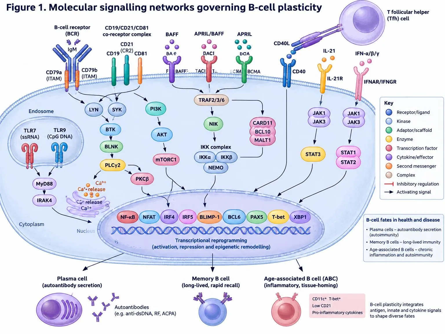
Figure 1. Molecular signalling networks governing B-cell plasticity.
BCR engagement (IgM with CD79a/CD79b ITAMs) and the CD19/CD21/CD81 co-receptor recruit LYN, SYK, BTK, BLNK and PLCγ2, generating Ca²⁺ flux and PKCβ activation, while PI3K–AKT–mTORC1 supports metabolic growth. BAFF-R, TACI and BCMA signal via TRAF2/3/6, NIK and the IKK complex; CD40–CD40L contact with Tfh cells, IL-21R and interferon receptors signal through JAK–STAT; endosomal TLR7/TLR9 act via MyD88–IRAK4. These inputs converge on NF-κB, NFAT, IRF4, IRF5, BLIMP-1, BCL6, PAX5, T-bet and XBP1, directing differentiation into plasma cells, memory B cells or age-associated B cells.
4. B-Cell Subpopulations in Autoimmune Diseases
Phenotyping studies, increasingly supported by single-cell transcriptomic and epigenomic analyses, have revealed that the circulating and tissue B-cell compartment in autoimmune disease is more heterogeneous than the classical naive, memory and plasma-cell scheme suggests. Several populations are enriched in patients and appear to represent distinct functional states reached through plastic differentiation. Table 1 summarises the principal subsets discussed below.
Table 1: Selected B-cell Subsets Implicated in Autoimmune Disease.
| Subset | Typical phenotype | Main drivers | Disease association | Refs |
|---|---|---|---|---|
| Double-negative (DN2) / atypical memory | CD27⁻ IgD⁻ CD11c⁺ T-bet⁺ FcRL5⁺ | TLR7, IFN-γ, IL-21, BCR | Active SLE, extrafollicular plasmablast precursors | [23–27] |
| Age-associated / T-bet⁺ B cells | CD11c⁺ T-bet⁺, class-switched | TLR7/9, IFN-γ, IL-21 | Lupus-like disease in models, SLE, RA | [25,26] |
| Regulatory B cells (Breg) | CD24ʰⁱ CD38ʰⁱ transitional-like; IL-10⁺, IL-35⁺ | CD40, TLR9, inflammatory cues | Reduced or impaired in SLE and RA | [28–32] |
| Long-lived plasma cells | CD138⁺ CD38⁺⁺, CD20⁻ | BAFF, APRIL, IL-6, niche signals | Persistent autoantibody production | [4,14] |
4.1 Double-negative and atypical memory B cells
A population of memory-like B cells lacking both CD27 and IgD, termed double-negative (DN) cells, is expanded in patients with active SLE and has been reported in other autoimmune conditions [23]. A subset of these cells, often called DN2, is characterised by high expression of CD11c, T-bet and FcRL5, low expression of CD21 and CD27, and a strong responsiveness to TLR7 and interferon-γ [24]. Jenks and colleagues showed that these cells are poised to differentiate into antibody-secreting cells through an extrafollicular route, and that their frequency correlates with disease activity, autoantibody specificities and nephritis in patients, particularly those of African ancestry [24]. IL-21 promotes the expansion and plasma-cell differentiation of autoreactive CD11c-high T-bet-positive B cells in lupus, linking this state to T-cell help from peripheral helper populations [26]. Atypical memory B cells with related features accumulate in chronic infection and ageing, suggesting that persistent stimulation, rather than an autoimmune-specific lesion, may drive this state, but in SLE the combination with nucleic-acid autoantigens makes it particularly pathogenic [27].
4.2 Age-associated and T-bet-expressing B cells
In mouse models, B cells that express T-bet and CD11c accumulate with age and in autoimmune-prone strains. Rubtsova and co-workers showed that T-bet expression in B cells is required for the development of lupus-like autoimmunity in a mouse model, and that these cells are a major source of autoantibodies of the IgG2a/c isotype [25]. The transcription factor T-bet in this context is induced by interferon-γ, TLR7 or TLR9 signals and cooperates with IL-21 to support differentiation into plasmablasts [25,26]. Taken together with the human DN2 data, these findings point to a conserved, interferon- and TLR-driven differentiation programme that is amplified in autoimmune hosts. A challenge for the field is that nomenclature differs between laboratories, and the same cells may be labelled DN2, atypical memory, age-associated or CD11c-positive B cells depending on the markers chosen [27].
4.3 Regulatory B cells
B cells can also restrain immunity. Mauri and Bosma reviewed evidence that B cells exert regulatory functions largely through IL-10, as well as through IL-35, TGF-β and direct cell contact [28]. Regulatory B cells (Bregs) do not constitute a single lineage; rather, regulatory capacity is a functional state that can be acquired by cells of several phenotypes, including transitional-like CD24-high CD38-high cells, plasmablasts and CD1d-high marginal-zone-like cells [29]. In healthy individuals, CD24-high CD38-high B cells suppress Th1 responses through IL-10, and this capacity is impaired in patients with SLE [30]. In experimental autoimmune encephalomyelitis, B-cell-derived IL-10 was needed for recovery, demonstrating that B cells can protect as well as harm [31]. IL-35-producing B cells, including plasmablasts, were also shown to be critical regulators in autoimmune and infectious disease models [32]. The clinical significance of these observations is evident: B-cell depletion removes regulatory as well as pathogenic cells, which may explain disease exacerbations or new autoimmune phenomena that occasionally follow anti-CD20 therapy [29,31].
4.4 Plasmablasts and long-lived plasma cells
Circulating plasmablasts expand during flares of SLE and other diseases and are enriched for autoreactive specificities, whereas long-lived plasma cells reside in bone marrow and inflamed tissues, supported by BAFF, APRIL, IL-6 and stromal contacts [4,14]. Because these cells express little or no CD20, they are largely unaffected by rituximab, which may account for the persistence of certain autoantibodies after successful B-cell depletion [4]. Targeting this compartment has therefore become a major theme in therapeutic development (Section 9).
5. Role of B Cells in Autoimmune Pathogenesis
5.1 Failure of tolerance checkpoints
A defining abnormality in many autoimmune diseases is the failure of tolerance checkpoints that normally limit autoreactive B cells. Yurasov and colleagues found that patients with SLE had a high frequency of autoreactive and polyreactive clones among new emigrant and mature naive B cells, reflecting defects at both the central and peripheral checkpoints [33]. Patients with RA showed a similar defect in early tolerance, with an increased proportion of autoreactive cells at the immature stage [34]. These defects precede, and may contribute to, the later appearance of disease-specific autoantibodies. Importantly, they are not simply restricted to patients with active disease and may be present in unaffected relatives, suggesting a heritable contribution [8,33].
5.2 Autoantibody-dependent mechanisms
Autoantibodies cause disease by forming immune complexes that deposit in tissues and activate complement and Fc receptors, by binding directly to cell-surface or extracellular targets, and by modulating receptor function [3]. In SLE, anti-dsDNA and anti-nucleosome antibodies contribute to glomerular injury; in RA, rheumatoid factor and anti-citrullinated protein antibodies are linked to joint destruction and may precede clinical onset by years; in myasthenia gravis, antibodies against the acetylcholine receptor interfere with neuromuscular transmission [3]. The pathogenic potential of an autoantibody depends on its affinity, isotype, glycosylation and the availability of its target, features that are determined by the differentiation pathway through which the producing cell passed.
5.3 Antibody-independent mechanisms
Several lines of evidence show that B cells contribute to autoimmunity independently of antibody secretion. The clinical benefit of B-cell depletion often precedes any decline in autoantibody levels [1,36]. B cells serve as antigen-presenting cells for autoreactive T cells, secrete pro-inflammatory cytokines including IL-6, TNF and lymphotoxin, and help organise lymphoid architecture. Barr and colleagues demonstrated that IL-6-producing B cells drive disease in a mouse model of EAE, and that the therapeutic effect of B-cell depletion was linked to the removal of these cells [36].
5.4 Ectopic lymphoid structures
In inflamed target tissues, B cells can organise into tertiary lymphoid structures with features of GCs. In rheumatoid synovium, such structures support local class switching, somatic hypermutation and production of autoantibodies, including those directed against citrullinated antigens [35]. Similar structures occur in the salivary glands of patients with Sjögren's disease and in the meninges in MS. By creating a privileged niche for local selection and differentiation, ectopic structures allow autoimmune responses to be sustained within the tissue itself, which may explain why peripheral blood B-cell depletion does not always clear tissue-resident B cells [4,35].
6. B-Cell–T-Cell Interactions
6.1 T follicular helper cells and the germinal centre
The collaboration between B cells and CD4⁺ T cells is essential for high-affinity, class-switched antibody responses. T follicular helper (Tfh) cells, defined by expression of CXCR5, PD-1, BCL6 and the cytokine IL-21, provide the signals through CD40L, ICOS and cytokines that GC B cells require for survival, proliferation and selection [37,38]. In autoimmune settings, excessive or misdirected Tfh activity can permit the survival of autoreactive B cells. Expanded circulating populations resembling Tfh cells have been reported in patients with severe SLE, with their frequency correlating with disease activity and autoantibody levels [40].
6.2 Follicular regulatory T cells
Foxp3-positive follicular regulatory T (Tfr) cells counterbalance Tfh activity within the GC. Linterman and colleagues showed that Tfr cells limit the size of the GC response and restrict the emergence of non-antigen-specific B-cell clones [39]. Reduced Tfr function, or an imbalance in the Tfh to Tfr ratio, may therefore contribute to the escape of autoreactive B cells from GC control, although human data remain less definitive than data from mouse models [38,39].
6.3 Peripheral helper T cells and extrafollicular responses
A further T-cell population that drives B-cell responses outside classical follicles has been identified. Rao and co-workers described PD-1-high CXCR5-negative T peripheral helper (Tph) cells that accumulate in the synovium of RA patients and secrete IL-21 and CXCL13, thereby providing B-cell help in inflamed tissue [41]. In SLE, Tph-like cells promote plasmablast differentiation through IL-21 and MAF-dependent mechanisms and correlate with the frequency of DN2 B cells [42]. This extrafollicular axis offers a plausible explanation for the rapid generation of autoantibody-secreting cells that often bypass GC-based quality control and thereby carry reduced selective pressure against self-reactivity [26,42].
6.4 Bidirectional communication
The interaction is not unidirectional. B cells present peptide antigen on MHC class II and provide co-stimulatory signals, cytokines such as IL-6 and lymphotoxin, and chemokines that shape T-cell differentiation. In turn, cytokines from T cells influence B-cell fate decisions, as seen with IL-21, interferon-γ and IL-4 [37,38]. Autoreactive B cells therefore recruit and instruct autoreactive T cells just as T cells select and expand B cells, establishing positive feedback loops that sustain chronic disease [1,36]. Breaking these loops, for instance by targeting co-stimulatory molecules such as CD40L, ICOS or CD28 signalling, remains an attractive therapeutic strategy, although clinical translation has been challenging.
7. Epigenetic and Metabolic Regulation of B-Cell Plasticity
7.1 Epigenetic programming of B-cell states
Transcription factor networks act on a chromatin landscape that determines which genes are accessible, and epigenetic modifications therefore provide a layer of memory and stability on top of plastic transcriptional programmes. DNA methylation, histone modification and chromatin accessibility change as B cells activate, divide and differentiate. Barwick and colleagues showed that plasma-cell differentiation is coupled to division-dependent DNA hypomethylation, with progressive loss of methylation at regulatory regions across successive cell divisions that facilitates expression of the plasma-cell programme [43]. This linkage between proliferation and epigenetic remodelling provides a mechanistic basis for the observation that the number of divisions a B cell has undergone strongly influences its fate and isotype.
In autoimmune disease, there is increasing evidence that these epigenetic landscapes are altered before overt differentiation. Scharer and colleagues profiled chromatin accessibility and gene expression in B-cell subsets from patients with SLE and reported that even naive B cells displayed epigenetic features resembling those of activated, plasma-cell-primed cells, with accessible chromatin at sites bound by factors that respond to interferon and promote differentiation [44]. These findings suggest that B cells in SLE are epigenetically poised to differentiate along extrafollicular routes, a state that could be maintained by persistent interferon exposure and would help explain the disease's chronic course [44].
Cytokines can imprint such programmes directly. Zumaquero and co-workers found that interferon-γ induces epigenetic programming of human T-bet-high B cells and promotes their TLR7/8- and IL-21-driven differentiation into antibody-secreting cells [45]. This type of cytokine-driven imprinting is a plausible mechanism by which a transient inflammatory environment can leave a durable mark on B-cell fate, and it suggests that epigenetic modifiers, such as inhibitors of histone methyltransferases or bromodomain proteins, might in future be used to reverse pathogenic programming, although such approaches remain experimental and carry a risk of broad off-target effects.
7.2 Metabolic reprogramming
B-cell activation imposes substantial metabolic demands. Resting B cells are metabolically quiescent, but on activation they increase glucose uptake, glycolysis, oxidative phosphorylation and biosynthetic activity to support rapid proliferation, and plasma cells must further sustain extremely high rates of protein synthesis and secretion [46,47]. The PI3K–AKT–mTOR axis connects BCR, TLR and cytokine signals to this metabolic response, and the balance between its activity and that of counter-regulatory factors determines whether activation leads to productive differentiation, anergy or death.
Caro-Maldonado and colleagues found that metabolic reprogramming is necessary for antibody production, that this reprogramming is suppressed in anergic B cells, and that it is exaggerated in B cells chronically exposed to BAFF [46]. This observation connects two arms of the picture developed above: the survival-factor environment that permits autoreactive cells to persist, and the cell-intrinsic metabolic capacity that permits those cells to differentiate. Boothby and Rickert reviewed how nutrient availability, hypoxia in GCs and the mTOR pathway regulate humoral responses, noting that the metabolic state of the cell shapes its decisions between GC and plasma-cell fates [47]. Metabolic pathways are thus attractive therapeutic targets: mTOR inhibition, for example, has been explored in lupus, and glycolysis and mitochondrial function represent additional candidate pathways, although specificity for pathogenic B cells over protective immunity is a major challenge [47].
7.3 Integration of epigenetic and metabolic cues
Epigenetic and metabolic regulation are closely linked, since many chromatin-modifying enzymes use metabolites such as acetyl-CoA, S-adenosylmethionine and alpha-ketoglutarate as substrates or cofactors. Changes in nutrient availability and metabolic flux may thus translate directly into altered chromatin states. This relationship is only beginning to be explored in autoimmune B cells but provides a conceptual framework in which environmental exposures, inflammation and genetic risk converge on the same cell-intrinsic regulatory machinery [44,47].
8. B-Cell-Mediated Inflammation and Autoantibody Production
8.1 Extrafollicular and germinal centre routes to autoantibody production
Autoantibodies can be generated through two broad routes. The germinal centre route, in which T-cell-dependent selection operates, generally produces high-affinity, class-switched, somatically mutated antibodies and long-lived plasma cells and memory cells. The extrafollicular route generates short-lived plasmablasts rapidly, often with less affinity maturation and less rigorous negative selection. In lupus, both routes appear to contribute, and extrafollicular responses driven by TLR7, interferon-γ and IL-21 are especially prominent in active disease [24,26]. Plasmablast expansions in acute SLE flares contain clones that have undergone extensive diversification and include cells of naive origin that have been recruited into the response [3,24]. The route of differentiation has consequences for therapy: extrafollicular plasmablasts are short-lived and might respond to approaches that interfere with their generation, while long-lived plasma cells would require agents directed against their survival niche or intrinsic biology [4,14].
8.2 Immune complexes, complement and Fc receptor activation
Once produced, autoantibodies exert effects through immune complex formation and engagement of effector pathways. Immune complexes containing nucleic acids can activate plasmacytoid dendritic cells through TLR7 and TLR9 to produce type I interferon, which in turn enhances B-cell differentiation, thereby generating a self-amplifying circuit [21,24]. Complement activation by immune complexes causes local inflammation and tissue injury, particularly in kidney, skin and the nervous system, while Fc receptors on myeloid cells and neutrophils amplify damage through cytokine release and effector functions [3].
8.3 Cytokine-mediated inflammation
B cells also drive inflammation through cytokines. IL-6, TNF and GM-CSF produced by B cells can act on T cells, myeloid cells and stromal cells, and the secretion of IL-6 by B cells in particular has been linked to pathogenic Th17 responses in experimental autoimmune encephalomyelitis [36]. In MS, the observation that anti-CD20 therapy rapidly reduces new inflammatory lesions even though it does not target antibody-secreting plasma cells supports an important role for antibody-independent mechanisms, including antigen presentation and cytokine production, in that disease [50]. Conversely, loss of IL-10- and IL-35-producing regulatory B cells removes a brake on inflammation [30,32]. The net inflammatory output of the B-cell compartment is therefore the sum of pathogenic and regulatory functions whose balance may differ between individuals, tissues and stages of disease.
8.4 Positive feedback and chronicity
The interplay of these mechanisms creates positive feedback loops that sustain chronic disease. Autoantibody-containing immune complexes stimulate interferon production, which enhances B-cell responsiveness to TLR ligands and supports T-bet-high B-cell differentiation; the resulting plasmablasts generate more autoantibodies; autoreactive B cells present antigen to T cells that provide further help; and ectopic lymphoid structures provide protected sites for the local selection and survival of these cells [24,35,45]. These loops explain why autoimmune diseases are self-perpetuating, and they identify nodes at which interruption may produce durable benefit: the BAFF survival signal, the TLR–interferon axis, T-cell help and the plasma-cell niche.
9. Emerging Biological Therapies Targeting B Cells
The recognition of plasticity as a driver of disease shapes current therapeutic strategy. Approaches fall into those that deplete B cells, those that block survival or activation signals, those that remove plasma cells or their products, and those that aim to reset the immune system. Table 2 summarises representative agents and their principal limitations.
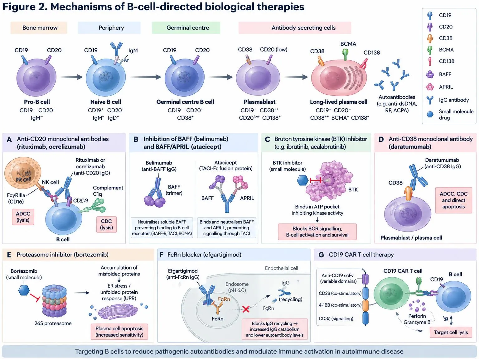
Figure 2. Mechanisms of B-cell-directed biological therapies.
(A) Anti-CD20 antibodies deplete B cells by ADCC and CDC. (B) Belimumab neutralises BAFF; atacicept (TACI-Fc) neutralises BAFF and APRIL. (C) BTK inhibitors occupy the ATP pocket and block BCR signalling. (D) Daratumumab targets CD38 on plasmablasts and plasma cells. (E) Bortezomib inhibits the 26S proteasome, triggering the unfolded protein response and plasma-cell apoptosis. (F) Efgartigimod blocks FcRn-mediated IgG recycling, lowering autoantibody levels. (G) CD19 CAR T cells (scFv, CD28/4-1BB, CD3ζ) lyse CD19⁺ B cells via perforin and granzyme B.
Table 2: Representative B-cell-directed Therapies in Autoimmune Disease.
| Target | Representative agent(s) | Evidence (examples) | Principal limitation | Refs |
|---|---|---|---|---|
| CD20 | Rituximab, ocrelizumab | RA, ANCA vasculitis, relapsing MS | Spares plasma cells; hypogammaglobulinaemia; infection | [48–50,60] |
| BAFF | Belimumab | SLE, lupus nephritis | Modest effect size; incomplete B-cell depletion | [51,52] |
| BTK | Evobrutinib | Phase 2, relapsing MS | Long-term safety and efficacy to be defined | [22,53] |
| CD38 / proteasome | Daratumumab, bortezomib | Refractory SLE (small series) | Hypogammaglobulinaemia; limited controlled data | [54,61] |
| FcRn | Efgartigimod | Generalised myasthenia gravis | Reduces IgG without removing producing cells | [55] |
| CD19 | Inebilizumab; CD19 CAR T cells | NMOSD; refractory SLE | Cost, toxicity, durability and access | [56–59] |
9.1 Anti-CD20 antibodies
Rituximab, a chimeric anti-CD20 antibody, depletes CD20-positive B cells through complement-dependent cytotoxicity, antibody-dependent cellular cytotoxicity and direct signalling effects. Randomised trials established its benefit in RA, where it improved clinical responses in patients with inadequate response to methotrexate [48], and in ANCA-associated vasculitis, where it was non-inferior to cyclophosphamide for remission induction [49]. In relapsing MS, ocrelizumab, a humanised anti-CD20 antibody, reduced relapse rate and new lesions compared with interferon beta-1a, producing a major shift in the treatment of that disease [50]. The success of anti-CD20 therapy in conditions in which autoantibodies are not clearly pathogenic underscores the importance of antibody-independent B-cell functions [50].
The limits of anti-CD20 therapy are equally informative. Plasma cells and some tissue-resident B cells are CD20-negative or poorly accessible and thus persist [4,35]. Depletion is incomplete in lymphoid tissue, so that residual cells may seed relapse. Moreover, repopulating B cells are initially dominated by transitional and naive cells, which may restore tolerance in some patients but not others [4,5]. Prolonged therapy is associated with hypogammaglobulinaemia and increased infection risk [60], and, as noted above, depletion of regulatory B cells may be detrimental in some contexts [29].
9.2 Targeting the BAFF/APRIL axis
Because BAFF supports survival of autoreactive cells, neutralising it is a rational approach. Belimumab, a monoclonal antibody against soluble BAFF, improved responses in patients with active SLE in randomised trials [51] and was subsequently shown to improve renal outcomes in lupus nephritis when added to standard therapy [52]. Effect sizes have been modest, consistent with the idea that BAFF blockade removes a survival advantage without eliminating established autoreactive memory cells and plasma cells [20,51]. Dual inhibition of BAFF and APRIL, and combination with anti-CD20 therapy, have been proposed as ways to increase efficacy by limiting both the entry of new autoreactive cells and the persistence of existing ones.
9.3 Bruton's tyrosine kinase inhibitors
BTK is required for BCR and TLR signalling in B cells and for Fc receptor signalling in myeloid cells, and its inhibition thus targets several pathogenic pathways at once [22]. In a phase 2 trial in relapsing MS, the BTK inhibitor evobrutinib reduced the number of enhancing lesions compared with placebo, providing proof of concept for oral B-cell modulation [53]. Because BTK inhibitors modulate rather than deplete B cells, they may preserve regulatory function and avoid prolonged immunoglobulin suppression, although the effects on plasma cells are limited, and long-term safety, including hepatic effects, requires careful evaluation [22,53].
9.4 Plasma-cell-directed therapy and neonatal Fc receptor blockade
The persistence of long-lived plasma cells has driven interest in agents developed for multiple myeloma. The proteasome inhibitor bortezomib depleted plasma cells and was associated with clinical improvement and reduction in autoantibody titres in patients with refractory SLE in a small series [61]. The anti-CD38 antibody daratumumab produced similar effects, with reductions in anti-dsDNA antibodies and in interferon signature in two patients with severe, refractory disease [54]. These observations are encouraging but come from small uncontrolled cohorts, and prolonged plasma-cell depletion carries the risk of hypogammaglobulinaemia and loss of protective serological memory [54,61].
An alternative approach is to reduce the concentration of pathogenic IgG without removing its source. Efgartigimod, an engineered Fc fragment that blocks the neonatal Fc receptor and accelerates IgG catabolism, improved outcomes in generalised myasthenia gravis in a randomised placebo-controlled trial [55]. This strategy is rapid and reversible but does not affect antibody production, and its role in diseases mediated by cellular mechanisms is likely to be limited.
9.5 CD19-directed therapy and CAR T cells
CD19 is expressed on a broader range of B-lineage cells than CD20, including plasmablasts and some plasma cells. Inebilizumab, an anti-CD19 antibody, reduced attack risk in neuromyelitis optica spectrum disorder in a randomised trial [56]. More radical is the use of autologous CD19-directed chimeric antigen receptor (CAR) T cells, which can achieve deeper depletion than monoclonal antibodies, including in lymphoid tissue. In a first-in-human series of patients with refractory SLE, CD19 CAR T cells led to rapid B-cell depletion, disappearance of autoantibodies and clinical remission [57]. Follow-up of a larger case series of patients with SLE, idiopathic inflammatory myositis and systemic sclerosis showed that most patients achieved remission and discontinued immunosuppression, with B-cell repopulation consisting predominantly of naive cells [58]. This pattern is consistent with a true immunological reset in which the repertoire is reconstituted without the autoreactive memory compartment [58,59].
Several caveats apply. Experience is limited to small numbers of patients with short follow-up, and cytokine release syndrome, neurotoxicity, prolonged cytopenias and infections have been documented, although they appear generally milder than in oncology [59]. Lymphodepleting chemotherapy, manufacturing cost and limited access constrain wide application. Allogeneic, off-the-shelf products, in vivo CAR T generation, and T-cell engagers that deplete B cells without cell manufacturing are in development and may widen access [59].
9.6 Combination and sequential strategies
Given the multiple routes by which pathogenic B-cell states are maintained, combination strategies are logical. Examples include depletion followed by BAFF blockade to limit repopulation by autoreactive cells, pairing anti-CD20 therapy with plasma-cell-directed agents, and use of co-stimulation blockade to interrupt T-cell help [5,20,59]. The optimal sequence, timing and patient selection remain to be defined and will probably depend on biomarkers that identify the dominant pathogenic B-cell state in each individual.
10. Challenges and Therapeutic Limitations
10.1 Heterogeneity and the absence of validated biomarkers
The most fundamental obstacle to precision B-cell therapy is heterogeneity. Patients with the same clinical diagnosis may differ markedly in the dominant pathogenic B-cell state, with some showing prominent extrafollicular DN2 expansion and interferon signatures and others dominated by GC-derived or tissue-resident populations [24,27,44]. Inconsistent nomenclature further complicates comparison across studies, as the same populations are described under different names and defined by different marker panels [27]. At present there are no widely validated biomarkers that predict which patient will respond to which B-cell-directed agent, and peripheral blood phenotyping may not reflect the situation in tissues such as the kidney, synovium or central nervous system [4,35].
10.2 Incomplete targeting of pathogenic compartments
No current agent reliably eliminates all pathogenic B-cell states. Anti-CD20 antibodies spare plasma cells and are incompletely effective in tissue [4,35]; BAFF blockade limits survival signals but has modest effects on established memory and plasma-cell populations [20,51]; plasma-cell-directed therapy is supported only by small series [54,61]; and even CD19 CAR T cells, though impressive, have been evaluated in small numbers of patients with short follow-up [58,59]. Long-lived plasma cells in protective niches remain a particular challenge, and the relative importance of CD19-negative or CD20-negative compartments for disease persistence remains to be established in most conditions [4].
10.3 Loss of protective immunity and regulatory function
Broad B-cell depletion carries the cost of infection risk and impaired vaccine responses. Hypogammaglobulinaemia occurs in a meaningful proportion of patients receiving repeated rituximab and is associated with serious infections [60]. Deeper depletion, whether by CAR T cells or plasma-cell-directed therapy, raises questions about loss of humoral memory against pathogens and vaccines, although early CAR T experience suggests that protective antibody titres to some prior vaccines may be preserved [58,59]. In addition, depletion of regulatory B cells, which are reduced and functionally impaired in some autoimmune settings, may eliminate protective mechanisms and has been proposed as an explanation for paradoxical disease flares or new-onset autoimmunity after anti-CD20 therapy [29,30].
10.4 Durability, access and cost
Most B-cell-directed therapies, including anti-CD20 antibodies and belimumab, require repeated administration, and disease often relapses when treatment is stopped or when B cells repopulate [4,5]. CAR T therapy offers the prospect of drug-free remission but requires specialised centres, lymphodepleting chemotherapy and substantial cost, which limits access and raises equity concerns [59]. Long-term safety, including the risk of secondary malignancy, reproductive effects and late infections, is incompletely characterised for newer modalities [59].
10.5 Translational limitations
Mouse models have been invaluable in defining mechanisms such as T-bet-dependent B-cell autoimmunity and regulatory B-cell function [25,31], but differences between mouse and human B-cell subsets, TLR expression patterns and lymphoid organisation limit direct extrapolation. Clinical trials in heterogeneous populations with conventional endpoints may fail to detect benefit in responsive subgroups, and several B-cell-directed agents have shown disappointing results in unselected cohorts despite strong mechanistic rationale [5,51]. More sensitive and mechanism-aligned endpoints, together with enrichment strategies based on B-cell biomarkers, would improve the efficiency of development.
11. Future Perspectives
Several developments are likely to shape the next decade of research and therapy. First, high-dimensional profiling, including single-cell RNA sequencing, chromatin accessibility assays, repertoire sequencing and spatial transcriptomics of tissue, will allow patients to be stratified by dominant B-cell state rather than by clinical label alone [27,44]. The epigenetic findings in SLE suggest that signatures of chromatin accessibility might eventually serve as biomarkers of disease activity and of treatment response [44,45].
Second, therapeutic approaches are likely to become more selective. Depleting all B cells is a blunt instrument, whereas targeting pathogenic states, such as T-bet-high atypical memory cells or autoreactive clones, might preserve protective immunity. Antigen-specific strategies, such as chimeric autoantibody receptor T cells that target cells bearing a specific autoreactive receptor, and approaches that restore tolerance rather than eliminate cells, represent attractive directions [59]. Modulating the inputs that sustain pathogenic states, including interferon signalling, TLR7 activation, IL-21 and metabolic pathways, offers a complementary route [24,26,47].
Third, CAR T cell therapy will probably evolve towards simpler, safer and more accessible forms. Allogeneic cell products, in vivo CAR generation and bispecific T-cell engagers directed at CD19 or BCMA are under investigation, and if safe and effective they could broaden access considerably [59]. Important questions concern the durability of remission, the extent of immune reconstitution and whether depletion of plasma cells by BCMA-directed approaches is required in some diseases [58,59].
Fourth, rational combination and sequencing strategies will be informed by mechanistic understanding. For example, anti-CD20 therapy followed by BAFF blockade is intended to prevent autoreactive cells from re-entering the repertoire, and co-stimulation blockade may add benefit by interrupting T-cell help [5,20]. Trials designed to test such combinations in biomarker-selected populations are needed.
Finally, deeper understanding of the epigenetic and metabolic basis of plasticity may open new therapeutic opportunities, but this field is at an early stage. A key priority is to determine whether pathogenic B-cell states are reversible, and whether early intervention before the epigenetic programme is consolidated can prevent progression to chronic disease [43–47].
CONCLUSION
B cells are central, versatile players in autoimmune disease whose pathogenic contribution reflects not only antibody production but also plastic transitions between effector, regulatory, antigen-presenting and long-lived antibody-secreting states. These transitions are controlled by transcriptional networks, BCR and innate signalling, survival factors, cytokines, T-cell help, and epigenetic and metabolic programmes, and in autoimmune disease they are biased towards extrafollicular and tissue-based pathogenic differentiation. Therapies targeting CD20, BAFF, BTK, plasma cells, FcRn and CD19 have each delivered clinical benefit, and CD19 CAR T cells have raised the prospect of immune reset in refractory disease. Nevertheless, current approaches remain incomplete, limited by persistent plasma cells and tissue-resident B cells, loss of protective immunity, uncertain durability and restricted access. Progress will depend on integrating high-resolution profiling with mechanism-based, biomarker-guided, and increasingly selective interventions that target pathogenic B-cell states while preserving protective and regulatory functions.
REFERENCES








Mohit Kasana, Aatif Ashfaque Pathan, Kanad Yerpude, Ashutosh Devatwal, Sudhanshu Ranjan, Mohammed Faizal, Gokul Varnan, Shivprasad Dhage, B-Cell Plasticity in Autoimmune Diseases: Pathogenic Mechanisms and Emerging Biological Therapies, Int. J. of Pharm. Sci., 2026, Vol 4, Issue 11, 451-467. https://doi.org/10.5281/zenodo.23923813
 10.5281/zenodo.23923813