

We use cookies to ensure our website works properly and to personalise your experience. Cookies policy
Krishna school of pharmacy and research.
Dementia is a progressive neurodegenerative syndrome marked by the deterioration of cognitive, behavioural, and functional abilities and represents a major global health challenge in aging populations. It comprises a heterogeneous group of disorders, including Alzheimer’s disease, vascular dementia, frontotemporal dementia, and dementia with Lewy bodies, each defined by distinct clinical trajectories and molecular pathologies. This review integrates current understanding of dementia subtypes, disease stages, and progression patterns, emphasizing the interconnected molecular and cellular mechanisms underlying cognitive decline, such as amyloid-? deposition, Tau pathology, neuroinflammation , mitochondrial dysfunction, synaptic failure, and vascular impairment. Recent advances in biomarker discovery are highlighted, spanning genetic and epigenetic markers, blood-based and cerebrospinal fluid biomarkers, neuroimaging modalities, and emerging digital health technologies. In particular, plasma phosphorylated tau, amyloid-? ratios, neurofilament light chain, and inflammatory markers demonstrate strong diagnostic and prognostic potential, even at preclinical stages. The convergence of multimodal biomarker panels with artificial intelligence-based analytics is poised to transform early diagnosis, patient stratification, and precision-based therapeutic development in dementia.
Dementia is a progressive neurodegenerative syndrome characterized by a decline in cognitive functions such as memory, language, reasoning, and executive abilities, severe enough to interfere with daily life and independence [1]. It represents one of the major global public health challenges, with prevalence rising rapidly due to increased life expectancy and aging populations worldwide [2]. According to the World Health Organization, dementia affects more than 55 million people globally, with Alzheimer’s disease accounting for approximately 60–70% of all cases [3]. Despite its growing burden, dementia remains incurable, highlighting the urgent need for improved diagnostic, prognostic, and therapeutic strategies [2,4]. Dementia is not a single disease but an umbrella term enclose several disorders with distinct pathological mechanisms, including Alzheimer’s disease, vascular dementia, frontotemporal dementia, and dementia with Lewy bodies [1,5]. These disorders differ in their clinical presentation, age of onset, rate of progression, and underlying molecular pathology [5]. Common pathological features include abnormal protein aggregation, such as amyloid-β plaques, tau neurofibrillary tangles, and α-synuclein inclusions, along with synaptic dysfunction, neuroinflammation, mitochondrial impairment, and progressive neuronal loss [6,7]. Collectively, these cellular and molecular abnormalities disrupt neural networks and lead to progressive cognitive decline [7]. Recent advances in biomarker research have significantly transformed the understanding and diagnosis of dementia [8]. Biomarkers obtained from cerebrospinal fluid, blood, neuroimaging, genetic testing, and digital health technologies provide insights into disease-specific pathological processes and enable earlier detection [8,9]. Blood-based biomarkers such as plasma phosphorylated tau and amyloidβ ratios, along with neuroimaging techniques including Magnetic Resonance Imaging (MRI) and Positron Emission Tomography (PET), have demonstrated high diagnostic accuracy even in preclinical disease stages [9,10]. In addition, emerging digital biomarkers—such as speech analysis, gait assessment, and eyetracking—offer non-invasive, scalable tools for large-scale screening and longitudinal monitoring [11]. This review outlines the major types of dementia, their stages and progression, and the molecular mechanisms underlying cognitive decline. It further discusses established and emerging biomarkers, recent clinical trials, and future directions aimed at integrating multi-biomarker panels and artificial intelligence-based approaches to enhance early diagnosis, personalized care, and therapeutic development in dementia [4,8].
3. Pathophysiological And Foundation Of
Dementia
3.1 Forms and Stages of Dementia
3.1.1 Types of Dementia
1. Alzheimer’s Disease:
Alzheimer's Disease (AD) is a common neurodegenerative disorder that is found to affect mainly older people, causing memory loss, confusion, and changes in behaviour. It is also the most common cause of dementia worldwide [14,16]. The disease advances slowly, with progressive damage and neuron loss in the brain [14,20].
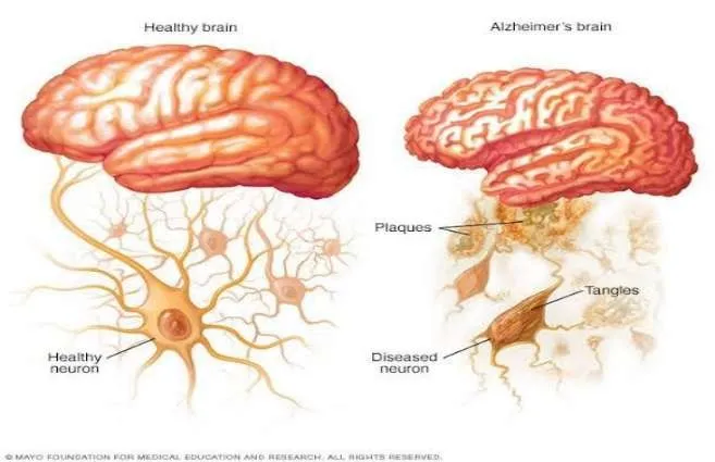
Figure 3.1: Alzheimer’s Disease
These abnormalities disrupt normal cell signalling and eventually lead to cell death. Factors that contribute to the disease are genetic predisposition, chronic inflammation, and oxidative stress [23,28]. Currently, AD has no cure, and treatments that exist are primarily aimed at symptom relief [14]. Research continues to identify the mechanisms behind it to create therapies that can prevent or halt disease progression [16,29].
2. Vascular Dementia
Vascular Dementia ranks as the second most common type of dementia after Alzheimer's disease and is induced by compromised blood supply to the brain, which reduces cognitive functions [17,18]. The symptoms will vary depending on the individual but include impairments in memory, concentration, decisionmaking, and organization.
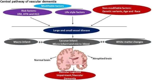
Figure 3.2: Vascular Dementia
The development and advancement of Vascular Dementia may be sudden or occur gradually, depending on the severity and character of the vascular damage [17]. The risk factors are common and include high blood pressure, diabetes, increased cholesterol levels, and smoking [17,18]. Despite there being no ultimate treatment, management focuses on regulating these vascular risks to slow disease advancement [17].
3. Frontotemporal Dementia
Frontotemporal dementia (FTD)* is a group of neurodegenerative disorders characterized by frontotemporal degeneration of the brain [19]. In contrast to Alzheimer's disease, FTD characteristically presents in its initial stages with personality change, behavioural changes, and language function impairment. Clinically, FTD can be divided into three main subtypes: the behavioural variant (bvFTD), the semantic variant of primary progressive aphasia (svPPA), and the non-fluent/agrammatic variant of primary progressive aphasia (nfvPPA).
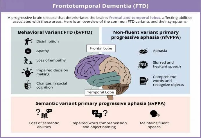
Figure 3.3: Frontotemporal Dementia
Onset usually precedes other dementias, generally at the age range 45-65 years [19]. No precise cure is currently available, and treatment options focus on relieving symptoms and improving quality of life. Diagnosis is still challenging because of the clinical heterogeneity.
4. Dementia with Lewy Bodies
Lewy Body Dementia (LBD) is a neurodegenerative disease characterized by the accumulation of alpha-synuclein protein inclusions, known as Lewy Bodies, in both cortical and subcortical regions of the brain [12]. It comprises two overlapping clinical syndromes:*Lewy Body Dementia (LBD)* and *Parkinson's Disease Dementia (PDD)*, which are distinguished mainly on the basis of whether cognitive impairment or motor symptoms first emerge [12]. The patient can also have autonomic impairment and increased sensitivity to antipsychotic drugs [12].
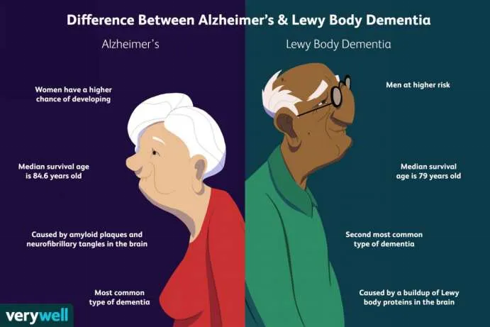
Figure 3.4: Dementia with Lewy Bodies
Accurate diagnosis is hindered by symptom overlap with other forms of dementia, and thus there is a critical need for enhanced imaging modalities and the identification of biomarkers. While no curative treatment has been found, therapy is focused on the control of symptoms through an inter-disciplinary approach to care.
3.1.2 Stages of Dementia
1. Initial Phase
Dementia is characterized by an extremely individualized course, and the early symptoms are often subtle and not always immediately apparent [13]. Most people in this early stage are still quite independent and only require occasional help with routine activities. The focus should be on the person's remaining abilities, minimizing the need for unnecessary intervention or takeover of tasks within their control. Poor orientation is also characteristic of early-stage dementia. One would no longer know where they are and therefore become lost, even in a familiar setting [13].
2. Middle Stage
In the moderate stage of dementia, clinical presentation becomes more pronounced, and the person will usually need more help to manage day-to-day activities [13]. At this point, additional support may be needed for some of their daily routines and additional help with some personal care activities like bathing and dressing—for example, having their clothes prepared in advance. How a person communicates or uses language will continue to decline at this stage also. It will get worse in the middle stage for problems with memory and thinking that are already present. A patient at this phase can also begin to experience and perceive things that do not exist (hallucinations) [13].
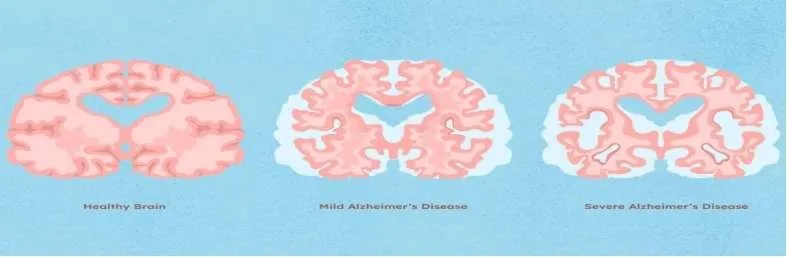
Figure 3.5: Stages of Dementia
3. Late Stage
At this later stage, dementia severely impacts almost every aspect of a person's functioning [15]. The individual will eventually need full-time care and full-time assistance with daily activities and personal hygiene, such as eating, bathing, and dressing. Though this type of support could be provided in a home setting, it is more typically provided in a care facility residency, walk slowly, with shuffle and less steadily – in the end they might spend more time in a chair or a bed [15].
3.2 Molecular and Cellular Mechanism Underlying Cognitive decline.
Cognitive impairment in dementia results from complex molecular and cellular processes that increasingly interfere with neuronal function and synaptic connectivity [29]. A number of interconnected pathways are involved in the decline of cognitive functions seen in the various dementias. A key mechanism involves the accumulation of aberrant proteins like β-amyloid (Aβ) plaques and hyperphosphorylated tau tangles [16,26].These aberrant proteins disrupt synaptic communication, trigger inflammatory reactions, and enhance neuronal apoptosiss [22,28]. In Alzheimer's disease, Aβ oligomers have been reported to impair longterm potentiation and trigger oxidative stress, finally leading to memory and learning impairments [21,23]. Likewise, tau pathology also shows a strong correlation with cognitive impairment severity, as it destabilizes microtubules and axonal transport [26]. Neuroinflammation is another key pathway in the pathogenesis of dementia [28].
Activated astrocytes and microglia react to protein aggregates by releasing reactive oxygen species (ROS) and pro-inflammatory cytokines, which are toxic to neurons [28]. Repeated neuroinflammation amplifies synaptic loss and neuronal degeneration, speeding cognitive deterioration further. Mitochondrial dysfunction is also a key player. Neurons are extremely energy-requiring, and mitochondrial damage results in decreased ATP production, augmented ROS formation, and calcium dyshomeostasis—all of which are contributory to synaptic dysfunction and cell death [24,27]. Synaptic loss is also one of the earliest and most reliable correlates of cognitive impairment in dementia [25].
Weakening and disappearance of synaptic connections, frequently induced by Aβ and tau pathology, precede neuronal loss and are strongly correlated with the extent of cognitive impairment [27]. defective proteostasis and autophagy pathways prevent the degradation of damaged proteins and organelles, and they accumulate and contribute to further cellular stress. Lysosomal and ubiquitinproteasome system deficiencies have been reported in different dementias such as Frontotemporal dementia and Parkinson’s disease dementia [19]. Vascular contributions like blood-brain barrier dysfunction and cerebral hypoperfusion can synergistically exacerbate neurodegenerative changes by restricting oxygen and nutrient supply to the brain, enhancing vulnerability to amyloid deposition and white matter lesions [18]. All together, these molecular and cellular aberrations constitute a nexus of complex pathological processes that combine to result in the progressive cognitive deterioration seen in dementia [29].
3.3 Progression Pattern of the Disease
Dementia is a chronic, progressive neurological syndrome that reflects impairments in several cognitive domains such as memory, reasoning language and executive function [15]. The disease progresses gradually over time, although the rate and pattern of progression are highly variable based on the underlying type of dementia, comorbidities, and individual differences. The course of dementia is usually gradual, starting with minimal cognitive impairments that are often undetected or ascribed to aging. The impairments become more pronounced over time, compromising one's ability to engage in ordinary activities and socially interact. The clinical course usually involves a widening disparity between cognitive function and functional autonomy [15].
Memory loss is the most common and often the first symptom in most people, especially in Alzheimer's disease [14]. Yet, some types, for example, frontotemporal dementia or Lewy body dementia, can start with language impairment, personality changes, visual hallucinations, or motor symptoms [12,19]. The decline can be linear and gradual or come in fits and starts, with rapid deterioration at times, particularly in vascular dementia, where there can be stepwise decline after cerebrovascular accidents [17]. Following progression following progression of the disease, neuropsychiatric symptoms like apathy, agitation, delusions, or depression can occur and increase. Behavioural and psychological symptoms of dementia (BPSD) are strong predictors of caregiver burden and are the most important determinants of institutionalization [13].
Functionally, dementia sufferers gradually lose the capacity to cope with complicated tasks, and then more routine activities of daily living. Eventually, the condition results in total dependency. Death tends to be precipitated by complications such as infections, malnutrition, or illnesses due to immobility, but not by the neurodegenerative process itself [15].
4. Biomarkers: Definition And Classification
Biomarkers are quantifiable biological markers that help in the timely detection, diagnosis, and monitoring of dementia [30]. They can be retrieved from body fluids, imaging, or genetic testing and offer information on important pathological features like amyloid deposition, tau impairment, and neurodegeneration [30]. Good biomarkers must be accurate, reproducible, and ideally non-invasive, facilitating more accurate diagnosis and personalized treatment options in dementia management [30].
4.1 Types
1. Genetic Biomarkers
Genetic biomarkers are informative about inherited pathogenesis and susceptibility [31,32]. The APOE ε4 allele is the best-characterized genetic risk factor for late-onset Alzheimer's disease and has a substantial effect on promoting amyloid plaque deposition and premature symptom onset [31]. Mutations in genes like APP (amyloid precursor protein), PSEN1, and PSEN2 underlie familial forms of dementia and cause early-onset Alzheimer's [31]. Moreover, risk loci identified by genome-wide association studies (GWAS) have suggested additional contributors, including TREM2, associated with neuroinflammation and microglial function [32].
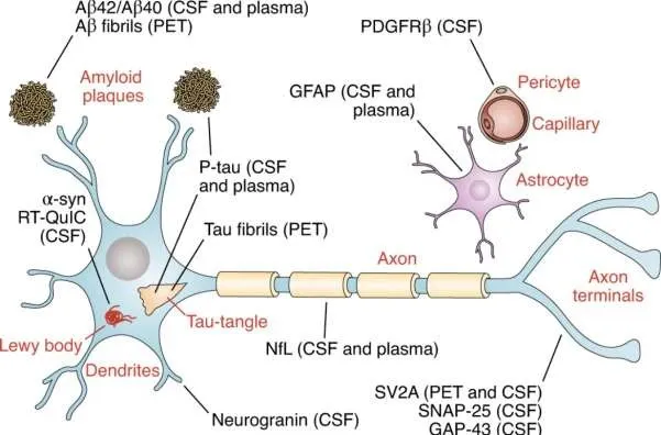
Figure 4.1: Genetic Biomarkers
2. Amyloid Biomarkers
Amyloid-β (Aβ) biomarkers represent the deposition of extracellular plaques, a fundamental aspect of Alzheimer's pathology [30]. Reduced cerebrospinal fluid (CSF) Aβ42 and lowered Aβ42/Aβ40 ratio reflect sequestration of Aβ within brain tissue [30]. Concomitantly, amyloid Positron Emission Tomography (PET) imaging with radiolabeled probes (e.g., ^11C-PIB, ^18F-florbetapir) makes possible direct visualization of amyloid deposits [33]. These biomarkers are now generally accepted in research and clinical diagnostic criteria for Alzheimer's disease [33].
3. Tau Biomarkers
Tau pathology is reflected by the presence of high levels of total tau (t-tau) and phosphorylated tau (p-tau181, p-tau217, p-tau231) in CSF, which are associated with the formation of neurofibrillary tangles and disease severity [30]. New tau PET imaging (e.g., ^18F-flortaucipir) allows for in vivo measurement of tau deposition and disease staging [33]. Importantly, p-tau isoforms in plasma are now being considered good, non-invasive markers for early diagnosis [35].
4. Neurodegeneration Biomarkers
These biomarkers indicate neuronal damage and structural loss of the brain [34].
Structural MRI usually demonstrates hippocampal atrophy, especially in Alzheimer's disease, whereas FDG-PET imaging emphasizes areas of decreased glucose metabolism, reflecting synaptic dysfunction [33]. In fluid biomarkers, neurofilament light chain (NfL) in CSF and plasma has proven to be a sensitive indicator of axonal damage and general neurodegeneration, non-specific for a type of dementia but an indicator of disease severity [34].
5. Neuroinflammation Biomarkers
Neuroinflammatory mechanisms are increasingly being identified as key to dementia progression [32]. Activated microglia and astrocytes produce cytokines and oxidative species that can be detected in CSF or blood (e.g., IL-6, TNF-α) [32]. TSPO PET imaging also identifies microglial activation in vivo but is so far of limited clinical application due to variability in binding affinity [33]. Genetic modifications like TREM2 mutations further emphasize the contribution of innate immunity in modulating dementia risk and progression [32].
5. Recent Developments In Molecular And Blood Based Biomarkers.
5.1 Amyloid beta, tau proteins
Amyloid beta (Aβ) and tau proteins remain at the forefront of the evolution of diagnostic biomarkers for Alzheimer's and other dementias [36]. Technical improvements in assay technologies and molecular profiling have made their measurement not just possible in cerebrospinal fluid (CSF) but increasingly even in peripheral blood, offering less invasive options for early diagnosis and monitoring [36,37].
1. Amyloid Beta (Aβ) as a Biomarker
More recent work has better defined detection of Aβ42 and Aβ40 isoforms in plasma with ultrasensitive technology like Single molecule array and mass spectrometry [37]. A decrease in the plasma ratio of Aβ42/Aβ40 has been associated with amyloid PET positivity and is proposed as a surrogate marker for cerebral amyloid burden [37,38]. Further, preclinical trials have shown plasma Aβ alterations can occur years ahead of cognitive impairment, providing promise for early detection in asymptomatic risk individuals for dementia [38]. A side from quantitative measurement, more recent methods aim to define the conformational states and aggregation tendencies of Aβ species, potentially providing greater specificity to differentiate Alzheimer's disease from other dementias [36].
2. Tau Protein as a biomarker
Tau proteins—particularly phosphorylated variants like p-tau181, p-tau217, and p-tau231—have emerged as strongly specific biomarkers [39]. Blood-based p-tau tests are now accurately able to detect early tau pathology, even before noticeable cognitive impairment [39]. It has been found that plasma p-tau217 shows superior performance in terms of predicting Alzheimer's pathology when compared with conventional CSF total tau or Aβ levels [40]. Simultaneously, progress in neuroimaging and fluid biomarker studies has enabled scientists to associate blood tau levels with local brain atrophy and tau PET uptake [39]. These findings indicate that blood-based tau assays not only can detect the presence of disease but potentially its course as well. In addition, attempts are being made to create panels of combined Aβ and tau markers with other protein biomarkers, including neurofilament light chain (NfL) and glial fibrillary acidic protein (GFAP), for enhanced diagnostic accuracy and prognostic utility in dementia subtypes [41].
5.2 Inflammatory markers
Neuroinflammation is central to the pathogenesis of many types of dementia, notably Alzheimer's disease (AD) and vascular dementia [42]. There is emerging evidence that peripheral and central inflammation is not merely secondary to amyloid and tau pathology but could actively contribute to disease progression [42]. As such, inflammatory markers are being increasingly investigated as diagnostic and prognostic biomarkers.
Molecular and Blood-Based Inflammatory Markers
The main pro-inflammatory cytokines include interleukin-6 (IL-6), tumor necrosis factor-alpha (TNF-α), and interleukin-1β (IL-1β) and have been found to be consistently elevated in dementia patients [42]. High levels of plasma IL-6 and TNF-α correlate with cognitive deterioration and cortical thinning and have been implicated as systemic markers of neurodegenerative processes [42]. Over the past decade sensitive multiplex immunoassays and proteomic platforms have made it possible to quantitate several cytokines, chemokines, and acute phase proteins simultaneously from plasma of serum [43]. Of note, YKL-40 (chitinase3-like protein 1), an astrocytic inflammatory marker, has attracted interest due to dual detectability in cerebrospinal fluid and plasma. It is linked to glial activation and disease progression in AD and frontotemporal dementia [43].
Additionally, triggering receptor expressed on myeloid cells 2 (TREM2), specifically its soluble form (sTREM2), has been identified as an appealing biomarker of microglial activation [44]. Increased sTREM2 levels in plasma and CSF have been correlated with early neuroinflammatory alterations and amyloid deposition, before tau pathology in longitudinal research [44].
Inflammatory Marker Panels and Biomarker Combinations
Instead of using solitary markers, current research highlights the value of inflammatory biomarker panels, which include IL-6, CRP, YKL-40, and sTREM2 to improve diagnostic specificity [42,43]. These biomarker clusters have demonstrated promise in discrimination of Alzheimer's disease from other neurodegenerative diseases and in predicting conversion from mild cognitive impairment (MCI) to dementia. In addition, integration of inflammatory markers with amyloid-beta, tau, and neurodegeneration markers (the AT (N) framework) is underway to create multi-dimensional biomarker signatures for capturing the heterogeneity of dementia pathophysiology [36].
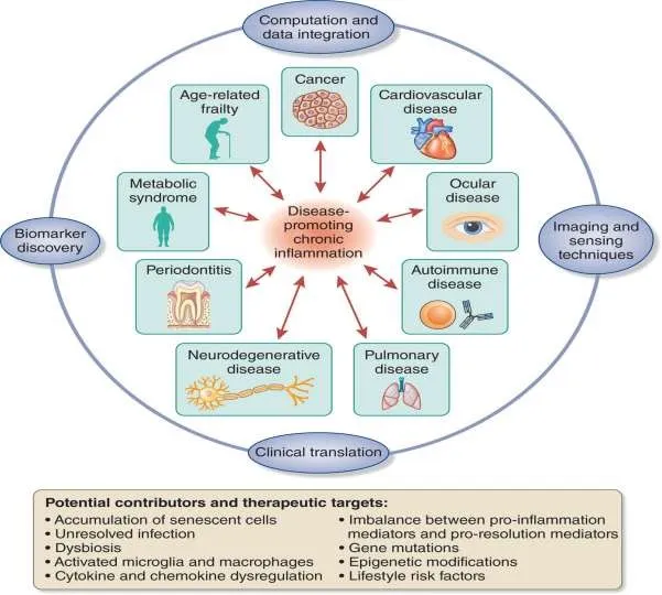
Figure 5.1: Inflammatory Markers
5.3 Protein and metabolite profiling in plasma
Over the past decade, the development of blood-based biomarkers for dementia has gained momentum, offering a minimally invasive and scalable approach to early detection, diagnosis, and monitoring of neurodegenerative diseases, especially Alzheimer’s disease (AD) [36,41]. Plasma protein and metabolite profiling—leveraging advances in mass spectrometry and immunoassays—has emerged as a promising frontier in this area.
1. Protein Profiling: Insights from Plasma Biomarkers
Plasma protein biomarkers have been of primary interest because of their ease of availability and association with neuropathological alterations [39]. Most importantly, plasma phosphorylated tau (p-tau) isoforms, especially p-tau181 and p-tau217, have proven to be highly specific and sensitive in differentiating AD from other neurodegenerative diseases and from normal aging [39,40]. The markers have been found to be concordant with PET imaging and cerebrospinal fluid (CSF) results, implying that plasma p-tau is a marker of brain tau pathology.
Another promising protein is neurofilament light chain (NfL), used as a general marker of neurodegeneration [41]. High plasma NfL levels are correlated with cognitive decline in various types of dementia, including frontotemporal dementia (FTD) and AD (Mattsson et al., 2017). Coupled with amyloid and tau markers, NFL can be used to stratify stage and track progression [41].
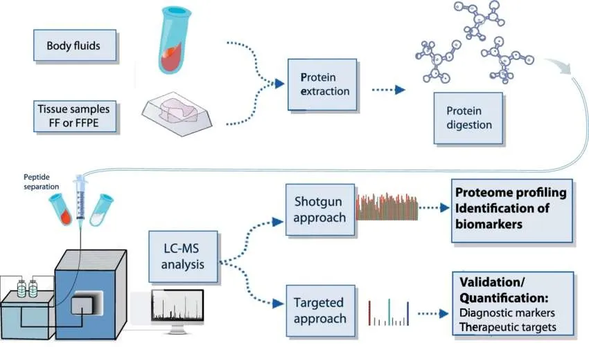
Figure 5.2: Protein Profiling in Plasma
2. Metabolite Profiling: Emerging Metabolic Signatures
Metabolomics has also yielded valuable information on dementia pathogenesis, especially via the identification of systemic metabolic alterations. Aberrant levels of lipids, amino acids, and energy metabolism-related metabolites have been documented in the plasma of individuals with AD and MCI in various studies. For instance, aberrations in sphingolipid and phospholipid metabolism, including reduced plasmalogens and elevated ceramides, have been linked with cognitive impairment and AD pathology [45].
Altered branched-chain amino acid levels, glutamate, and kynurenine pathway metabolites have also been implicated, suggesting dysregulation of neurotransmitter production and neuroinflammation. In addition, more recent metabolomics analyses by nuclear magnetic resonance (NMR) and LC-MS platforms have revealed metabolite signatures that are predictive of conversion from MCI to AD and highlight their value in early risk stratification [45].
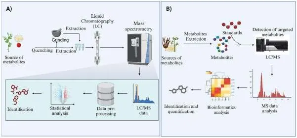
Figure 5.3: Metabolite Profiling in Plasma
5.4 Recent advances and clinical trial
Over the last few years, a significant amount of progress has been achieved in the identification and validation of molecular and blood-based biomarkers for dementia, especially Alzheimer's disease (AD) [36,41]. They propose to facilitate early diagnosis, enhance stratification of patients in clinical trials, and track disease progression or therapeutic response. Improved technologies in proteomics, metabolomics, and ultrasensitive immunoassays have significantly favored detecting pathological alterations in readily available fluids like blood [36].
Advances in Molecular Biomarkers
Molecular biomarkers in dementia studies mainly mirror pathological features like amyloid-β (Aβ) accumulation, tau pathology, and neurodegeneration [36]. Of these, plasma phosphorylated tau (p-tau), particularly p-tau181, p-tau217, and ptau231, has emerged. These isoforms are now established as good markers of tau pathology in the brain and have exhibited strong diagnostic accuracy in differentiating AD from other dementias and cognitively normal controls [39,40].
In addition, neurofilament light chain (NfL), an axonal damage marker, is also universally increased across different neurodegenerative conditions, such as AD, frontotemporal dementia (FTD), and vascular dementia [41]. Increased plasma NfL concentrations have also been correlated with cognitive impairment and brain atrophy longitudinally, implicating it’s role as a universal marker of neurodegeneration. Additionally, emerging evidence points to glial fibrillary acidic protein (GFAP) as a promising marker of astrocytic activation and neuroinflammation [41]. Recent studies have shown that plasma GFAP levels may rise even before clinical symptoms emerge, correlating with amyloid pathology and future cognitive decline.
Clinical Trials Incorporating Blood Biomarkers
Current clinical trials have increasingly incorporated blood-based biomarkers as inclusion factors, as outcome measures, or as exploratory endpoints [46]. The AHEAD 3-45 trial, for example, a preclinical AD prevention trial, employs plasma Aβ42/40 and p-tau217 to select amyloid-positive participants before being confirmed by PET. This not only lessens the dependence on risky lumbar puncture or expensive PET scans but also optimizes trial efficiency by enrolling subjects at the earliest phase of pathology [46]. Another instance is the TRAILBLAZER-ALZ 2 trial, where the anti-amyloid monoclonal antibody donanemab was tested [47]. Blood biomarkers including plasma p-tau and NfL were employed to monitor treatment effect and relate with amyloid clearance and cognitive alterations based on imaging. Outcomes showed that blood measures may be capable of acting as early markers of treatment response [47].
Concurrently, exploratory studies are examining multi-omics biomarkers using proteomic, metabolomic, and transcriptomic information from plasma to improve knowledge of disease heterogeneity. The EMIF-AD Multimodal Biomarker Discovery study is one such endeavor, with large datasets available for machine learning-based biomarker development [48].
6. Cerebral Imaging Biomarkers.
6.1 Structural imaging.
Structural neuroimaging, or magnetic resonance imaging (MRI), has become a standard tool in the evaluation of dementia [49]. It offers non-invasive information regarding anatomy of the brain and is particularly valuable when detecting patterns of regional atrophy that correspond to various forms of neurodegenerative disorders [49,50]. Progress in imaging resolution and computational methods has enriched the clinical and research use of structure imaging biomarkers for the diagnosis and prognosis of dementia [50].
Key Structural Imaging Biomarkers
The most reliably reported structural feature of Alzheimer's disease (AD) it’s medicinal temporal lobe atrophy, with specific involvement of the hippocampus, entorhinal cortex, and para-hippocampal gyrus [51]. Volumetric MRI has demonstrated hippocampal volume reduction not only to be a diagnostic indicator, but also to be highly correlated with severity and progression of disease and cognitive impairment over time [51]. Automated methods such as voxelbased morphometry and surface-based cortical thickness mapping have increased the precision of hippocampal atrophy as a biomarker [50].
On frontotemporal dementia (FTD), structural imaging shows atrophy mainly in the frontal and anterior temporal lobes, which may vary according to the clinical variant [52]. For example, behavioral variant FTD displays thinning of the frontal lobe, whereas semantic dementia demonstrates asymmetric temporal lobe involvement [52]. Vascular dementia tends to show widespread white matter hyperintensities (WMHs), lacunar infarcts, and cortical microinfarcts, which are visible on T2-weighted and FLAIR images [53]. Measurement of WMHs has become important in the differential diagnosis between vascular and neurodegenerative causes of dementia and is becoming more incorporated into multi-modal diagnostic ratings [53].
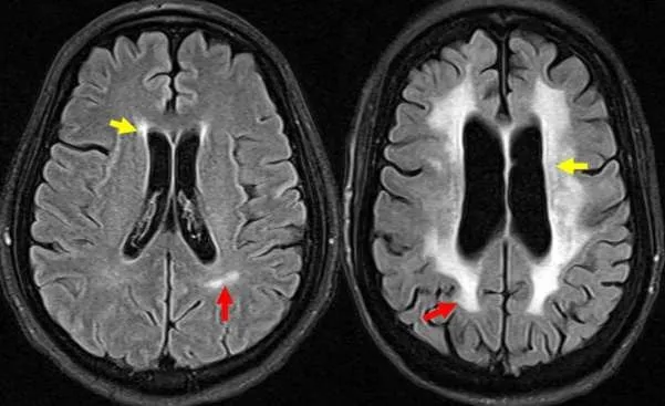
Figure 6.1: Structural Imaging
6.2 Functional imaging.
Functional imaging methods offer important information on brain function and neural network abnormalities that are antecedent or concomitant with brain structural changes in dementia [54]. Modalities identify changes in cerebral blood flow, metabolism, and connectivity—characteristics usually apparent before overt atrophy is visible on structural imaging. Thus, functional imaging biomarkers have drawn significant interest for the purposes of early diagnosis, differential diagnosis, and tracking of disease progression [54].
Key Functional Imaging Modalities in Dementia
One of the most well-established functional imaging modalities in dementia clinical practice and research is [^18F]-fluorodeoxyglucose positron emission tomography (FDG-PET). The modality quantifies regional cerebral glucose metabolism and indicates synaptic function. In Alzheimer's disease (AD), FDGPET is characteristically associated with hypometabolism in the posterior cingulate cortex, precuneus, and temporoparietal cortex, with relative preservation of the primary sensorimotor cortices [55]. These metabolic profiles are associated with cognitive impairment and may be used to differentiate AD from other forms of dementia.
In frontotemporal dementia (FTD), FDG-PET shows hypometabolism in the frontal and anterior temporal lobes, predominantly, compared to posteriorpredominant hypometabolism in AD [52]. Such regional differences in metabolic signatures enhance diagnostic specificity, especially in atypical or overlapping cases.
Resting-state functional MRI (rs-fMRI), a non-invasive measure of spontaneous brain activity through blood oxygen level–dependent (BOLD) signals, has joined the list of critical functional biomarkers [56]. Abnormal connectivity in the default mode network (DMN), specifically between the posterior cingulate and medial prefrontal cortex, is a recurring finding in early and preclinical AD [56]. Frontoparietal and salience networks are similarly disrupted in FTD and other dementia syndromes, justifying the increasing application of rs-fMRI in delineating network-level impairment.
6.3 Strength and limitation of early diagnosis.
Early diagnosis of dementia with cerebral imaging biomarkers provides several clinical and research benefits but also imposes pragmatic, ethical, and technical constraints [49,54]. With ongoing developments in neuroimaging methods, their utility in diagnosing disease processes before the development of maximal clinical symptoms is increasing, especially in Alzheimer's disease and associated disorders [54].
Strengths
1. Improved Diagnostic Accuracy
Cerebral imaging biomarkers—especially structural MRI, FDG-PET, amyloid PET, and tau PET—enable more precise discrimination between dementia subtypes [49,55]. Early recognition of certain patterns like hippocampal atrophy, temporoparietal hypometabolism, or amyloid deposition can facilitate accurate diagnosis, even at the mild cognitive impairment (MCI).
2. Detection of Preclinical and Prodromal Stages
Imaging biomarkers can identify disease-related alterations prior to the onset of overt cognitive symptoms. This is especially useful in Alzheimer's disease, where amyloid PET and structural MRI can disclose pathology years ahead of clinical diagnosis, thereby providing an opportunity for early intervention and lifestyle changes [57].
3. Monitoring Disease Progression and Treatment Response
Imaging allows longitudinal tracking of neurodegeneration and metabolic changes, enabling clinicians and researchers to monitor disease progression or responses to pharmacologic and non-pharmacologic treatments [50].
4. Patient Stratification in Clinical Trials
Cerebral imaging biomarkers are increasingly being applied to select and stratify patients in clinical trials, particularly trials that focus on specific pathological pathways like amyloid or tau [57]. This enhances trial efficiency and the enrollment of correct patient subgroups.
Limitations
1. High Cost and Limited Accessibility
Advanced neuroimaging methods like PET scans are costly and not equally accessible, especially in low-resource countries [54]. Even MRI, although more general, can sometimes not be applied quantitatively in everyday clinical practice. 2. Radiation Exposure (in PET Imaging)
Although generally safe, multiple exposure to the radioactive tracers involved in amyloid or FDG-PET imaging poses problems, especially in asymptomatic subjects or in longitudinal follow-up [55].
3. Definitive Predictive Value
While imaging biomarkers can show increased risk or early alterations, they are not necessarily predictive of an individual's likelihood of developing clinical dementia. For instance, amyloid PET can be positive for cognitively normal older people who never develop the condition [57].
4. Ethical Considerations
Early diagnosis in the absence of efficacious disease-modifying therapies raises ethical concerns, for example, psychological injury to patients or impact on employment and insurance [58]. Patients can also be given ambiguous prognostic results that are hard to interpret without further context.
5. Inter-individual and Technical Variability
Inter-individual variability arises due to differences in image acquisition protocols, scanner calibration, and analysis pipelines, which translates into variability in biomarker interpretation and requires standardization and validated normative datasets [50].
7. DNA -Based And Epigenetic Markers.
7.1 Risk Alles.
Dementia is highly dependent on genetic susceptibility, with many risk alleles having been discovered through genome-wide association studies (GWAS) and candidate gene methods [59]. Of DNA-based markers, the APOE ε4 allele is the most established genetic risk factor for Alzheimer's disease (AD), linked to accelerated amyloid-beta deposition and a younger age at onset [60]. These alleles are responsible for polygenic risk and are increasingly used in the development of predictive genetic models and personalized risk estimation [61].
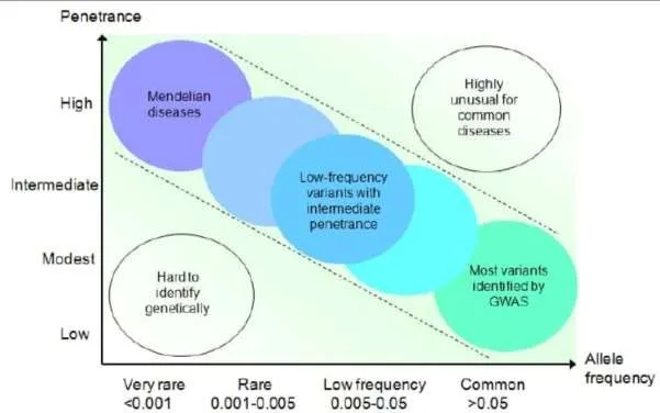
Figure 7.1: Risk Alles
In addition to static DNA variants, epigenetic alterations like DNA methylation also demonstrate gene-environment interactions impacting dementia risk [62]. Methylation patterns in genes such as ANK1, ABCA7, and BIN1 have consistently been associated with dementia, especially in cortical brain areas [63]. These epigenetic alterations can occur before overt clinical presentations and be used as early biomarkers [62,63]. In addition, risk alleles of methylation—genetic variations that affect methylation at certain CpG sites (termed as meQTLs) -are being investigated to close the gap between genetic susceptibility and epigenetic control [64]. Merging risk alleles with epigenetic information strengthens knowledge on dementia pathogenesis and could enhance early diagnosis, prognosis, and therapeutic intervention.
7.2 Polygenic Risk Score.
Polygenic risk scores (PRS) have now become useful markers for quantifying the aggregate genetic liability for complex conditions such as dementia, with a special emphasis on Alzheimer's disease (AD) [61]. PRS combines the impact of numerous single nucleotide polymorphisms (SNPs) along the entire genome, with every SNP having a very small effect size, to predict an individual's overall genetic risk [61]. Genome-wide association studies (GWAS) have found many
SNPs that have been associated with dementia risk, including APOE, CLU, PICALM, and BIN1 variants, that are incorporated into PRS models to enhance disease prediction and stratification [59]. APOE ε4 is still the strongest single genetic risk factor, but PRS augments risk estimation even in non-carriers of APOE ε4, lending credence to its applicability to early at-risk person identification [61].
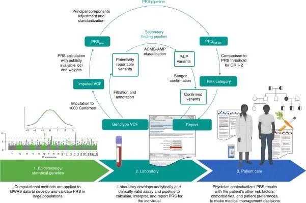
Figure 7.2: Polygenic Risk Score
Additionally, epigenetic alterations—like DNA methylation and histone acetylation—are becoming increasingly integrated into risk models to further improve prediction [65]. Methylation risk scores (MRS), based on differentially methylated regions (DMRs) of dementia, introduce a dynamic and environmentally modulated component to the highly static DNA-centric PRS [65]. Novel evidence points to incorporating epigenetic markers into PRS as a way of achieving greater predictive value for dementia onset to predict both the inherited and acquired risk factors [65]. This integrated strategy not only improves biomarker-based screening strategies but also allows for personalized interventions according to a person's genetic and epigenetic profile. Nevertheless, greater validation in various populations and longitudinal studies are needed to make PRS and MRS standard in clinical practice.
7.3 Epigenetic Modification and DNA Methylation Profiles.
Epigenetic changes, in the form of DNA methylation, are important in the regulation of gene expression without changing the fundamental DNA sequence and are becoming increasingly recognized as central to the pathogenesis of dementia [62]. DNA methylation is most commonly found at CpG dinucleotides and can be involved in silencing or activation of genes that are involved in neuronal function, synaptic plasticity, and inflammation [62]. In AD, global changes in DNA methylation have been seen in brain tissue, particularly in genes such as ANK1, ABCA7, BIN1, MEF2C, and SLC24A4, also identified genetic risk loci [63]. These methylation events tend to happen in temporal and frontal cortex—territories that have been implicated in cognitive dysfunction—and potentially before neuropathological alterations, making them promising early disease progression biomarkers [63].
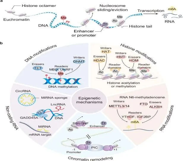
Figure 7.3: Epigenetic Modification.
Environmental influences like age, nutrition, stress, and chemical exposure can trigger epigenetic modifications that interact with genetic susceptibility to influence dementia risk [66]. Recent studies also describe the promise of such peripheral tissues as blood and saliva in recapitulating brain-specific methylation alterations, providing a non-invasive pathway for biomarker development [67]. Notably, methylation patterns are not only dynamic but also reversible, and thus therapeutic possibilities exist [66]. Integrating DNA methylation data and genetic risk variants provides a broader insight into the regulation of dementia, and aids the construction of epigenetically aware risk models and therapy targeting.
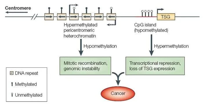
Figure 7.4: DNA Methylation Profiles.
8. Digital Health And Minimally Invasive Biomarkers.
8.1 Speech And Language Pattern Changes.
Speech and language changes are emerging as non-invasive digital biomarkers for dementia [68]. Typical changes are slower speech, increased pauses, diminished pitch variation, difficulties with word-finding, and less complex or less coherent sentences [68,69]. By applying AI-based analysis of continuous speech tasks like picture description or story retelling, researchers can separate mild cognitive impairment and Alzheimer's disease from normal aging with reasonable accuracy [69]. A subset of speech features also relates to biological markers like amyloid levels and cognitive test scores, implying true diagnostic utility [69]. Although speech biomarkers are appealing for being cost-effective, scalable, and amenable to remote monitoring, issues like cross-language variation, recording quality, and the demand for consistent methodologies persist [68].
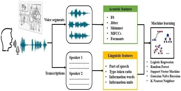
Figure 8.1: Speech and Language Pattern Changes.
8.2 Gait, Eye Movement And Pupillometry.
Motor and ocular activities are now researched more than ever as digital biomarkers of dementia since they can be recorded unobtrusively and manifest early neural alterations [70]. Gait – Slight changes in walking velocity, stride length, variability, and dual-task ability occur in prodromal Alzheimer's disease and other types of dementias [70]. Gait monitoring with wearables and smartphone accelerometers can differentiate patients from healthy elderly individuals and monitor progression [71].
Eye movements – Saccade abnormalities, smooth pursuit difficulties, and fixation instability correlate with attention and executive function deficits. Eye-tracking of reading or visual search performance shows slowed saccades and decreased accuracy in dementia [72]. Pupillometry – Alterations in pupil size dynamics, including decreased dilation response to cognitive demand, are linked to locus coeruleus and cholinergic impairment. Handheld pupillometry provides a minimally invasive assessment of cognitive effort and initial disease burden [73].
Such assessments are appealing for being low-cost, non-invasive, and remotely or clinic-friendly [71]. Their clinical translation, however, needs to wait for larger longitudinal cohorts, harmonized methods, and comparison with proven biomarkers [68].
9. Recent Clinical Trial And Approvals.
9.1 FDA Approved Blood Test.
In May 2025, the FDA approved the Lumi pulse G pTau217/β-Amyloid 1-42 plasma ratio test (Fujirebio Diagnostics), the first blood test cleared to aid Alzheimer's disease diagnosis. It is being used for adults aged over 55 with mild cognitive symptoms [74]. Validation in almost 500 patients revealed approximately 92% accuracy for amyloid plaque detection and 97% for exclusion compared with PET or CSF biomarkers [75].

Figure 9.1: FDA Approved Blood Test.
This authorization presents a less invasive and less expensive alternative to PET scans or lumbar punctures and potentially directs eligibility for anti-amyloid treatments such as lecanemab and donanemab [76]. It is advocated, though, as a diagnostic assist rather than an independent test, and more research will be required to establish its long-term value and real-world use [74].
9.2 Ongoing Studies.
1. Read-Out & Blood Biomarker Trials in the UK
The Read-Out study (Real-World Dementia Outcomes) is one of the Blood Biomarker Challenge projects, a large UK-wide study. The goal is to roll out blood-based biomarker testing for Alzheimer's and other dementias across NHS memory clinics by 2029. Almost 4,000 volunteers are participating to identify the most accurate markers. Later stages will assess the impact of blood tests on diagnosis, care provided, patient awareness, and quality of life. In a similar vein, the Blood Biomarker Challenge involves Alzheimer's Society, Alzheimer's Research UK, and NIHR in piloting blood tests for dementia diagnosis in the NHS over the next few years [77].
2. Pharma-Led Therapeutic Trials
Donanemab: An anti-amyloid monoclonal antibody that, in Phase III trial, reduced Alzheimer's progression by about 35% over 76 weeks in early cognitive symptom patients with biomarker-evident amyloid pathology [78]. Emerging Therapeutics: Some new investigational drugs are in active development [79].
3. Emerging Biomarker-Based Diagnostic and Monitoring
Technologies
AD Marker: A federated, multi-modal learning platform using sensors in everyday life to detect passively early Alzheimer's digital biomarkers. In a first four week trial (n = 91), it reached ~94% accuracy in detecting biomarkers and 89% for AD classification early [80].
9.3 Applications.
Lecanemab (Leqembi): In January 2023, FDA approved it through their accelerated process for early-stage Alzheimer's. This monoclonal antibody markedly decreases amyloid plaques and curbs cognitive decline in combination with baseline assessment in diagnosis and treatment tracking [81].
Donanemab: Phase III trials (e.g., TRAILBLAZER-ALZ 2) currently in progress aim to target amyloid plaques in early Alzheimer's, with a focus on future approval and inclusion in treatment algorithms [78].
Varoglutamstat (PQ912): As a new small-molecule drug capable of preventing toxic amyloid aggregation, it is being tested in the VIVA-MIND and VIVIAD Phase 2 trials among patients with early Alzheimer's and mild cognitive impairment (MCI)—showing promise for potential early intervention strategies [82].
Remternetug: There is currently an ongoing Phase 3 anti-amyloid trial (TRAILRUNNER-ALZ1) assessing its capacity to clear plaques and define its place in therapeutic pipelines [83].
Trontinemab: An intriguing new medication with outstanding amyloid reduction (91% of the participants amyloid-negative at 28 weeks) in Phase 2 trials, greatly surpassing current treatments [84]. Phase 3 trials are being organized, indicating the possibility of rapid clinical rollout.
10. Future Direction And Research.
10.1 Multi Biomarkers Panels And Machine Learning.
Recent studies underscore the utility of integrating multiple biomarkers—such as plasma p-tau isoforms, amyloid ratio, neurofilament light chain (NfL), and measures of neuroinflammation—into combined panels [85]. These panels give a more stable impression of disease state than single assessments can and perhaps more adequately capture the heterogeneity of dementia syndromes [85,86].
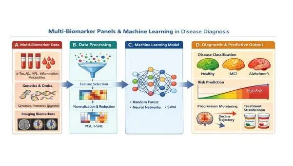
Figure 10.1: Multi Biomarker Panels and Machine Learning.
Concurrently, machine learning (ML) and artificial intelligence (AI) are increasingly used to biomarker data, neuroimaging, voice patterns, and digital health measurements. These models have the capability to identify faint, nonlinear patterns and produce predictive scores for premature diagnosis, progression tracking, and treatment response [87]. Notably, ML-based methods can process multimodal datasets and provide greater accuracy than conventional statistics [87].
Future studies will increasingly be concerned with the creation of standardized, scalable panels of biomarkers and AI-based diagnostic devices that can be translated to clinical practice and telemonitoring [86,88]. Challenging yet promising issues remain in terms of reproducibility, external validation, and ethical use of patients' data [88].
10.2 Multi Based Screening.
Screening in community settings is emerging as a viable method of early detection of dementia outside of specialist clinics. Strategies involve quick cognitive assessments, digital devices (e.g., drivers or gait analysis apps), and blood-based biomarker tests that can be used in primary care, pharmacies, or senior centers [89]. These methods seek to detect at-risk individuals earlier, minimize health disparities, and refer patients to specialist assessment or clinical trials more effectively.
Next-generation research is headed towards low-cost, scalable models that merge digital health information with minimally invasive biomarkers [85,87]. The convergence of community screening with telemedicine and artificial intelligence-based triage systems may improve dementia diagnosis to be more accessible and equitable, particularly in low-resource environments [89]. But issues like standardization, privacy, and false positives must be addressed with care before vast implementation [88].
REFERENCES

Siddhi Shah, Dhairyashri Kher, Nishit Patel, Siddhi Rathod, Parth Patel, Early Detection of Dementia: Identifying Biomarkers for Early Intervention., Int. J. of Pharm. Sci., 2026, Vol 4, Issue 11, 411-433. https://doi.org/10.5281/zenodo.23923626
 10.5281/zenodo.23923626