

We use cookies to ensure our website works properly and to personalise your experience. Cookies policy
Shree Santkrupa College of Pharmacy, Ghogaon, Tal-Karad, Dist-Satara, 415111.
Exosomes, nano-sized extracellular vesicles ranging from 30 to 150 nm, have emerged as extremely promising targeted drug delivery systems (DDSs) due to their poor immunogenicity, organic biocompatibility, and unique ability to cross rigid Barriers include the barrier between the blood and the brain.This review presents a complete analysis of exosomes as movable nanocarriers, defining their structural components (proteins, lipids, and nucleic acids) and categorizing them as natural, modified, or manufactured. We evaluate current isolation methods , such as size-exclusion chromatography, ultracentrifugation, and microfluidics, as well as endogenous and exogenous drug-loading methods including sonication and electroporation. Additionally, exosome-based DDSs' medicinal properties are being examined in a range of disease settings, including oncotherapy, neurological disorders, cardiovascular diseases, and bacterial infections, along with the clinical administration routes. Eventually, we examine crucial translational issues such as low purification yields, structural heterogeneity, and scale-up standardization in accordance using GMP (Good Manufacturing Practice) regulations. Finally, this study examines the evolution of modified exosomes from laboratory inventions to next-generation clinical therapies.
Exosomes are microscopic small sized extracellular vesicles generated by cells that transport RNA, lipids, proteins, and various other bioactive substances to aid in the body's pathological and physiological functions [1].Exosomes are 30–150 nm in diameter [2].They affect both physiological and pathological processes [3].The exosome are remarkable and superior drug delivery vehicles for application in a wide range of disorders and cancer therapy because of their characteristics, which include biocompatibility, targeted tumor tracking, variable targeting efficiency, and reliability [4].Similarly, all eukaryotic cells and bacteria can secrete exosomes, which are the primary extracellular communication mediators and comprise fats, proteins, nucleic acids ,other active ingredients [5].They are mostly present in a variety of bodily fluids, including serum, saliva, urination, and cerebrospinal fluid (CSF). Similarly, they are present in various types of cells, such as platelets, dendritic cells, T cells and B cells These EVs might offer a novel approach for diagnosing and treating gut-related illnesses [6].Exosomes have been isolated using a variety of standard techniques. These include immunoaffinity separation, size exclusion, precipitation, ultrafiltration, and differential and unstable gradient centrifugation. The most popular technique for removing exosomes from bodily fluids and media for cell culture is differential and floating gradient centrifugation [7].One of the most desirable aspects of exosomes as carriers for medication delivery is their natural origin and instinctive biocompatibility. Exosomes, which are generated by the body's natural cells, are often well tolerated through immunity, decreasing the risk of immunogenicity that is often associated with synthetic delivery methods [8].One of the primary benefits of EVs is their ability to function as targeted delivery vehicles, allowing for the selective transfer of therapeutic material to certain tissues while limiting off-target effects and lowering toxicity for health organs.However,EVs may avoid immune surveillance, thereby shielding their cargo from premature degradation, and they can also be programmed to release their contents in response to certain physiological or pathological stimuli [9].The three main categories of EVs are:(i) Apoptotic Bodies (Abs)(ii) Shedding Micro Vesicles (MVs) and (iii) Exosomes (EXs)) [10].
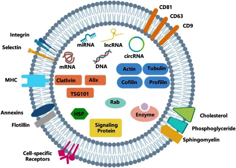
Figure 1.1: Structure of Exosome [11]
1. Apoptotic Bodies (Abs):
Apoptotic bodies, which are between 50 and 5000 nm in size and have different kinds of shapes, are released after cell death (apoptosis). They are made of the plasma membrane and include messenger molecules, DNA, RNA, and histones [12].
2. Shedding Micro-Vesicles (MVs):
Micro-vesicles are generated by breaking down the cell membrane while simultaneously absorbing cytosolic proteins, and their Diameters between 20 to 1000 nm, based on the origin cells and the method used to isolate them from cell medium [13].Ca2+ influx, phosphate esters, ATP, and Other elements may all be involved in their creation [14].
3. Exosomes (EXs):
Exosomes are more similar (in shape and size) than microvesicles, with diameters between 50 and 120 nm.During their synthesis, the plasma membrane buds inward, resulting in the formation of intermediate endosome-vesicles called multicellular bodies [15].
Types of Extracellular Vesicle :
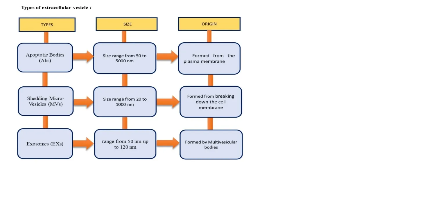
Figure 1.2: Types ,Size and Origin of Extracellular Vesicle [14]
ADVANTAGES
1. Good Biocompatibility, Low Immunogenicity
Because they are natural, cell derived structures, they have excellent biocompability, low immunogenicity, and low inherent toxicity compared to synthetic carriers like liposomes [15].
2. Versatile Cargo Capability:
They can naturally carry or be exogenously engineered to load a wide variety of therapeutics ,including RNA, proteins, and small molecule medications [16].
3. Intrinsic Targeting:
The intrinsic targeting refers to the natural, inherent ability of unmodified exosome to navigate through the body and bind to specific recipient cells or tissues [15].
4. Barrier Penetration:
The barrier penetration refers to the unique biological ability of exosome to cross tightly sealed protective boundaries in the body that normally block most drugs [17].
5. Cargo Protection and Stability :
It refers to the exosomes ability to act as a highly secure, natural shield for the therapeutic molecules (like RNA, DNA, proteins, or drugs) it carries inside [16,17].
DISADVANTAGES
1. Low and variable production yield :
Natural exosome production is often relatively low, making it difficult to sufficient quantities for therapeutic application [17].
2. Difficult and time-consuming isolation and purification
Exosome isolation is technically challenging because exosomes coexist with proteins, lipoproteins, protein aggregates, and other extracellular vesicles [17,18].
3. Limited targeting specificityAlthough exosomes possess natural cell-targeting properties, their biodistribution is not always sufficiently specific [18].
4. Storage and stability problems
Exosome characteristics and biological activity can be affected by storage conditions, temperature, freeze–thaw cycles, and formulation [18,19].
COMPOSITIONS
Exosome formation and production changes depending upon the parent cells, physiological conditions, and external triggers. Biochemical and molecular studies of exosomes revealed the presence of lipids, protein molecules, RNA, and various components [20]. Exosomes of various animals and cell types contain 4563 proteins, 194 lipids, 1639 mRNAs, and 764 miRNAs [21].
Lipid :
Lipids are essential components of all kinds of organism and is broadly distributed in EVs. Several research have comprehensively reported on the lipidic profile of exosomes produced from different origins,as well as the enrichment of specific lipid categories linked with producer cells [20].In this condition, B lymphocyte-derived exosomes contain up to three times as much cholesterol (CHOL) as the cell membrane [21].Lipids are deemed to be the major constituent. Exosomes have higher concentrations of cholesterol, diglycerides, phospholipids, sphingolipids, glycerophospholipids, and ceramides than parent cells [20].Exosomes include a variety of bioactive lipids like as prostaglandins and leukotrienes, and also enzymes such as phospholipase C [22].
Proteins :
Exosomes contain significant numbers of proteins involved in vesicle production and cell signalling. Exosomes contain more protein and are more representative than mRNA [24].Tetraspanins, including CD9, CD63, and CD81, help to synthesize exosomes and other vesicles. Exosome proteins perform a distinct role in biogenesis and transport pathways [25].
Nucleic acid:
Nucleic acids found in exosomes contain mRNA as well as non-coding RNAs, and miRNA [24]. High concentrations of [miRNAs] and ncRNA [non-coding RNA] are seen in exosomes, along with double-stranded DNA sometimes [25].Transferring these RNAs to target cells allows for their translation or use in controlling gene activity [26].miRNA packaging may follow sequence-specific patterns, indicating active organizing rather than passive loading [27].
CLASSIFICATION OF EXOSOME
Exosomes are categorized into three categories: natural exosomes, modified exosomes, and synthetic exosomes [28].
1) Natural exosomes
Natural exosomes are classified into animal-derived and plant-derived exosomes [28].Plant-based exosome-like nanoparticles (ELN) present in food products such as a ginger, grapes, carrots, and grapefruit mimic mammalian exosomes and have the ability to prevent liver disease and preserve intestinal health through anti-inflammatory activities [27,28].Exosomes are formed under normal and malignant settings; animal-derived exosomes are further classified as Malignant and regular exosomes [27].Exosomes are produced spontaneously by many cells, such as epithelial, endothelial, mesenchymal stem, macrophage, dendritic, and immunological cells [28,29].
2) Modified exosomes
Modified exosomes are naturally produced exosomes that have been modified for more effective and enhanced therapeutic applications such as drug loading, targeted delivery, biodistributions, and others [29].They can be modified in two ways: first, by changing the exosome's internal structure, and secondly, by changing the exosome's outer surface [30].
3) Synthetic exosomes
Synthetic exosomes are generated using two fundamental methods: cell-based approach and lipid membrane biomembrane production technique [30,31]. Exosomes are generated in nearly every normal cell, including humans gastrointestinal veins endothelial cell types, the mesenchymal stem cells (MSC), B cells, macrophages, T cells, dendritic cell lines (DC), and natural killer, or NK, cells [32].
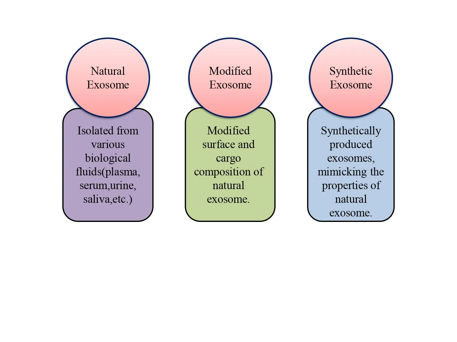
Figure 1.3: Classification of Exosome [32]
ISOLATION TECHNIQUES FOR EXOSOME
To produce ultrapure exosomes, they must be separated from cell fragments and other interfering particles [33].Exosomes can be isolated from cell culture or cellular fluids using various approaches dependent on the dimension and origin.Exosomes can be isolated using many procedures, including differential centrifugation, filtration, SEC, or size exclusion chromatography,and polymer precipitation [34].
1. Ultracentrifugation:
Ultracentrifugation is the traditional method for separating high-dose sample components according to density and size class, as well as extracting exosomes [34].In general, ultracentrifugation is used with a sucrose density gradient to separate different densities of particles and also protein-protein clumps [33,34].
2. Ultrafiltration:
Ultrafiltration is typically performed during ultracentrifugation or the final step of chromatography using ordinary conventional membranes with defined size exclusion limits [34,35].The other two types of ultrafiltration technologies are direct flow filtration and tangential or crossflow filtration [35].Ultrafiltration is often employed as a step after
ultracentrifugation or as the final step in chromatography. The standard ultrafiltration process is as follows: deadened filtration to remove floating cells and cell detritus using the residue of a typical cellular culture [36].
3. Size Exclusion Chromatography (SEC):
In SEC,Large molecules and other particulate debris are separated in SEC using a porous stationary phase that varies in size according to the exosome [36,37].Exosomes isolated from the SEC have been shown to retain their structure, as shown by transmission electron microscopy. Furthermore, unlike the centrifugal approach, shear force will not impact the shape or integrity of the exosomes during separating from the SEC [37].The SEC are used to segregate exosomes according on hydrodynamic radii. It maintains biological activity and integrity without requiring any additional processing [38]
4. Microfluidic Technique :
In recent years, microfluidics-based isolation technologies have become a viable approach.to exosome separation [39].These approaches rely on the difference in exosomes' physical and biochemical features, such as antibody immunoaffinity, thickness, and size [40].Purification and separation methods using microfluidics-based isolation techniques can be divided into three categories: ways incorporating immunoaffinity for exosome trapping, sieving approaches, and procedures involving exosomes adsorbed into the porous structure [39].These technologies can collect and analyses exosomes from small sample volumes, often resulting in high purity and fast processing times [41].
5. Immunoaffinity- chromatographic (IAC):
Immunoaffinity chromatographic procedure is a purification or separation process that uses the particular binding of antigens and agonists to separate desired molecules from heterogeneous mixtures [39,41].The binding efficiency is intrinsically linked to biological affinity pairs, extraction conditions, and Polymer carriers [41].When compared to ultracentrifugation, IAC produces comparable findings with a smaller sample volume [41,42].It can also be used to analyses exosomes qualitatively and quantitatively. When using magnetic beads, it has excellent specificity,high sensitivity, purity, and yield and does not limit the initial sample volume [42,43].
6. Polymer Precipitation:
Polymer precipitation is typically performed using polyethylene materials as a solvent, and exosomes are recovered using centrifugation to reduce their solubility [44].This approach was initially used to isolate viruses [45].The polymer precipitating technique is generally straightforward to apply, needs minimal analytical period, and can handle large amounts of
Material [44, 45].However, both purity and rate of recovery are rather poor, and inaccurate results may occur, and the polymer formed is difficult to remove, making it unsuitable for further functional experimental examination [46].
DRUG LOADING METHODS
• Endogenous / Active Loading Method:
The process of endogenous or active loading is an experimental technique that depends on primary cells. This approach uses direct transfection and co-incubating donor cells to introduce target molecules [47].The endogenous loading method may load a wide variety of cargoes, the most common of which being RNAs, proteins, Nucleic acid or microscopic molecules. Transfection and cell co-incubation are two extensively utilized endogenous drug-loading methods for delivering monomeric chemicals or nucleic acids into donor cells [48].
1. Cell co-incubation :
Cell co-incubation, also known as pre-secretion loading, is an endogenous drug loading method in which therapeutic drugs are introduced directly to the culture medium of live donor cells [48,49].Parental cells are cultivated with a higher concentration of the medication or tiny compounds.The medication is absorbed by the cells via endocytosis and then secreted within the outgoing exosome [49].
2. Transfection :
Transfection is a method of inserting pharmaceuticals into exosomes using transfection agents [48].This method is more efficient and has higher molecular stability than other methods, however the efficiency is not constant [49].Transfection agents may influence gene expression in exosomes derived from donor cells, influencing the nucleic acid medicines and biological activity carried by exosomes [50].
• Exogenous / Passive Loading Methods:
Exogenous loading is the using barrier penetration methods to directly load medications into pre-separated exosomes [48,51]. It can be utilized to carry tiny molecule medicines, proteins, and even nanomaterials[52].Exogenous loading has less technical requirements and greater possibilities than endogenous loading methods, so it is now the most frequent method for creating exosomes to carry pharmaceuticals [51,52].Exogenous loading methods typically used include drug incubation, sonication, electroporation, saponin, freeze-thaw processes, and extrusion [50,52].
1. Incubation:
Two different incubation procedures have been designed [52,53]. The first involves direct incubation of medicines and exosomes. The efficacy of this approach is dependent on the drug's hydrophobicity, which allows it to bind with the lipid layer of the exosome membrane [53]. The second kind involves incubating medications with exosome donor cells and then isolating drug-carrying exosomes, which is mostly employed for small-molecule chemical therapies with low cytotoxicity [53,54].This procedure is a simple, and non-invasive, and non-invasive, and non-invasive, and non-invasive [52]. It keeps exosomes intact, which is essential for preserving their original structure and functionality [54].
2. Sonication:
Once the exosomes were combined with medicines, ultrasonic treatment was performed using a homogenizer probe [55].During the initial step of ultrasound treatment, the mechanical shear stress disrupted The stability of the exosomes membrane, allowing the medication to enter the exosome during membrane deformation [56].After ultrasound, exosomes were incubated to restore membrane integrity [55,56].Ultrasound had the greatest efficiency, and exosomes were efficiently taken by target cells, however their sizes and shapes unavoidably changed[56].This approach has a high loading efficiency and is adaptable, accommodating both hydrophilic and hydrophobic medicines, which increases its therapeutic potential Furthermore, the approach may modify surface indicators or the biochemical action of exosomes, affecting targeting ability and safety profiles [57].
3. Electroporation:
Electroporation is a regularly used method for loading siRNA into exosomes [58].The approach employs an electric field to create microscopic pores in the phospholipid bilayer, allowing the payload to enter the vesicle [57,58].Electroporation is simple to produce and It has been widely used to encapsulate siRNA, microRNA, and miRNA; nevertheless, it may result in RNA precipitation or exosome aggregation, lowering drug loading efficiency [59,60].
4. Saponin:
Saponins are effective membrane-penetrating compounds that form combinations with membrane cholesterol, creating holes/pores that improve cell membrane permeability [62]. Saponins have little effect on exosome morphology and, in general, increase encapsulation rate. However, because of the haemolysis action, saponins have restricted in vivo use. Thus, exosomes should be isolated after being incubated with saponin [62,63].
5. Freeze-thaw cycles :
The freeze-thaw strategy includes incubating exosomes with a targeted medication at ambient temperature for a set amount of time before rapidly freezing or freezing in liquid nitrogen [64].The mixture is then warmed to room temperature [65].To improve drug encapsulation, freeze-thaw cycles are conducted at least three times. This process has a lesser drug loading capacity than sonication or extrusion [61,65].
6. Extrusion :
The extrusion approach involves combining exosomes are loaded with molecules of drugs and passed through a nanofiltration-based membrane-like filter, resulting in cracks in the exosome forming membranes for drug delivery [66].The technique involves dissolved drugs in a suitable solvent and gradually adding them into exosomes.To encapsulate pharmaceuticals in exosomes, the mixture is extruded using a manual extrusion or microgrinder [66,67].Finally, surplus solvent and unencapsulated medicines are removed via centrifugation or filtration, yielding pure drug-loaded exosomes [67].
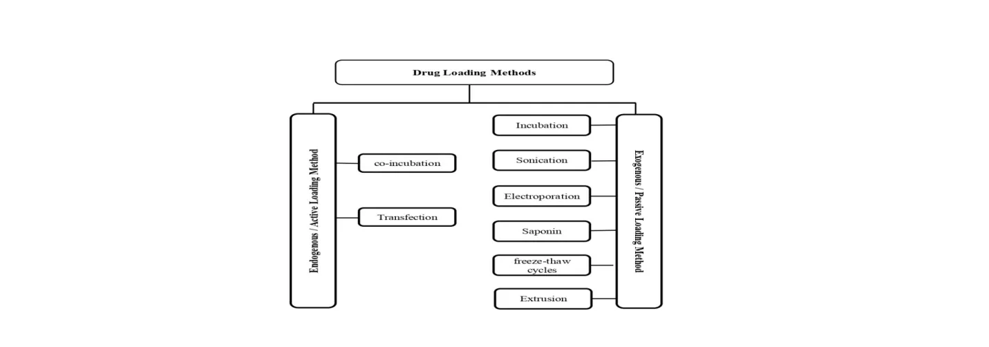
Figure 1.4: Drug Loading Methods [47,67]
EXOSOME BASED DDSs IN TREATMENT OF VARIOUS DISEASES:
1. Exosome in Asthma :
Asthma is a non-infectious condition that can arise from allergies.It is distinguished by bronchial hyperreactivity, mucosal edema,and airflow obstruction [68].Exosomes produced by eosinophils and innate immune cells play a vital role in controlling and promoting asthma pathogenesis [69].Asthma pathology is caused by the stimulation of structural lung cells and airway remodelling, both of which include enzymes such as eosinophil cationic protein, eosinophil peroxidase, and major basic proteins that cause epithelial damage.Aside from that, the exosomes contain chemicals nitric oxide (NO), lipid mediators, and radicals are the triggers for inflammation [68,69].Previous research has confirmed that patients with asthma have a higher concentration of exosomes containing those enzymes than healthy persons [69].T cells, which have crucial roles in the inflammatory response in asthma, have also been shown to produce exosomes [69,70].
2. Exosomes in Bacterial Infections :
Exosomes may transmit infection by transporting bacterial compounds involved in pathogenesis [68].The exosomes serve an important, bidirectional function in bacterial infection by acting as nanoscale communication bundles between host cells and pathogens [72].During an infection, the term exosome refers to both the vesicles released by human host immune cells and those released by bacteria themselves [73].In Mycobacterium TB, exosomes from infected macrophages carry mycobacterial proteins and lipids that trigger TNF-α and T cell responses. They also aid in recruitment of CD4+ and CD8+ T lymphocytes are antigen-specific, improving bacterial clearance [72,73].
3. Exosome in Neurological Disorder :
Exosomes mediate their activity in the CNS by passing through the BBB and carrying the therapeutic substance to the desired location [70].When exosomes enter the central nervous system,they can interact with neurons, astrocytes, microglia, and oligodendrocytes via receptor-ligand interactions, fusion of the membranes, or endocytosis [70,71].Their cargo, which includes proteins, lipids, mRNA, microRNA, and a variety of regulatory substances, can alter gene expression, influence synaptic plasticity, regulate inflammatory responses, and aid in neuroprotection or regeneration [71].Exosomes are important drivers of neurological illnesses because they operate as vehicles for spreading harmful proteins, promoting neuroinflammation, and degrading the blood-brain barrier [72].
4. Exosome in Cancer Treatment :
Exosome-based cellular carriers are attractive therapeutic vehicles in oncology, owing to recent advancements and their inherent ability to transport drugs to cancerous cells [74]. Exosome-based vehicles have intrinsic homing ability and are clinically more stable than a number of other cutting-edge drug delivery methods often employed in cancer treatment [73,74].Exosomal-based systems are derived from donor cells, have a larger circulation capacity, and are non-immunogenic and biocompatible [75].Compared to recombinant viral vectors, exosomes are safer, less dangerous, and do not carry the risk of insertion causing tumor formation [73,75]. Additionally, the surface of exosomes may be altered to improve patient and cell-specific delivery [75,76].In a recent experiment, it was examined whether exosome-based drug carriers were capable of transporting the chemotherapeutic medicament doxorubicin to the target tumor cells [76].
5. Exosome in Rheumatoid Arthritis :
The rheumatoid arthritis is a frequent autoimmune disorder for which effective therapies are currently lacking [75,77]. Exosomes, on the other hand, offer a viable approach for the targeted delivery and treatment of various illnesses because they can carry anti-inflammatory chemicals [77,78].According to research, exosomes generated from mesenchymal stem cells can spread bioactive substances and are essential for immunomodulation and regeneration [77]. Exosomes' potential as a cutting-edge treatment approach for autoimmune disorders [78].
6. Exosome in Cardiovascular Diseases :
In relation to cardiovascular diseases, growing research suggests that these nanovesicles play a role in cardioprotection, which could lead to their future use as a therapeutic tool for a variety of cardiovascular disorders such as cardiac artery disease, coronary heart failure, cerebrovascular disease, and others [79,80].Exosomes have been proven to promote communication among several cell types in cardiac tissue cells [79]. Exosomes produced from stem cells have been considered to be essential for heart healing [80].
7. Exosome in Epilepsy :
Exosomes are extracellular mediators that carry a molecular cargo capable of altering the receiving cellular gene expression and phenotype [80,81].A brief RNA sequence was utilized to identify variations in microRNA cargo after exosomes from epileptogenic and non-epileptogenic tuberous complex (TSC) tubers were separated [81].They found a variety of microRNAs that are significantly more prevalent in epileptogenic tubers and include genetic patterns that activate Toll-like signals and initiate an inflammatory neural pathway activated genetic tissue [80].These discoveries add to our knowledge of how exosomes and non-coding RNA cargo function in the epileptic neuroinflammatory cascade, which may help develop novel biomarkers and therapies for drug-resistant epilepsy [81,82].
8. Exosome in Renal Diseases :
The kidney is one of the most important organs in the human body because it regulates and maintains homeostasis [82,83]. Exosomes' significance in renal function has been explored both in vivo and in vitro. Several studies have shown exosomes originating from diverse regions of the neuron [83].Along from their role in renal physiology, urinary exosomes act as immune effectors, shielding the urine tract from bacterial infection [83,84].
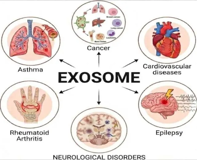
Figure 1.5: Exosome in Various Diseases [11,84]
ROUTES OF ADMINISTRATION
The administration of exosomes method can be administered via a variety of routes, including intravenous, intraperitoneal, intratumoral, nasal, and oral. The method of medicine delivery is intrinsically linked to treatment.efficacy of many disorders [85].
1. Intravenous Route :
The most common method of administering exosomes to the body is through an IV. Some disorders are targeted by the IV method [84,85].
Targeted Diseases : include stroke, Parkinson's disease, acute kidney injury, and anticancer therapy [84,85].
2. Intraperitoneal Route :
It gives permission for storing of higher EV dosages [85].
Targeted Diseases : include dysplasia and chronic type 1 diabetes [85,86].
3. Oral Route :
It is a simple and convenient route of administration for patients [86].
Targeted Diseases: Facilitates the resolution of colitis and arthritis [85,86].
4. Intranasal Route :
Appropriate for EV delivery into the brain, by crossing the blood-brain barrier [87]. Targeted Diseases: Treatment for Parkinson's and brain cancer [87,88].
5. Intratumoral Route :
Due to the increased EV retention in tumors, it is a more successful technique for anticancer therapy [87,88].
Targeted Diseases : Antitumor therapies [89].
CHARACTERIZATION OF EXOSOME
Exosome characterization is specialized and identical, thus various characterization indexes are required to establish whether the isolated components are exosomes [90]. In general, we consider the identification of isolated exosomes at three levels, including TEM Exosome shape detection, NTA size identification, and Western Blot detection of exosomes ,surface protein markers, ELISA [90,91].
1. Transmission Electron Microscopy (TEM):
The electron microscopy examines particle form and structure using an accelerated photon beam [91].The TEM can study the interior structure and provide data on particle size distribution. The scientists devised the "Cryo-TEM" technique to overcome the limits of previous methods [90,91].Cryo-TEM is a complex technology for removing sample preparation effects. In rare situations, an electron beam can harm biological material [90].When examined with TEM, isolated exosomes exhibit a cup-shaped structure, whereas freezing exosomes have circular forms [91,92].
2. Scanning Electron Microscopy (SEM):
SEM detects very low-energy electrons released from the sample surface [93].It can provide useful sample information such morphology, composition, texture of the surface, and roughness. SEM can reveal exosome surface properties like as size, shape, and morphology [90,93].It collects electrons from deeper below the sample's surface than dispersed electrons, providing information on the composition and structure [94].
3. Enzyme-Linked Immunosorbent Assay (ELISA):
Plate-based enzyme-linked immunosorbent assays can identify and measure proteins, peptides, hormones, and antibodies [94,95].This assay, which uses ELISA, is intended to yield accurate cancer exosome counts. It offers great specificity and quick detection [95].It can assess marker proteins qualitatively and quantitatively, making it suited for high-throughput study. It also detects exosomes in plasma, serum, and urine using various antibodies [95,96].
4. Fluorescence Correlation Microscopy (FCM):
Microfluidic-Independent Fluorescence Correlation Microscopy (FCM) involves collecting exosomes with a specific antibody, labeling them with a fluorescent dye, and analyzing them using a plate reader [96].FCM can help create a microfluidic chip for immunocapture and quantitative exosome analysis [97].
5. Colorimetric Detection:
Calorimetry detects particles based on the color intensity of a chromogenic substance [98]. Microfluidic technology was used to capture exosomes, which were then detected and quantified using ELISA. Researchers have successfully used this approach to detect exosomes in cancer cells [97,98].
6. Dynamic Light Scattering (DLS) :
The DLS determines the size of exosomes [98].The lower limit of measuring is 10 nm. It is not suited for evaluating complicated exosome samples with a exosome concentration cannot be measured within this size range and it is difficult to separate contaminated proteins from exosomes [99].
APPLICATION OF EXOSOME
In contrast to previous reviews of transformed exosomes, which largely focus on exosome synthesis, isolation, purification, and disease therapeutic applications.Exosomes contain several biomarkers for illness diagnosis and prediction [100].They are mostly used in cancer treatment, but they have also made success in cardiovascular, TB, and diseases of the central nervous system [100, 101].Regarding the potential benefits of synthesized exosomes as entirely novel drug delivery systems, we outline the essential prospects for studies, which include improving exosome purification and isolation methods, standardizing exosome characterization and identification methods, clarifying exosome absorption and drug transport processes, creating an efficient exosome loading approaches, integrating a functional exosome surface with targeting ligands, enhancing exosome stability, and testing exosome toxicity [102].
CHALLENGES WITH EXOSOME-BASED DRUG DELIVERY
A exosomes provide numerous benefits for medication delivery, but there are many challenges to overcome[103].The fundamental limitation of employing exosomes as a medicine is the difficulties in extraction and separation, resulting in low yield [100,103].To use exosomes for medication administration on a commercial scale, it's important to isolate and separate enough high-quality exosomes [103].Exosomal formulations must follow GMP rules, which cover quality control protocols, conventional cell culture techniques, downstream exosome isolation, and exosome source selection. A further challenge emerges from exosome heterogeneity, which is caused by differences in size, composition, function, and cellular origin, complicating their classification [103].
CONCLUSION
In conclusion, exosomes constitute an evolution in the field of targeted medication administration, filling the gap between synthetic nanoparticles and natural cellular communication vehicles. As discussed throughout this review, their inherent biocompatibility, stability in circulation, and exceptional ability to cross tightly sealed protective boundaries this makes them suitable applicants for treating complex pathologies ranging from to drugs cancers to central nervous system disorders. Meanwhile, before based on exosome drug delivery systems can make the successful transformation from laboratory platforms to commercial, bedside clinical applications, significant technological and regulatory challenges must be overcome. The principal obstacles are poor and variable manufacturing yields, time-consuming separation techniques, and inherent structural variability caused by various cell sources. Future study should focus on the optimization and standardization of high-purity isolation techniques, exact quantitative characterization methods, and scalable formulation pipelines that closely follow to Good Manufacturing Practice (GMP) requirements. Advancing surface engineering methodologies and resolving mass-production restrictions will be critical in unlocking the full therapeutic and diagnostic capacity of these natural nanovesicles, paving the door for highly effective, patient-specific tailored therapies.
REFERENCES



Sanika Patil, Vidya Kapurkar, Mayuri Patil, Samruddhi Patil, Harshad Bhoi, Shantanu Khedakar, Exosome-Mediated Targeted Drug Delivery: Engineering Approaches, Therapeutic Potential and Pharmaceutical Challenges, Int. J. of Pharm. Sci., 2026, Vol 4, Issue 11, 434-450. https://doi.org/10.5281/zenodo.23923750
 10.5281/zenodo.23923750