
We use cookies to ensure our website works properly and to personalise your experience. Cookies policy
Dr. Rajendra Gode College of Pharmacy, Amravati, Maharashtra, India.
Cancer remains a major global health burden, with conventional treatment modalities such as chemotherapy, radiotherapy, and surgery often limited by inadequate targeting, systemic toxicity, and unfavorable pharmacokinetic profiles. To overcome these drawbacks, nanotechnology-based drug delivery systems have been widely explored due to their ability to enhance drug solubility, enable controlled and sustained release, and improve tumour localization via passive and active targeting mechanisms. Biomimetic nanocarriers have recently emerged as an advanced strategy to address these limitations by integrating biological components with engineered nanomaterials. By replicating natural biological architectures such as cell membranes, extracellular vesicles, and virus-like structures, these systems demonstrate superior biocompatibility, prolonged circulation, reduced immune clearance, and enhanced targeting capability. This review summarizes the fundamental principles underlying biomimetic nanotechnology and describes various platforms, including cell membrane-coated nanoparticles, exosome-based systems, protein- and peptide-functionalized carriers, hybrid nanostructures, and virus-inspired nanoparticles. It further outlines key targeting approaches such as the enhanced permeability and retention effect, ligand–receptor interactions, and stimuli-responsive drug release.Recent progress in fabrication techniques, characterization methods, and preclinical investigations has shown improved therapeutic efficacy in both in vitro and in vivo models. Additionally, diverse applications in cancer treatment—including chemotherapy, immunotherapy, gene therapy, and photothermal and photodynamic approaches—are discussed, with emphasis on combination therapies. Overall, biomimetic nanocarriers represent a highly promising platform for precision cancer therapy, supporting the advancement of personalized medicine, although further research is necessary to facilitate their successful clinical adoption
Cancer is still one of the top causes of sickness and death worldwide because it is very diverse, spreads easily, and traditional treatments like surgery, radiation, and chemotherapy can cause serious side effects. Standard chemotherapy often struggles with problems like not targeting the cancer specifically, leaving the body too quickly, spreading to healthy tissues, and harming normal cells. Because of these challenges, researchers are working on new drug delivery methods that can target tumours more accurately, reduce side effects, and make treatments safer and more effective.
Since it has made it possible to create nanoscale carriers (such as liposomes, polymeric nanoparticles, dendrimers, and mesoporous silica) that can encapsulate medications and enhance their solubility, stability, and pharmacokinetic profile, nanotechnology has become a game-changing tool in oncology. These nanosystems increase accumulation in tumour tissue and decrease systemic toxicity by taking advantage of surface functionalization techniques for active targeting and the enhanced-permeation-and-retention (EPR) effect in tumours. Nevertheless, biological obstacles like poor tumour penetration, limited long-term circulation, and quick clearance by the mononuclear phagocyte system still affect synthetic nanocarriers despite tremendous advancements. The idea of biomimetic drug delivery systems has drawn more attention in an attempt to address these issues. The structure, makeup, or functionality of biological components—such as cell membranes, extracellular vesicles, or particles derived from pathogens—are mimicked by biomimetic nanocarriers. Enhancing circulatory half-life, active tumour targeting, immunological evasion, and biocompatibility.1
The methods used to produce BMNPs result in a more uniform and refined end product. Furthermore, when given at equivalent doses, these nanoparticles exhibit better biological activity and internalization efficiency than EVs. Although BMNPs have a lot of potential, their full clinical applications won't be possible until a few technological obstacles are resolved. In order to maximize cargo loading efficiency and standardize production procedures in the development of BMNPs, it is first critical to ensure the integrity and functionality of the biomimetic coatings. To enable their widespread use, BMNP production's scalability and cost-effectiveness must also be improved.2
In this review, we thoroughly investigate the state of biomimetic drug delivery systems based on nanotechnology for targeted cancer treatment. We go over the fundamentals of biomimetic carrier design, manufacturing techniques, functionalization methods for tumour targeting, and the ways in which they improve drug delivery to tumours. We also examine these systems' shortcomings, translational difficulties, and prospects for the future, paying particular attention to immune interactions, scalable manufacturing, and microenvironmental cues unique to cancer. In presenting this overview, we hope to draw attention to the potential and significant challenges of biomimetic nano-drug delivery in targeted cancer therapy, as well as to pinpoint important avenues for further study and clinical application.
2. Fundamentals of Drug Delivery Using Nanotechnology
Targeted drug delivery has undergone a revolution thanks to the application of nanotechnology in medicine, which has created new chances to increase therapeutic efficacy while reducing systemic side effects. In order to take advantage of special physicochemical characteristics like large surface area, adjustable size, and surface functionalization potential, nanotechnology manipulates materials at the nanometer scale (1–100 nm).3 Compared to traditional formulations, these properties allow nanoparticles (NPs) to more precisely and controllably encapsulate, protect, and deliver therapeutic agents, such as small molecules, peptides, and nucleic acids.4
2.1 Properties of Nanocarriers
Materials with a diameter of 1–100 nm are known as nanocarriers, and they have the ability to transport several medications and/or imaging agents. For targeting purposes, a high ligand density on the surface can be achieved because of their high surface-area-to-volume ratio. By carrying the drug inside and releasing it under control when attached to the targets, nanocarriers can also be used to raise local drug concentrations. Nowadays, clinically approved formulations of lipids and natural and synthetic polymers are commonly employed as drug delivery vehicles. Polymer conjugates, polymeric nanoparticles, dendrimers, carbon nanotubes, lipid-based carriers like liposomes and micelles, and gold nanoparticles, including nanoshells and nanocages, are all members of the nanocarrier family.5
2.2 Types of Nanocarriers in Dug Delivery
For cancer treatment, several classes of nanocarriers have been created, each with unique structural and functional properties:
• Liposomes: These are spherical vesicles made of phospholipid bilayers that contain hydrophobic medications in the lipid membrane and hydrophilic medications in the aqueous core. PEGylated liposomes, like Doxil® (liposomal doxorubicin), are revolutionary in nanomedicine because they provide longer circulation and less cardiotoxicity. 6
• Polymeric nanoparticles: Made from biodegradable polymers (like chitosan, PEG, and PLGA), these carriers give exact control over the rate of degradation and release kinetics. Amphiphilic block copolymers self-assemble to form polymeric micelles, which are especially useful for administering hydrophobic medications.
• Dendrimers: Precise drug conjugation and multivalent interactions with target cells are made possible by highly branched polymeric structures with distinct architecture and multiple surface functionalities.
• Inorganic nanoparticles: Due to their distinct optical or magnetic characteristics, gold, silica, iron oxide, and quantum dots are all appropriate for theranostic applications that combine imaging and therapy.
• Lipid–polymer hybrid nanoparticles (LPHNs): These offer enhanced stability and sustained release profiles by fusing the structural integrity of polymeric nanoparticles with the biocompatibility of liposomes.7
2.3 Drug Loading and Release Mechanisms
Covalent attachment, adsorption, or physical entrapment are the three ways that drugs can be encapsulated into nanoparticles.8 Nanocarrier systems are characterized by controlled and stimuli-responsive release, which allows for site-specific delivery. There are two types of stimuli: external (like temperature, magnetic field, light, or ultrasound) and internal (like pH, redox potential, or enzymatic activity). For instance, enzyme-sensitive polymers selectively break down in cancerous tissues, whereas acid-labile linkers can facilitate drug release in the acidic tumour microenvironment.9
2.4 Considerations for Pharmacokinetic and Biodistribution
Drugs' pharmacokinetic profiles can be dramatically changed by nanocarriers. Circulation time, biodistribution, and cellular uptake are determined by variables like size, hydrophobicity, surface charge, and shape. Renal filtration quickly removes particles smaller than 10 nm, while the reticuloendothelial system frequently sequesters particles larger than 200 nm. Surface modification using polyethylene glycol (PEG) or natural biomolecules (albumin, polysaccharides) is used to avoid immune recognition, resulting in "stealth" nanoparticles with an extended half-life. However, new data suggests that because of high interstitial pressure and variable vascularization, the EPR effect by itself is not enough for efficient tumour targeting in human cancers. Therefore, to increase tumour specificity and therapeutic index, active targeting techniques like conjugating ligands (antibodies, peptides, aptamers) are frequently required. 10
2.5 Limitations of Conventional Nanocarriers
Despite their promise, conventional nanocarriers still face challenges that limit clinical translation. Major obstacles include,11,12
These challenges highlight the need for biomimetic approaches that integrate natural biological interfaces and mechanisms into nanocarrier design to improve compatibility and targeting efficiency. This concept forms the foundation for the next generation of biomimetic nanomedicine, discussed in the following sections.16
3. Overview of Biomimetic Nanotechnology
The design of next-generation nanomedicines has been significantly impacted by the idea of biomimicry, which is the imitation of biological structures, functions, and processes. The goal of biomimetic nanotechnology in cancer treatment is to overcome the drawbacks of traditional synthetic systems by fusing the adaptability of engineered nanocarriers with the complexity of natural biological systems. These systems preserve the physicochemical tunability of synthetic nanoparticles while improving biocompatibility, immune evasion, and tumour targeting by imitating cellular components like membranes, exosomes, or particular biomolecules. 17
By using a biomimetic technique, nanoparticles can act as "disguised" biological entities that can move covertly, engage with tumour cells only, and effectively deliver therapeutic payloads. 18 Common strategies include coating nanoparticles with cell membranes, integrating natural vesicles (like exosomes), or functionalizing them with proteins and peptides that replicate cellular recognition mechanisms.These approaches have shown remarkable promise in improving pharmacokinetics, biodistribution, and tumour accumulation.19
4. Types of Biomimetic nanocarriers for targeted cancer treatment
In order to improve therapeutic specificity and reduce immune recognition, biomimetic nanocarriers for cancer therapy are designed to mimic natural biological entities, including blood cells, immune cells, exosomes, and viruses. Depending on its biological origin, each type of biomimetic nanocarrier offers distinct functional advantages. The main categories, production methods, workings, and therapeutic uses of biomimetic nanocarriers utilized in targeted cancer treatment are covered in detail in this section.20
4.1 Cell Membrane-Coated Nanoparticles
Cell- and cell membrane-based NPs possess multifunctional abilities, which make them ideal in NP-based cancer therapies. Cell membrane-coated NP (CMCNPs) have been increasingly studied for their mimicry of cell surface functionality, which can aid in reducing the immune responses of synthetic NPs in vivo and introduce the ability to combine both natural and synthetic materials. Cell- and cell membrane-based drug carriers exhibiting intrinsic properties of in vivo biology have been shown to overcome the challenges faced by synthetic NP-based drug carriers and achieve acceptable toxicity and better biocompatibility than their synthetic counterparts. Major advantages of using cell- and cell membrane-based drug carriers include provision of immune escape and specific tumour targeting imparted by the cell membrane proteins leading to improved EPR in cancer therapies, and an ability to generate desired cytotoxic immunomodulatory effects via cell surface engineering, leading to tumour regression. 21

Fig 1. The cancer cell membrane-coated nanoparticle (CCNP). Schematic representation of CCNP fabrication and two potential applications. Cancer cell membrane along with its associated antigens is collected from source cancer cells and coated onto polymeric nanoparticle cores made of poly (lactic-co-glycolic acid) (PLGA) polymer. The resulting CCNPs can then be used to deliver tumour-associated antigens to antigen presenting cells or to homotypically target the source cancer cells.21
• Nanoparticles coated with the membrane of red blood cells (RBCs)
With a cycle life of up to 120 days, red blood cells are the most prevalent cellular component in the human body. Since red blood cells are the most basic cells and have virtually no organelles, it is simple to rupture and purify them in order to gather cell membranes. Self-identification markers, such as complement regulatory molecules and CD47, are highly expressed on the membrane surface of red blood cells.22
• Nanoparticles coated with platelet membranes
PLTs are a class of acaryotic cells that are widely distributed in the bloodstream and are crucial for controlling coagulation. PLTs are frequently utilized as membrane sources in the production of MCNPs, giving them the capacity to evade the immune system and retain information for longer.
• Nanoparticles coated with immune cell membranes
Dendritic cells (DCs), natural killer (NK) cells, neutrophils, T cells, and macrophages are examples of immune cells. One of the traits of tumours is the presence of persistent inflammation. Numerous immune cells, such as NK cells, macrophages, granulocytes, and lymphocytes, frequently infiltrate the tumour tissue and control the tumour microenvironment.23
• Nanoparticles coated with the membranes of stem cells and tumour cells
The homing effect is a unique phenomenon that occurs between tumour cells and stem cells. Consider cancer cells as an example. Over time, these cells developed the ability to adhere to one another homologously and to evade the immune system, which allowed them to form stable tumour foci within the body. Numerous adhesion glycoproteins, including integrin protein, N-cadherin, and epithelial cell adhesion molecule, are expressed on the surface of tumour cell membranes and contribute to the homologous adhesion ability of tumours. It is possible to give MCNPs coated with membranes derived from stem cells or cancer cells the ability to mimic tumour cells, including the ability to target tumours. Furthermore, the coated tumour cell membrane expressed a number of tumour-associated antigens that can be used as vaccines to restore the depleted tumour-associated microenvironments and activate the cell-mediated immune effect. These days, autologous cancer cell membranes can be extracted by dispersing human-sourced tumour tissues that have been surgically removed into cells. These membranes can greatly increase the immune response against tumours when paired with an immunologic adjuvant.24
4.2 Exosome-Mimetic and Exosome-Based Delivery Systems
Different cell types secrete extracellular vesicles, or exosomes. Proteins, lipids, deoxyribonucleic acid (DNA), small interfering ribonucleic acid (siRNA), messenger RNA (mRNA), microRNA (miRNA), and other signaling molecules are among the diverse bioactive cargoes found in these vesicles, which normally have a diameter of 30 to 150 nm. These lipid bilayer-packed cargoes have the ability to enter recipient cells, changing their behavior and being essential for cell-to-cell communication. Over the past ten years, exosomes have drawn more attention as promising drug delivery vehicles on the basis of these theoretical underpinnings. Compared to traditional drug delivery methods like liposomes, dendrimers, micelles, and polymeric nanoparticles, exosomes offer a number of benefits.25
Numerous cell types can produce exosomes, and the composition, bioactivity, and translational potential of these exosomes are significantly impacted by the donor source selection. Mesenchymal stem cell (MSC)-derived exosomes are the most extensively studied among mammalian sources because of their scalable culture systems, low immunogenicity, and regenerative and anti-inflammatory qualities.26 Numerous cancer vaccine platforms are based on immune cell-derived exosomes, especially those from dendritic cells and T cells, which are prized for their capacity to present antigens and to stimulate or modulate immune responses. Tumour-derived exosomes, on the other hand, have a high intrinsic loading of oncogenic and signaling molecules and a natural affinity for tumour tissues. Although they can be used as biomarkers or delivery models, prospective immunosuppressive and pro-tumourigenic effects limit their clinical applicability. Exosomes derived from epithelial and endothelial cells offer high yields and repeatable secretion, making them useful for vascular and barrier-related delivery.27
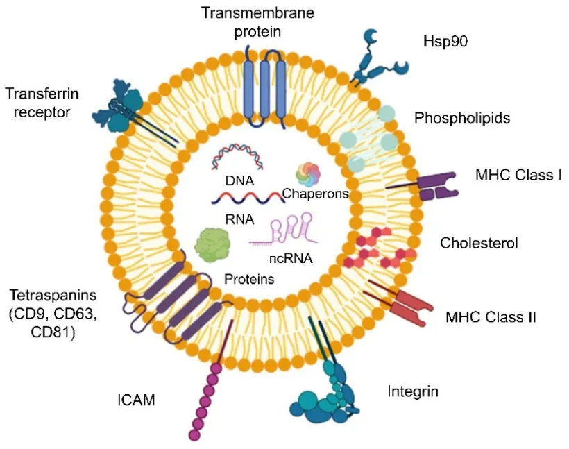
Fig 2. Shows the composition of exosomes schematically. The lipid bilayer structure, which is enriched with cholesterol, sphingomyelin, and ceramide, the surface proteins, such as integrins, adhesion molecules, and tetraspanins (CD9, CD63, and CD81), as well as the enclosed cargos, which include nucleic acids, proteins, and metabolites.28
4.3 Functionalized Nanoparticles of Proteins and Peptides
Another approach to biomimetic design is the functionalization of nanocarriers with proteins or peptides that replicate biological interactions. Because of their receptor-mediated uptake pathways and tumour tropism, proteins like albumin, lactoferrin, and transferrin are frequently used.For example, paclitaxel can be delivered clinically using albumin-bound nanoparticles (like Abraxane®), which take advantage of albumin's inherent transport capabilities.29
Compared to whole proteins, peptide-based biomimetic modifications have greater specificity and easier synthesis. Cell-penetrating peptides (CPPs) like TAT improve intracellular delivery, while peptides like RGD (arginine–glycine–aspartic acid) can target integrins that are overexpressed on the tumour vasculature. To accomplish multi-level targeting and controlled release, hybrid biomimetic designs have been developed recently. These designs combine synthetic nanocores with multiple biomimetic coatings, such as cell membranes functionalized with targeting peptides. These hybrid systems combine the advantages of engineering adaptability and biological selectivity, marking a significant advancement in precision nanomedicine.30
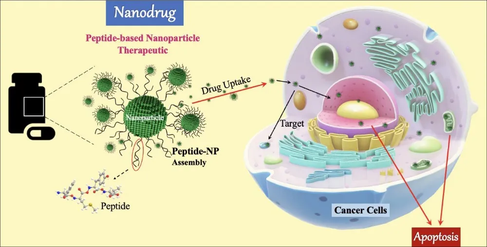
Fig 3. Schematic representation of the peptide-functionalized nanodrug mediating targeted anticancer activity.31
4.4. Hybrid Biomimetic Nanocarriers
Many biomimetic hybrid membrane-based nanoplatforms (BHMNs) are currently being developed for drug delivery, detoxification, cancer detection, and cancer vaccines. Red blood cells (RBCs), platelets, tumour cells, immune cells (like macrophages), and bacteria are the primary sources of membranes used in BHMNs. A hybrid membrane could combine the advantages of the original membranes, including active tumour targeting, immune escape, half-life extension, and adherence to tumour cells, in contrast to a monotypic cell membrane. Furthermore, through lymph node homing, the hybrid membrane presents tumour antigens to antigen-presenting cells (APCs), among other innovative functions not possible with the original membranes. Additionally, by decreasing the adhesion of unrelated natural cells (like white blood cells), hybrid membranes can enhance the tissue distribution of nanoparticles or specifically enhance the accumulation of nanoparticles in organs like the liver and spleen. Because of these clear benefits, combining several cell membranes to create hybrid membranes has demonstrated significant promise for use in bacterial detoxification and cancer treatment. Either the two cells can be fused before membrane extraction, or the membranes of each cell can be extracted before membrane fusion to create the hybrid membranes.32

Fig. 4. The synthesis of RBC-platelet hybrid membrane-coated nanoparticles ([RBC-P]NPs) is shown schematically . RBC and platelet membranes fuse to form membrane materials. [RBC-P]NPs are then created by coating poly(lactic-co-glycolic acid) (PLGA) polymeric cores with the resultant hybrid membrane.33
5. Targeting Mechanisms in Biomimetic Drug Delivery Systems
One important aspect of biomimetic nanocarriers' efficacy in treating cancer is their ability to selectively accumulate at tumour sites while lowering systemic exposure. Passive, active, and stimuli-responsive mechanisms are typically combined in the targeting process to increase therapeutic precision and efficacy.
5.1 Enhanced Permeability and Retention (EPR) Effect-Based Passive Targeting
The pathophysiology, anatomy, and theory underlying increased permeability and retention of tumour vasculature Angiogenesis is triggered when tumour cells proliferate, group together, and enlarge to a size of 2-3 mm in order to meet the tumour's constantly rising nutritional and oxygen needs. In terms of microscopic anatomical architecture, this neovasculature is very different from normal tissues. For example, the tumour's endothelial cells are ill-aligned or disorganized with large fenestrations, and the blood vessels are dilated, irregularly shaped, leaky, or defective. Additionally, the vascular wall usually lacks or has abnormalities in the smooth-muscle layer, basement membrane, and perivascular cells. While tumour tissues have inadequate lymphatic drainage, tumour vessels have a large lumen. Blood plasma components, including macromolecules, nanoparticles, and lipidic particles, extensively leak into the tumour tissue as a result of this anatomical defect and functional abnormalities.34
Furthermore, macromolecules are kept in the tumour while extravasation into the tumour interstitium continues due to the slow venous return in tumour tissue and the inadequate lymphatic clearance. We first described this phenomenon, which we call the EPR effect, nearly 20 years ago. It serves as the foundation for the selective delivery of macromolecular medications to the location of solid tumours. Within 1-2 days, very high local concentrations of polymeric drugs can be achieved at the tumour site, for example, 10–50 times higher than in normal tissue. This mechanism has been the basis for polymer conjugates, micellar or liposomal anticancer drugs, and antibody conjugates in more recent times. The EPR effect is quickly emerging as the gold standard for these drug designs. It's interesting to note that low-molecular-weight medications are exempt from the EPR effect due to their quick diffusion into the bloodstream and subsequent renal clearance.35
5.2 Active Targeting via Ligand–Receptor Interactions
Active targeting enhances tumour selectivity by decorating nanocarriers with ligands that bind specifically to overexpressed receptors on cancer cells or the tumour microenvironment. Examples include folate, transferrin, RGD peptides, and antibodies against HER2 or EGFR. In biomimetic systems, the biological membrane coating itself often provides natural targeting capabilities. For instance, cancer cell membrane-coated nanoparticles demonstrate homotypic binding, where surface adhesion molecules such as E-cadherin, N-cadherin, and galectin mediate recognition of homologous cancer cells. Similarly, platelet membrane-coated nanoparticles leverage P-selectin to bind CD44 receptors on tumour cells. These inherent recognition mechanisms reduce the need for artificial ligand conjugation while maintaining biocompatibility.36
5.3 Targeting Based on Stimuli
The fate of nanocarriers inside biological systems may be impacted by external stimuli, primarily thermal, magnetic, electronic, ultrasound, and light fields. It makes it easier to enhance the accumulation of nanocarriers in specific areas using external forces (like a magnetic field), controlled release, intracellular drug delivery, activated imaging, and therapy. Using external stimuli to deliver drugs to tumours has the following benefits: It could precisely regulate the location and strength of specific external stimuli (such as laser irradiation or magnetic fields); the external stimuli could be added or removed based on the needs of the treatment; multiple external stimuli could be superimposed to achieve multifunction in cancer theranostics; and it could offer multiple times or continuous (for instance, a few hours or days) stimuli for medication administration and treatment. However, when the location of the metastatic lesions is unknown, externally directed triggers would be impractical for accessing and treating them.37
5.4 Targeting Techniques in Combination
Multiple targeting mechanisms are increasingly incorporated into modern designs. At the same time, biomimetic nanocarriers can display stimuli-triggered release (from responsive polymers), active homotypic recognition (from tumour cell membranes), and prolonged circulation (from RBC or platelet membranes). By maximizing drug accumulation at tumour sites and minimizing off-target toxicity, these hybrid targeting strategies establish biomimetic nanotechnology as a significant breakthrough in customized cancer treatment.
6. Preclinical data and in vitro/in vivo evaluation
Numerous in vitro and in vivo studies have confirmed the therapeutic efficacy of biomimetic drug delivery systems. In vitro tests frequently use cancer cell lines that are identical to the coating membrane's source to assess characteristics like cellular uptake, cytotoxicity, and targeting specificity. When compared to uncoated particles, cancer-cell membrane-coated nanoparticles, for example, show improved homotypic targeting, allowing preferential internalization into the parent cancer cells and increased drug potency.38 Similarly, nanocarriers coated with erythrocyte and platelet membranes exhibit better immune evasion and longer stability in serum, as demonstrated by higher cell viability retention and decreased macrophage uptake. 39
These findings support the notion that cellular recognition and the effectiveness of selective drug delivery are directly correlated with surface protein retention.The translational potential of biomimetic systems is further demonstrated by in vivo evaluation using orthotopic tumour models or murine xenografts. When compared to traditional formulations, preclinical reports show a significantly improved antitumour efficacy, a longer circulation half-life, and increased tumour accumulation through homologous adhesion . Li et al., for instance, found that doxorubicin-loaded cancer-cell membrane-coated nanoparticles inhibited 4T1 tumour growth more efficiently than PEGylated nanoparticles, with negligible organ toxicity off-target . The need for design optimization and scale-up reproducibility for clinical translation is highlighted by systematic reviews that confirm that tumour delivery efficiency is still limited (less than 5% of injected dose) despite superior targeting and safety.40
Table 1. Representative in vitro and in vivo evaluations of biomimetic drug delivery systems for cancer therapy.38-40
|
Study / System |
Model Type |
Assay / Endpoint |
Outcome |
|
Cancer-cell-membrane-coated PLGA nanoparticles (DOX-loaded) |
In vitro (4T1 cells) |
Cellular uptake, cytotoxicity |
Enhanced homotypic uptake, increased apoptosis vs. control |
|
RBC-membrane-coated nanoparticles |
Invitro (RAW264.7 macrophages) |
Phagocytosis and serum stability |
Decreased macrophage uptake, prolonged stability |
|
Cancer-cell-membrane-coated ZnO@siSurvivin |
In vivo (4T1 xenograft mouse model) |
Tumour inhibition, survival rate |
~80 % tumour volume reduction, prolonged survival |
|
Platelet-membrane-coated nanocarriers |
In vivo (CT26 tumour-bearing mice) |
Biodistribution, pharmacokinetics |
Increased tumour accumulation, longer circulation |
7. ‘Biomimetic Nanocarriers' Therapeutic Uses in Cancer Treatment
New therapeutic paradigms in oncology have been made possible by the incorporation of biomimetic design into nanocarriers, which offer improved precision, decreased toxicity, and the capacity to overcome drug resistance. Numerous therapeutic modalities, such as immunotherapy, gene therapy, chemotherapy, and photothermal/photodynamic therapy (PTT/PDT), have investigated these systems.
7.1 Chemotherapy
Systemic toxicity, rapid clearance, and multidrug resistance (MDR) continue to be the limitations of traditional chemotherapy. These issues are resolved by biomimetic nanocarriers, which offer targeted, controlled delivery. In breast cancer models, for instance, doxorubicin-encapsulated RBC membrane-coated nanoparticles showed extended circulation and decreased off-target effects.Similarly, compared to non-coated systems, cancer cell membrane-coated nanoparticles (CCMNPs) show homotypic targeting, which increases the accumulation of chemotherapeutic drugs in tumour tissue.41
7.2 Immunotherapy
By improving antigen presentation and adjusting immune responses, biomimetic nanocarriers have also improved cancer immunotherapy. As a type of nanovaccine, for instance, cancer-cell membrane-coated nanoparticles exhibit tumour-associated antigens that can activate both innate and adaptive immunity. These nanoparticles produce strong cytotoxic T-cell responses and long-term immunological memory against recurrent tumours when co-loaded with immune adjuvants.
By targeting inflammatory tumour sites and interacting with immune cells in the tumour microenvironment to increase immune activation, platelet- and leukocyte-mimicking nanoparticles further aid immunotherapy.42
7.3 Gene Therapy
Because of their instability and low cellular uptake, genetic materials like siRNA, mRNA, or CRISPR-Cas9 components are still difficult to deliver. Biomimetic nanocarriers, especially exosome-based systems and virus-mimetic nanoparticles (VMNs), provide effective nucleic acid delivery with low immunogenicity. In hepatocellular carcinoma models, for instance, virus-like nanoparticles modified with tumour-targeting peptides have demonstrated a high gene transfection efficiency with negligible off-target effects.
Similarly, siRNA can be delivered by exosome-mimetic nanovesicles made from tumour cells to inhibit oncogenic pathways like KRAS or MYC, thereby effectively suppressing tumour growth in vivo.43
7.4 Photodynamic and Photothermal Therapy (PTT/PDT)
Biomimetic nanocarriers have been used in light-triggered cancer treatments, which use photodynamic or photothermal processes to kill tumour cells. When exposed to near-infrared radiation, RBC or tumour-membrane-coated nanoparticles containing photothermal agents such as indocyanine green (ICG) show significant tumour accumulation and effective photothermal conversion.
These systems ensure safety and effectiveness by reducing overheating in healthy tissues in addition to improving tumour targeting. In preclinical studies, PTT/PDT approaches in combination with immunotherapy or chemotherapy have shown notable synergistic effects. 44
7.5 Combination Treatments and Future Clinical Opportunities
Because of their adaptability, biomimetic nanocarriers can be used in combination therapies, combining gene therapy, immunotherapy, and chemotherapy onto a single platform. For better therapeutic results, for example, hybrid RBC–platelet membrane-coated nanoparticles can deliver medications and immune modulators at the same time.
A number of biomimetic systems have advanced into early-phase trials because of their scalable production and biocompatibility, despite the fact that clinical translation is still in its infancy. Clinical deployment will require ongoing improvements in membrane isolation, sterilization, and large-scale synthesis.45
8. Difficulties and Prospects for the Future
Although biomimetic nanocarriers have made remarkable strides in preclinical cancer treatment, a number of obstacles stand in the way of their clinical application. These include long-term stability, regulatory complexity, biosafety and immunological compatibility, and production scalability. To fully realize the therapeutic potential of biomimetic systems driven by nanotechnology, these limitations must be addressed.
8.1 Manufacturing and Scalability The ability to reproduce
The scalable and repeatable production of biomimetic nanocarriers is a significant clinical development bottleneck. Complex and time-consuming procedures like electroporation, extrusion, and ultracentrifugation are needed to extract biological membranes or exosomes. [69] It is still difficult to maintain membrane functionality and integrity on a large scale while maintaining sterility and uniformity. Potential solutions for large-scale production include bioreactor-based membrane generation and automated microfluidic systems.
To guarantee consistent performance across manufacturing batches, it is also essential to standardize quality control parameters such as size distribution, zeta potential, protein composition, and surface antigen integrity.46
8.2 Issues with Immunogenicity and Biosafety
The use of biological materials (such as tumour cell membranes or exosomes) raises biosafety concerns like pathogen transmission, immune activation, or unintentional delivery of carcinogenic components, even though biomimetic coatings increase biocompatibility. Clinical safety depends on making sure that any remaining cellular DNA, nucleic acids, and endotoxins are eliminated.
Furthermore, to evaluate the possible accumulation or delayed toxicity of biomimetic nanoparticles, long-term in vivo investigations are required. These risks could be reduced by employing techniques like the use of synthetic or engineered membranes that replicate natural components without the use of live cells.47
8.3 Regulatory and Ethical Challenges
For biomimetic nanomedicines, the regulatory pathway is still unclear and intricate. Existing frameworks for assessing nanodrugs do not completely account for hybrid systems that contain genetic materials or biological membranes. The FDA and EMA, among other regulatory agencies, will have to create new guidelines to address the immunogenicity profiles, long-term impacts, and environmental safety of these hybrid bio-synthetic platforms.
Transparent donor consent, traceability, and adherence to biobanking ethics are also necessary to address ethical concerns surrounding the sourcing of biological materials, especially from human or tumour-derived tissues.48
8.4 FUTURE PROSPECTIVES
In order to create intelligent, programmable nanocarriers, next-generation biomimetic systems are anticipated to combine synthetic biology, AI-driven design, and microfluidic engineering. These will have enhanced targeting specificity, tailored payload delivery, and adaptive responses to the tumour microenvironment. Future cancer nanotherapy platforms will probably be dominated by hybrid systems that combine stimuli-responsive polymers with cell-mimetic membranes.49
The establishment of scalable manufacturing, regulatory clarity, and long-term safety validation through multicenter trials will ultimately be necessary for successful clinical translation. With these advancements, biomimetic nanocarriers have the potential to revolutionize cancer treatment by moving from a broad cytotoxic approach to targeted, patient-specific medication.50
9. SUMMARY & CONCLUSION
By fusing the functional complexity of biological systems with the benefits of nanotechnology, biomimetic nanocarriers offer a revolutionary approach to targeted cancer therapy. In comparison to traditional nanomedicines, these systems have shown superior tumour accumulation, increased therapeutic efficacy, and decreased systemic toxicity through passive and active targeting, stimuli-responsive release, and homotypic recognition.
Large-scale production, biosafety, immunogenicity, and regulatory compliance are still major obstacles to clinical translation, even in the face of encouraging preclinical results. These constraints should be addressed by developments in synthetic biology, microfluidics, and hybrid biomimetic design, opening the door to highly accurate and individualized cancer treatments.
In summery, biomimetic drug delivery systems provide a cutting-edge oncology platform that connects precision medicine and traditional therapies. To reach their full potential in enhancing patient outcomes, more interdisciplinary research and thorough clinical validation are required.
REFERENCES


Prachi Tale, Nikita Bochare, N. Wakchaware, Dr. H. Sawarkar, Biomimetic Drug Delivery System for Targeted Cancer Therapy: A Comprehensive Review, Int. J. of Pharm. Sci., 2026, Vol 4, Issue 6, 2243-2258, https://doi.org/10.5281/zenodo.20609142
 10.5281/zenodo.20609142