

We use cookies to ensure our website works properly and to personalise your experience. Cookies policy
Nalanda Institute of Pharmaceutical Sciences, Siddharth Nagar, Kantepudi, Sattenapalli, Guntur, Andhra Pradesh, India 522438
Improved understanding of the immune system and its role in cancer development and progression has led to impressive advances in the field of cancer immunotherapy over the last decade. Whilst the field is rapidly evolving and the list of drugs receiving regulatory approval for the treatment of various cancers is fast growing, the group of PD1- PDL-1 inhibitors is establishing a leading role amongst immunomodulatory agents. PD1- PDL-1 inhibitors act against pathways involved in adaptive immune suppression resulting in immune checkpoint blockade. Within the last four years two PD-1 and three PD-L1 inhibitors have been utilized in clinical practice against a variety of malignancies. Focus was initially placed on targeting cancers considered immunogenic such as melanoma, renal and lung cancers but subsequently the application expanded to include amongst others Hodgkin Lymphoma, urothelial as well as head and neck cancer. This article provides a comprehensive review of the early and late phase trials that led to the regulatory approval of all five PD1- PDL-1 inhibitors in the corresponding cancer types. It presents available data on the combinations of PD1- PDL-1 inhibitors with other therapies (immunotherapy, targeted therapy and chemotherapy), the toxicity profile of the PD1- PDL-1 inhibitors and ongoing trials testing the efficacy of these agents in cancer types beyond those that have been addressed already. Finally, current and future challenges in the application of PD-1 and PD-L1 inhibitors are discussed with emphasis on the role of predictive biomarkers
Cancer immunotherapy has revolutionized oncology by leveraging the immune system’s ability to recognize and eliminate tumor cells. Among the most impactfuadvances are immune checkpoint inhibitors that target the programmed death-1 (PD-1) receptor and its ligand PD-L1, which can restore T cell activity and generate durable responses across several cancer types. However, their effectiveness is limited by both primary and acquired resistance, restricting long-term benefit to a subset of patients. This review highlights current progress in cancer immunotherapy and explores emerging strategies beyond PD-1/PD-L1 blockade. Novel inhibitory and co-stimulatory immune checkpoints such as lymphocyte activation gene-3 (LAG-3), T cell immunoglobulin mucin-3 (TIM-3), T cell immunoreceptor with Ig and ITIM domains (TIGIT), OX40, CD137 (4-1BB), and CD40 are examined for their potential to enhance anti-tumor immunity. The rationale for combining immunotherapy with chemotherapy, radiotherapy, targeted therapies, oncolytic viruses, and neoantigen vaccines is discussed in the context of overcoming resistance. Additional emphasis is placed on modifying the tumor microenvironment, targeting tumor metabolism, and overcoming stromal barriers. Advanced delivery systems such as chimeric antigen receptor (CAR) T cells, bispecific antibodies, and nanoparticle-based platforms offer improved specificity and reduced toxicity. Biomarker-guided precision immuno-oncology using tumor mutational burden, microsatellite instability, and immune gene signatures is advancing clinical decision-making. Gut microbiota modulation and artificial intelligence-based tools are also emerging as critical components in optimizing therapeutic outcomes. Collectively, this review proposes a multidimensional and personalized immunotherapy paradigm aimed at broadening clinical efficacy and overcoming resistance beyond conventional PD-1/PD-L1 inhibition.
1. Structure of PD-1 and PD-1 ligands :
Human PD-1 (CD279), encoded by the PDCD1 gene, is a member of the immunoglobulin gene superfamily. This factor was named programmed cell death protein 1, because its expression was shown to be enhanced by apoptotic stimuli in two different cell lines (2B4.11 and LyD9t), and it participates in apoptosis
PD-1 is a type I transmembrane glycoprotein of 50–55 kD that contains a single extracellular IgV domain, a hydrophobic transmembrane domain and a cytoplasmic tail structure domain. The IgV domain consists of 20 amino acids separated from the plasma membrane and exhibits 23% homology with CTLA-4. The cytoplasmic tail contains two tyrosine motifs, an immune receptor tyrosine-based inhibitory motif (ITIM) and an immune receptor inhibitory tyrosine-based switch motif (ITSM). Studies have shown that ITSM is necessary to exert the immune suppressive function of PD-1 on active T cells.
PD-L1 (B7-H1, CD274) and PD-L2 (B7-DC, CD273) belonging to the protein B7 family, are the ligands of PD-1. PD-L1 and PD-L2 are type I glycoproteins containing IgV and the IgC structure domains, a hydrophobic transmembrane domain and a cytoplasmic tail structure domain. The genes encoding these ligands are both located on chromosome nine, and their sequences are highly conserved. Interaction between PD-1 and PD-L1 occurs in the tumor microenvironment. Briefly, PD-1 is highly expressed on active T cells, and the ligand, PD-L1, is expressed on some types of tumor cells and antigen presenting cells (APCs). Interaction between PD-1 and PD-L1 results in the phosphorylaton of tyrosine residues in the PD-1 cytoplasmic region of the ITSM structure domain, which causes recruitment of Src homology 2 domain-containing protein tyrosine phosphatase-2 (SHP-2). This in turn causes the downstream proteins spleen tyrosine kinase (Syk) and phospholipid inositol-3-kinase (PI3K) to become phosphorylated, which subsequently inhibits downstream signaling and T cell biological functions, including lymphocyte proliferation, cytokine secretion, and cytotoxic T lymphocyte (CTL) cytotoxicity. This interaction results in tumor-specific T cell exhaustion and apoptosis, which enables tumor cells to evade immune surveillance by T cells.
2. Expression and functions of PD-1 and PD-L1 in tumors :
Similar to other inhibitory co-receptors, PD-1 is expressed on activated T cells, B cells, monocytes, dendritic cells (DCs), regulatory T cells (Tregs), and natural killer T cells (NKT) . PD-1 expression is defined as a hallmark of T cell exhaustion, which is well-defined in chronic virus infection and cancer. In many types of cancers, PD-1 is expressed on a large proportion of tumor infiltrating lymphocytes (TILs). Among CD4+ TILs, enhanced PD-1 expression is always observed on Treg cells, which may reflect their activation status, whereby the presence of actived Treg cells indicates that the tumor microenvironment (TME) is in an immunosuppressive state. For CD8+ TILs, increased PD-1 expression may reflect an anergic or exhausted T cell state, indicating a loss of CTL function. A recent study found that both mouse and human tumor associated macrophages (TAMs) express PD-1, which decreased their phagocytic capacity against tumor cells; conversely blockade of PD-1/PD-L1 increases phagocytosis and inhibits tumor growth.
PD-L1 is commonly upregulated in tumor cells, both in solid tumors and hemangiomas. PD-L1 is also expressed on T cells, B cells, macrophages, DCs, bone marrow-derived mast cells and some non-immune cells. Type 1 and type 2 interferon can increase expression of PD-L1 on tumor cells and APCs. In contrast, PD-L2 expression is greatly limited, as it is mainly expressed on activated macrophages and DCs. In addition to tumor cells, PD-L1 is expressed on TAMs, myeloid-derived suppressor cells (MDSCs) and DCs in the TME. Moreover, PD-L1 expression levels on TAMs have been associated with high CD4+ and CD8+ TIL levels in head and neck squamous cell carcinoma, and increased PD-L1 expression on MDSCs reportedly maintains their suppressive ability on T cell activation in colon cancer. In multiple myeloma (MM), PD-L1 is expressed on both plasma cell (PC) and DC subpopulations, and PD-L1+ PCs and CD141+ mature DCs inhibit the antitumor T cell response, which is the rationale for using anti-PD-1/PD-L1 antibodies to treat MM patients.
The PD-1/PD-L1 pathway plays an important role in autoimmune diseases, virus infection, transplantation immunology, and tumor immunity. Under normal conditions, the PD-1/PD-L1 pathway induces and maintains peripheral immune tolerance and has a positive effect on preventing excessive tissue inflammation and autoimmune disease. However, with the occurrence and during the development of tumors, the combination of PD-1 and PD-L1 inhibits the host’s antitumor immunity, leading to tumor immune escape by 1) inhibiting TIL activation and inducing their apoptosis, 2) inhibiting CTL granular enzyme and perforin production, 3) decreasing the secretion of inflammatory cytokines, such as IFN-γ, IL-2, TNF-α, and promoting the secretion of the immune inhibitory cytokine IL-10, 4) stagnating the T cell cycle, leading to accumulation of cells in G0/G1 phase, and 5) promoting tumor cell epithelial materialization, tumor metastasis and infiltration- (Summarized ). Based on the molecular mechanisms of the PD-1/PD-L1 pathway, various types of anti-PD-1/PD-L1 antibodies have been applied to cure tumors, though this treatment has produced successful, durable, and long-lasting responses in only a fraction of patients.
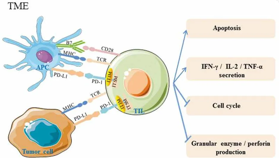
Fig:1: Expression and function of PD1 and PD-L1
3. Clinical application of PD-1/PD-L1 blockade therapies :
3.1. Anti-PD-1 antibodies :
Many anti-PD-1 antibodies have been produced to date. Nivolumab (Opdivo), pembrolizumab (Keytruda) and compliably (Libtayo) were approved by the FDA in September 2014, December 2014 and September 2018, respectively . Pidilizumab, AMP-224, AMP-514 and PDR001 are still in the experimental phase of development.
3.1.1. Nivolumab :
Nivolumab (brand name: Opdivo, also known as BMS-936558 and MDX1106) is a humanized IgG4 anti-PD-1 monoclonal antibody that has a high affinity for PD-1, blocking the binding of PD-1 to its ligand PD-L1. In December 2014, the FDA accelerated the approval of nivolumab for treating unresectable or metastatic melanoma, And in March 2015, the FDA approved nivolumab to treatment of metastatic squamous non-small cell lung cancer (NSCLC) In addition to melanoma and NSCLC, nivolumab has been demonstrated to be effective in a number of other malignancies, including, Hodgkin’s lymphoma and hepatocellular carcinoma The objective response rate (ORR) of nivolumab was found to be 23.7% in patients with NSCLC, with progression-free survival (PFS) of 91 days The overall survival (OS) of patients with Hodgkin’s lymphoma was approximately 80% at three years, and median the PFS ranged between 12 and 18 months.
3.1.2. Pembrolizumab :
Pembrolizumab (brand name: Keytruda, also known as MK-3475 and lambrolizumab) is a humanized IgG4 kappa anti-PD-1 antibody with high affinity. In 2014, the FDA approved pembrolizumab to treat metastatic melanoma. A phase I clinical trial showed that pembrolizumab was safe and effective for the treatment of melanoma, and a phase II clinical trial showed that the curative effect of pembrolizumab for advanced melanoma was obvious when compared with that of ipilimumab (anti-CTLA-4 antibody). The ORR of pembrolizumab in patients with advanced melanoma was reported to be 33%. In 2015, advanced NSCLC patient with no previously treatment were using pembrolizumab treatment and got the ORR as 18% Afterwards, pembrolizumab was approved by the FDA in May 2017 for locally advanced or metastatic urothelial carcinoma. Indeed pembrolizumab improved overall survival compared with the combined performance of influencing, docetaxel and paclitaxel in patients with locally advanced or metastatic urothelial carcinoma Furthermore, early clinical data showed that pembrolizumab also has great potential for the treatment of other tumors. The ORR of pembrolizumab in non-Hodgkin’s lymphoma was 53%, and in head and neck squamous cell carcinoma was found to be 19%.
3.1.3. Cemiplimab :
Cemiplimab (brand name: Libtayo) is a high-affinity anti-PD-1 antibody that was the first checkpoint treatment specifically designed for advanced cutaneous squamous cell carcinoma (CSCC); it was very recently approved by the FDA. The result of a phase 1 study of cemiplimab treatment in advanced CSCC patients showed a durable response, as no disease recurrence was found more than 16 months after the treatment. In an expansion phase I study, a 50% response was observed, and the response was durable. Moreover the results of the phase II study showed an objective response of 47%, with adverse events that were similar to other PD-1 inhibitors.
4. New insights regarding PD-1/PD-L1 blockade for cancer treatment :
4.1. Predictive biomarkers :
When using anti-PD-1 or PD-L1 antibodies to treat cancer, some patients with low PD-L1 expression might be poor responders. Therefore, to personalize treatments and obtain an optimal treatment effect, biomarkers need to be identified. In patients with thymic epithelial tumor, those with a PD-1-positive microenvironment had a shorter mean estimated survival time than did negative control and in ovarian cancer and hepatocellular carcinoma, patients with high PD-L1 expression had significantly worse outcomes than did patients who had low or lacked PD-L1 expression. Moreover, increased soluble PD-L1 (sPD-L1) has been associated with a low 3-year overall survival rate in patients with diffuse large B-cell lymphoma (DLBCL)..
Although the FDA has approved a PD-L1 immunohistochemistry (IHC) test as a companion diagnostic for anti-PD-1 antibody treatment in advanced NSCLC, the expression pattern of PD-1 and PD-L1 is not a good predictive biomarker for diagnosing all cancer types because of the following reasons: 1) biopsy specimens from patients may not accurately identify PD-L1 expression; 2) PD-L1 expression levels might differ among patients; 3) biopsy collection times might be too long or involve poor storage conditions, and thus PD-L1 levels might not represent the real PD-L1 status; 4) inappropriate tissue handling or IHC techniques for assessing PD-L1 expression might lead to PD-L1 epitope degradation before fixation; 5) not all antibodies used for PD-L1 are suitable; 6) only membrane PD-L1 is functional, as it can interact with PD-1 on T cells, and it is unclear whether the amount of cytoplasmic PD-1 expression is associated with cancer progression; and 7) PD-L1 can be expressed by different cells in the TME, and it is also remain unclear whether its expression on different cells is related to cancer progression. Given the complexity of T cell regulation, which can be controlled by multiple signaling pathways, other components of the TME may be useful as predictive biomarkers together with PD-1/PD-L1 expression to help doctors select patients for optimal treatment
4.2. Combination therapy:
Currently, an increasing number of researchers are realizing that responses to anti-PD-1/PD-L1 antibody treatment and other immunotherapies are dependent on the TME, which can be distinguished as an immunogenic (hot) TME or a non-immunogenic (cold) TME1. Large amounts of TILs and cytokines and high PD-L1 expression occur in hot tumors, whereas cold tumors show almost no PD-L1 expression and no T-cell infiltration. Therefore, some of the reasons why almost combination treatments with anti-PD-1/PD-L1 antibodies may provide better clinical results are as follows: 1) it will create a hot TME, for example, increasing PD-L1 expression or increasing TIL infiltration, which can enhance antitumor activity; 2) it will target other cell types in the TME, such as MDSCs, and provide an additive effect to the entire microenvironment; or 3) it may reduce the dosage or treatment time when compared with a single treatment, which can decrease the side effects of drugs. Overall, there are many combination therapy methods and these combination therapies have synergistic effects, which can improve the efficacy of therapies in a variety of cancers.
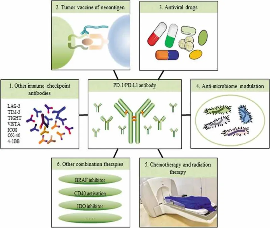
Fig 2: Combination therapy
4.2.1. Combination with other immune checkpoint antibodies :
CTLA-4 and PD-1 inhibit T cell activation in different ways. The first patient to use an immune checkpoint combination was treated with anti-PD-1 and CTLA-4 antibodies in 2009. In metastatic melanoma patients, the response rate for PD-1 and CTLA-4 blockade combination therapy was approximately 60% in phase II and phase III trials when compared with PD-1 blockade alone. Notably, a slightly higher 3-year survival rate was observed, but a higher frequency of toxicity was also found2. During 2018, three papers were published on lung cancer treatment with PD-1 plus CTLA-4 blockade. Showing a greater clinical benefit in high-tumor mutational burden patients when compared with low-tumor mutational burden patients, including improved objective responses, durable benefits, and progression-free survival rates Therefore, the tumor mutational burden may be useful as a genomic determinant to identify patients who may benefit from PD-1 and CTLA-4 blockade combination immunotherapy.
Additional clinical trials and pre-clinical tumor model experiments evaluating other checkpoint proteins combined with anti-PD-1/PD-L1 antibodies are also being conducted. LAG-3, TIM-3, T cell immune receptor with Ig and ITIM domains (TIGIT), and V-domain Ig suppressor of T-cell activation (VISTA) are all checkpoint proteins in T cells. An investigational study using anti-LAG-3 with and without anti-PD-1 antibodies is currently recruiting patients to test its safety, tolerability and effectiveness for the treatment of solid tumors Of note targeting TIM-3, TIGIT and VISTA combined with PD-1 pathway targeting improved antitumor immune responses. Inducible T cell costimulator (ICOS), TNF receptor superfamily member 4 (OX-40), and TNF receptor superfamily member 9 (4-1BB) are co-stimulatory molecules in T cells, and antibodies against these molecules combined with anti-PD-1/PD-L1 antibodies resulted in an enhanced immune response in tumor model. The use of low-dose combination checkpoint inhibition also appears to be an optimal approach for enhancing clinical benefits.
Unfortunately, patients may develop dermatitis, colitis, hepatitis, pancreatitis, pneumonitis and hypophysis after anti-CTLA-4 or anti-PD-1/PD-L1 antibody treatment. In fact, checkpoint combination therapies have notably high toxicity and side effects. For example, most melanoma patients experience immune-related adverse events during anti-PD-1/PD-L1 and anti-CTLA-4 antibody combination therapy. Therefore, further studies investigating the mechanisms of these checkpoint therapies may be required to ameliorate these toxicities.
4.2.2. Combination with neoantigen tumor vaccines :
Several studies have shown that genomic information, especially a high mutation burden, helps to determine the response of PD-1/PD-L1 blockade. As it has many mutations that can be recognized as neoantigens, melanoma is a well-known hot tumor, and checkpoint immunotherapy has remarkable clinical effects. Whole-exome sequencing has been applied in NSCLC and mutations in its genomic landscape helped to determine response to anti-PD-1 antibodies. In colon cancer, tumors with mismatch repair deficiency are highly sensitive to anti-PD-1 antibody treatment, and this deficiency indicate many mutant neoantigens, which are created by tumor cells because of mutations, and can be recognized by T cells. Although tumor vaccines can be prepared from cancer cells, parts of cancer cells, or pure tumor antigens, they always contain neoantigens from a tumor. However, questions regarding tumor vaccines from neoantigens have been raised, such as 1) How can neoantigen prediction be optimized?2) What is a good time for vaccine acquisition?3) What is the optimal burden and vaccine injection schedule?4) How do we select patients who will benefit most? Among melanoma patients using vaccines that targeted personal tumor neoantigens, those who had recurrent disease were then treated with anti-PD-1 therapy, and complete tumor regression and neoantigen-specific T cell expansion were found after this treatment. These studies provide strong rationale for the further development of this combination approach, though more clinical tests and comprehensive studies identifying mechanisms are required.
4.2.3. Combination with antiviral drugs :
Some human cancers are driven by oncogenes from integrated viruses, such as EBV in Burchett’s lymphoma, Hodgkin’s lymphoma, gastric carcinoma, HPV in cervical cancer and head and neck cancer, HBV and HCV in hepatocellular carcinoma, and Merkel cell polyomavirus (MCPyV) in Merkel cell carcinoma (MCC) and chronic lymphocytic leukemia (CLL). No clinical trial data for PD-1/PD-L1 blockade combined with antiviral drugs are yet available; however, some preliminary studies indicate promise. MCC is a skin cancer, among which approximately 80% are associated with MCPyV, and high ORRs, prolonged durable responses, and good tolerability were found when using anti-PD-1 therapy. In addition, IHC and cytometry by time-of-flight have been applied to examine the immune microenvironments in HBV-associated hepatocellular carcinomas, as more PD-1+ Tregs were found in HBV-related HCC than in non-viral-related HCC.
4.2.4. Combination with anti-microbiome modulation :
In addition to the tumor itself, host factors such as the gastrointestinal microbiome can influence responses to checkpoint immunotherapy. Published in 2018, three notable studies have shown a relationship between healthy gut flora and anti-PD-1 treatment. The authors reported that in melanoma and epithelial cancer, “good” or “favorable” gut bacteria were required for a patient to respond to PD-1 blockade treatment and that antibiotic use could inhibit clinical benefits. Therefore, for anti-PD-1/PD-L1 antibody treatment, therapeutic responses of tumor patients may be improved through host gastrointestinal microbiome modulation.
4.2.5. Combination with chemotherapy and radiation therapy :
Chemotherapy kills tumor cells by inducing DNA damage, cell cycle arrest and ultimately apoptosis, whereas radiation therapy activates the type I IFN pathway in DCs to effectively prime tumor-specific T cells. After more mechanistic studies, it appears that these methods may help turn cold TME into hot TME, as they can create neoantigens of tumors during treatment. Therefore, the response has been encouraging when these two methods have been combined with anti-PD-1/PD-L1 antibodies. In NSCLC, promising results were shown in combination chemotherapy with anti-PD-1/PD-L1 antibodies, including a high overall response and long overall survival but not fewer irAEs. In metastatic melanoma, major tumor regression occurred when radiation and PD-L1 and CTLA-4 dual checkpoint blockade was used77. Although these antitumor results are remarkable, a considerable amount of work regarding the dosage, time and sequence is still needed in future clinical trials.
4.2.6. Other combination therapies :
There are still many other combination therapies that are under development for application, and most are being tested in melanoma patients. For instance, a BRAF inhibitor used for the treatment of melanoma was combined with anti-PD-1 and anti-CTLA-4 antibodies, and the results showed that BRAF inhibitor therapy may affect subsequent clinical responses to checkpoint immunotherapy. CD40 activation can upregulate APC function, and convert cold tumors into hot tumors. In a pancreatic carcinoma mouse model treated with anti-PD-1 antibodies combined with anti-CD40/chemotherapy, the activity and durability of the response were extended when compared with anti-CD40/chemotherapy treatment alone. Recently, IDO inhibitor and PD-1 antibody combination therapy exhibited a good antitumor response in hepatocellular carcinoma because checkpoint immunotherapy increased IDO induction, which can cause adaptive resistance in some patients. In another phase 3 trial in advanced melanoma, the same IDO inhibitor plus an anti-PD-1 antibody had no increased benefit compared to the use of the anti-PD-1 antibody alone, indicating that combination therapy may not be necessary at all times; in particular no evidence to date from extensive trials supports the use of combination therapy.
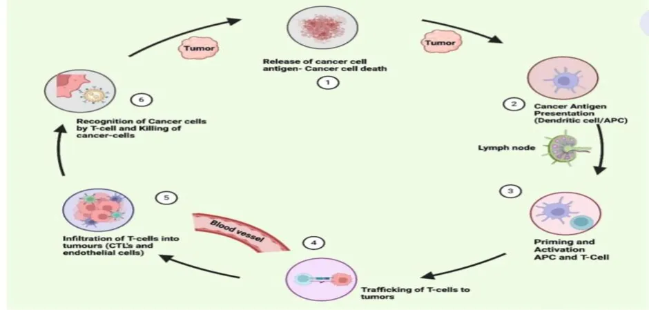
Fig-3
CONCLUSION:
Recently, cancer immunotherapy targeting PD-1 or PD-L1 has proven effective in causing durable antitumor immune responses with less toxicity in many types of tumors, We believe that PD-1/PD-L1 blockade therapy will be the major cancer immunotherapy method in the next few years, even though there is still much to be learned about this signaling pathway. Key questions remaining to be resolved include: 1) How to select PD-1/PD-L1-positive patient groups? What are their features of these patiens, and what efficient clinical detection method should be used? 2) How can the abundance of tumor infiltrating CD8+T cell be increased in TME? Especially the particular CD8+T cell that display tumor-reactive intratumoral TCR repertoires, but not bystander CD8+TILs that can not kill the tumor cell. 3) What is the mechanism by which PD-1 regulats on CTLs and Tregs, and what is the mechanism by which PD-L1 acts on tumor cells and APC cells in TME? Can we find more efficient inhibitors based on the mechanism? 4) Do we have any good treatment for PD-1/PD-L1- negative patients? Can other therapies or combination therapies with an anti-PD antibody approach be optimal for these patients? With a deeper understanding of personal genomic information, personalized markers in guiding anti-PD therapy alone or with other targets will be critical to achieve clinical results of such therapies, and more work needs to be performed to achieve clarity regarding these key questions. Similar to the tip of an iceberg, PD-1/PD-L1 blockade antitumor immunotherapy opens a new era of cancer treatment, and further work on safety and efficiency will be needed.
REFERENCES


Dr. D. Rama Bramha Reddy, K. Malleswari, Bandlamudi Abhishek, Cancer Immunotherapy Beyond PD1/PD-L1, Int. J. of Pharm. Sci., 2026, Vol 4, Issue 8, 2287-2296. https://doi.org/10.5281/zenodo.21927065
 10.5281/zenodo.21927065