

We use cookies to ensure our website works properly and to personalise your experience. Cookies policy
Yashodeep College of Pharmacy Saralgaon-Nagaon Road, Saralgaon, Tal-Murbad, Dist-.
Spinal muscular atrophy (SMA) type 1 is a serious inherited neuromuscular condition that primarily affects infants, leading to progressive muscle weakness as a result of motor neuron loss. Spinal muscular atrophy mainly develops when both copies of the SMN1 gene are deleted or altered, resulting in a reduced amount of functional SMN protein in the body. Infants affected by this condition typically experience severe hypotonia, delayed motor development, difficulty feeding and swallowing, and respiratory issues. Previously, SMA type 1 was linked to low survival rates and few available treatment choices. However, the emergence of disease-modifying therapies has significantly enhanced both the management and outcomes for affected children. This review highlights recent progress in treating SMA type 1, with particular attention to nusinersen, onasemnogene abeparvovec, and risdiplam. Nusinersen and risdiplam boost the production of functional SMN protein by altering SMN2 gene splicing, whereas onasemnogene abeparvovec supplies a functional copy of the SMN1 gene via gene replacement therapy. Clinical evidence indicates that these therapies can enhance motor development, respiratory function, and survival, particularly when treatment begins early. Even with these developments, issues such as side effects, drug administration, accessibility, long-term efficacy, and treatment response continue to be key considerations. Emerging strategies, such as next-generation gene therapies, myostatin inhibitors, gene-editing techniques, and combination treatments, are also under investigation. Early detection and prompt treatment may improve the chances of preserving motor function and achieving better long-term outcomes in children with SMA type 1.
Spinal muscular atrophy (SMA) is an inherited autosomal recessive disease that affects the lower motor neurons. It is mainly caused by the deletion or mutation of the SMN1 gene on chromosome 5q13. This leads to the loss of motor neurons in the anterior horn of the spinal cord, causing gradually worsening muscle weakness, muscle wasting, and difficulty with movement. SMA affects roughly 1 in 10,000 to 20,000 live births, and the carrier rate in the general population is estimated at about 1 in 40 to 1 in 70. Traditionally, SMA is classified into clinical types according to the age at onset and the severity of symptoms. Of these, SMA type 1 is the most frequent and severe form, representing about 50–60% of SMA cases.(1,2) Spinal muscular atrophy (SMA) is an inherited autosomal recessive neurodegenerative disease caused by the loss or malfunction of both copies of the SMN1 gene. This results in a reduced amount of survival motor neuron (SMN) protein, leading to the gradual loss of motor neurons in the brainstem and spinal cord. As the disease progresses, patients develop muscle weakness and wasting (atrophy), which affect their ability to move. (3) The SMN2 gene can partly compensate for the loss of SMN1 by producing a small amount of functional SMN protein. Generally, individuals with more copies of the SMN2 gene tend to have less severe symptoms, although disease severity can also depend on other factors. Disease onset ranges from infancy to adulthood, and muscle weakness usually worsens with age, possibly resulting in paralysis in the most severe cases.(4) Most SMA type 1 patients face complications of respiratory failure and dysphagia that require interventions (e.g., nasogastric tube or gastrostomy tube placement and initiation of NIV or intubation leading to tracheostomy, respectively), to prolong patient survival.(3) Natural history studies show that in patients older than 12 months, 100% required nutritional or combined nutritional and ventilation support, (3) and 61% of SMA type 1 patients required ≥1 intubation within the first year of life.(9) Despite supportive care, SMA patients have high hospitalization rates; infants with SMA1 were reported to have 4.2 to 7.6 hospitalizations/year compared to 1.7 hospitalizations/year in non-SMA patients.(3)
History of SMA
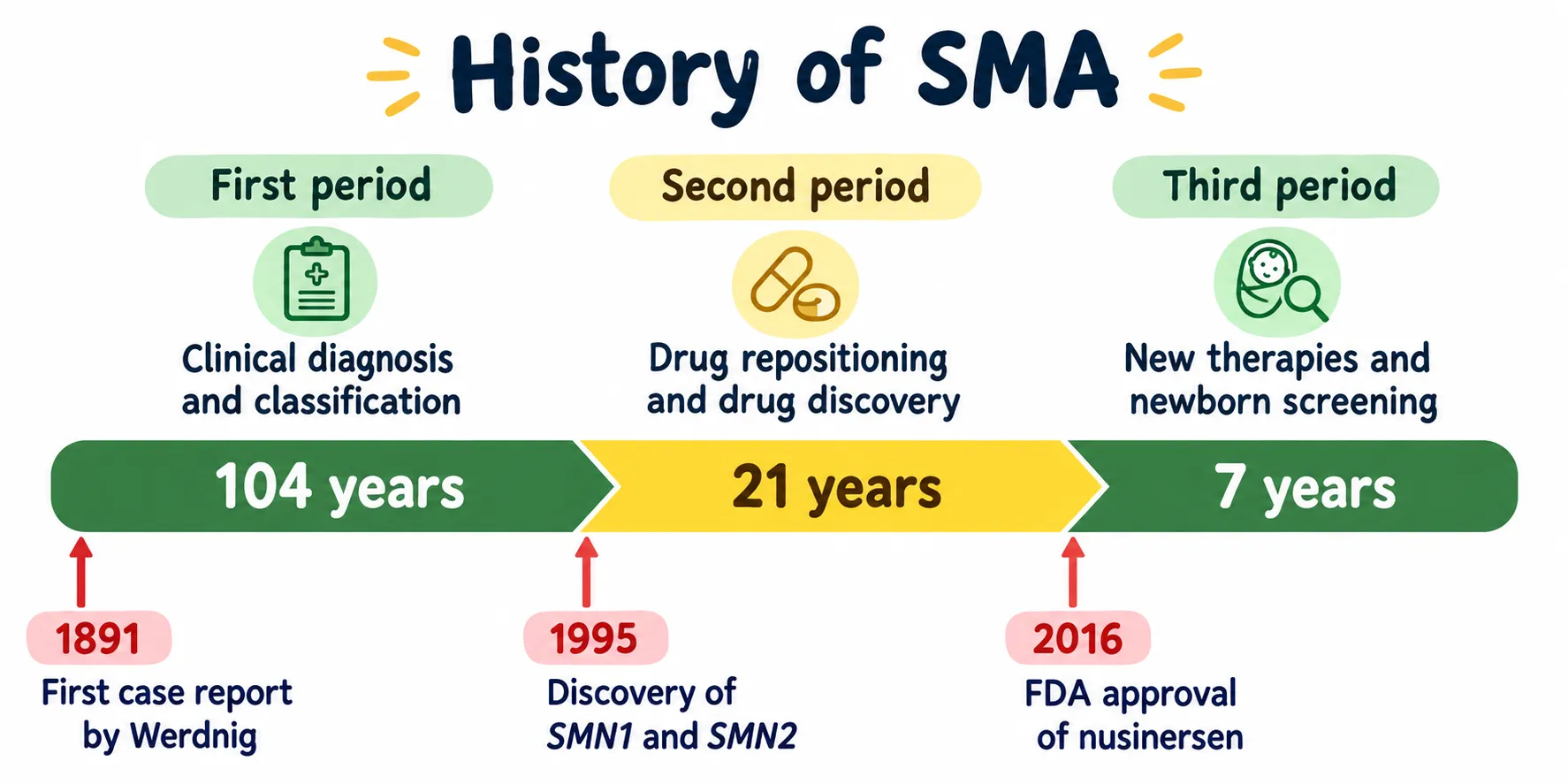
Three Periods in the History of SMA Research
Research on spinal muscular atrophy (SMA) can be divided into three main periods. In 1891, Werdnig described the first two cases of SMA. Over time, researchers identified multiple forms of the disease with differing clinical features. In 1995, two key genes, SMN1 and SMN2, were discovered. The SMN1 gene is responsible for SMA, whereas SMN2 functions as a modifier gene and affects disease severity. This finding enabled researchers to create new treatments for SMA. In 2016, nusinersen became the first drug approved by the US FDA for treating SMA. Afterwards, two further treatments received approval: onasemnogene abeparvovec in 2019 and risdiplam in 2020. These breakthroughs represented significant progress in the treatment and management of SMA.(2)
Classification
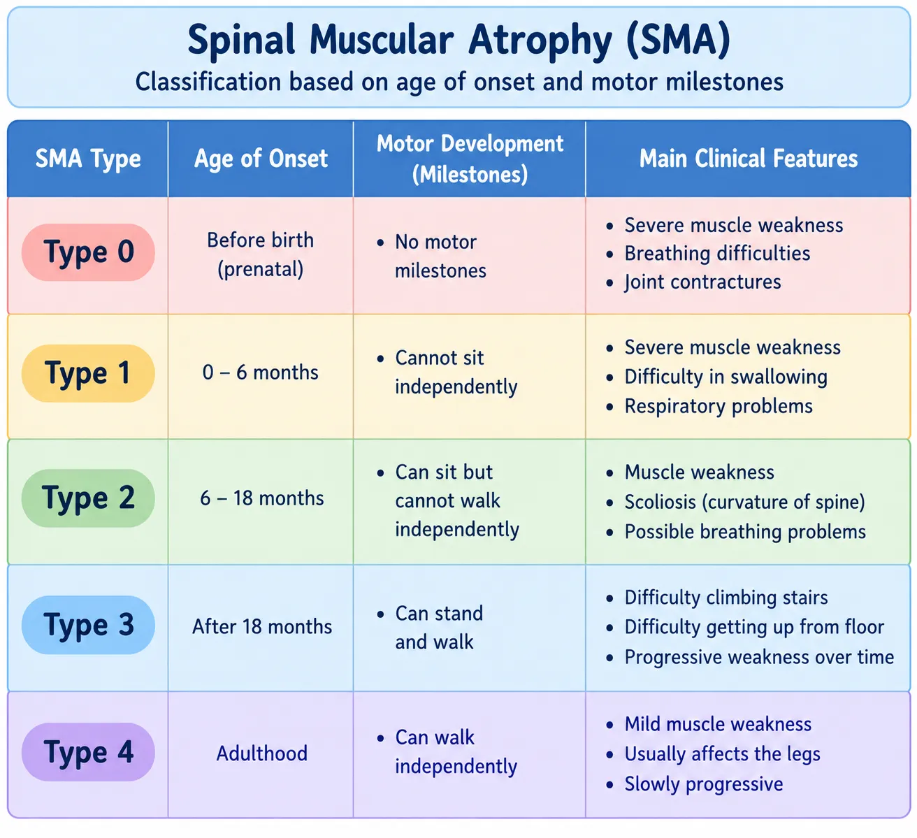
Table 1: Emerging Therapeutic Strategies for Spinal Muscular Atrophy: Ongoing Clinical and Preclinical Trials.(5)
2. Clinical features
SMA is divided into three primary phenotypes according to the age when symptoms or signs first appear and the highest level of motor function attained. Nevertheless, certain SMA patients fall outside the typical range at either extreme of the phenotypic spectrum. Subclassification has likewise been suggested for SMA types 1 and 3, and occasionally for the type 2 phenotype. (5) SMA Type 1 predominantly impacts the nervous and respiratory systems. Patients may present with symptoms including drooling, decreased muscle tone (hypotonia), trouble swallowing (dysphagia), acid reflux, and malnutrition. For this reason, a multidisciplinary healthcare team is essential to address the patient's neurological, musculoskeletal, respiratory, swallowing, and nutritional needs.(6) Type 1 SMA patients represent over 50% of SMA cases. These patients are referred to as non-sitters, who never attain major motor developmental milestones such as sitting, standing, and walking during their lifetime. (5)
SMA: Clinical Variability
Nearly all types of SMA result from
Bi-allelic (frequently homozygous) deletions of
SMN1
– These mutations are inactivating
– The lack of SMN protein is lethal
• Abnormal processing of SMN2 pre-mRNA results in an SMN1 transcript (“two
Wrongs make a right”).(7)
Clinical description
Spinal muscular atrophy (SMA) is a hereditary neuromuscular condition marked by worsening muscle weakness and wasting. It results from the degeneration and loss of motor neurons, especially the anterior horn cells of the spinal cord and certain brainstem nuclei. The age at which symptoms begin varies greatly, from before birth to adulthood. The muscle weakness is typically symmetrical and affects the muscles nearer the center of the body (proximal muscles) more severely than those farther out (distal muscles).(8)
Signs and Symptoms
1. Severe muscle weakness – Pronounced weakness is a key clinical feature of SMA Type 1. (2)
2. Generalized hypotonia – Affected infants usually show notably reduced muscle tone. (2)
3. Poor motor abilities – Infants experience severe delays in motor development and generally cannot sit unsupported. (2)
4. Reduced or absent reflexes (areflexia) – Deep-tendon reflexes are often absent or diminished. (2)
5. Feeding and swallowing difficulties – Weakness can impair sucking, swallowing, and feeding. (2)
6. Breathing difficulties – Weakness of the respiratory muscles can lead to impaired breathing. (2)
7. Bell-shaped chest and abdominal breathing – Weakness of the intercostal muscles combined with relative preservation of the diaphragm gives rise to characteristic respiratory patterns. (2)
8. Tongue fasciculations – Fasciculations affecting the tongue may be observed. (2)
3. Epidemiology
Spinal muscular atrophy (SMA) is a rare genetic neuromuscular disorder caused by the progressive loss of lower motor neurons, which leads to muscle weakness and atrophy. The condition is passed from parents to their child through an autosomal recessive pattern of inheritance. SMA occurs in approximately 1 in 10,000–20,000 live births, while the carrier rate in the general population is estimated at roughly 1 in 40–70 people. It is also considered a major genetic contributor to infant mortality. SMA has a wide spectrum of clinical manifestations and is categorized into distinct types according to the age when symptoms begin and the highest motor milestone achieved. SMA is grouped into five principal types, spanning type 0 through type 4. SMA type 1 is an early-onset form, with symptoms appearing in infancy, and affected infants usually never learn to sit without support. (2)
4. Pathophysiology
Biallelic SMN1 loss/pathogenic variants (2,9)
↓
Marked reduction of functional SMN protein (2,9)
↓
Because exon 7 is predominantly excluded, SMN2 offers only limited compensation (2)
↓
Not enough functional SMN protein (2-10)
↓
Degeneration and dysfunction of lower motor neurons (2,9)
↓
Loss of anterior horn cells within the spinal cord (9,11)
↓
Decreased neural stimulation reaching the skeletal muscles (9)
↓
Progressive muscle weakness, along with hypotonia and muscle atrophy (2,9)
↓
Marked weakness in proximal and axial muscles (9)
↓
Impaired motor development / inability to sit without support (9)
↓
Weakness of the respiratory and bulbar muscles (9,12)
↓
Ineffective cough, respiratory insufficiency, and difficulties with feeding/swallowing (9,12)
5. Diagnosis
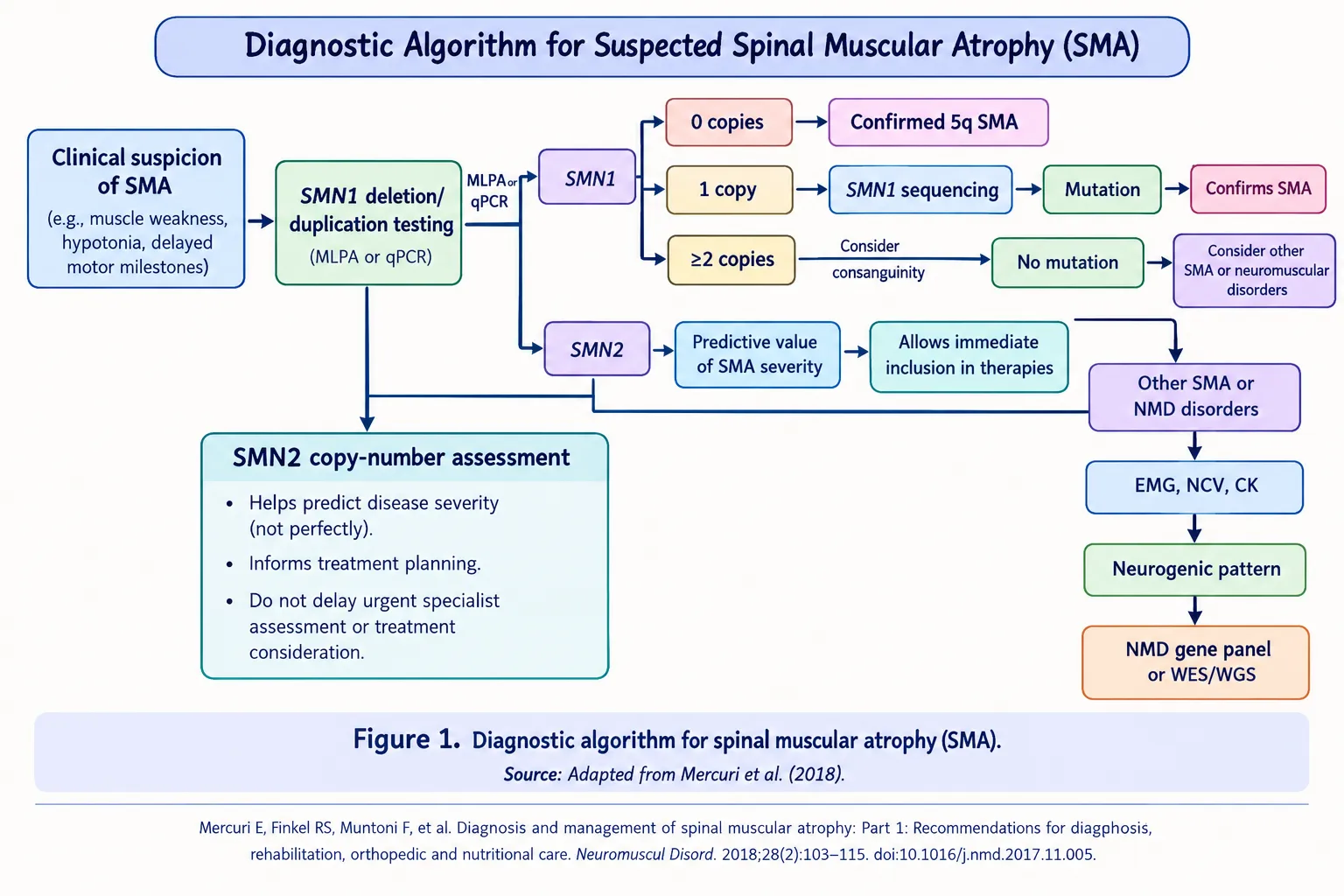
Figure 1: Diagnostic Algorithm for Spinal Muscular Atrophy (9).
1. Clinical evaluation
Spinal muscular atrophy (SMA) Type 1 is suspected in infants who show symptoms before reaching 6 months of age. Typical clinical features consist of severe muscle weakness, hypotonia, poor head control, delayed motor development, a weak cry, feeding difficulties, and weakness of the respiratory muscles. Infants affected by the condition are usually unable to sit independently in the absence of treatment. (9,8) SMA Type 1 is typically suspected in infants presenting with pronounced hypotonia and symmetrical weakness, especially of the proximal muscles. Deep-tendon reflexes are frequently diminished or absent owing to involvement of the lower motor neurons. Infants affected by the disease generally show delayed motor development and cannot sit without assistance. Respiratory function is assessed as well, since weakness of the respiratory muscles can lead to a weak cough, recurrent respiratory infections, and difficulty breathing. Sucking, swallowing, and feeding capacity are evaluated to detect possible involvement of the bulbar muscles. Motor function can be evaluated using standardized tools, such as the Children’s Hospital of Philadelphia Infant Test of Neuromuscular Disorders (CHOP INTEND). These clinical findings give rise to suspicion of SMA, which is then confirmed by molecular genetic testing of the SMN1 gene (9-13).
1.1 Neurological examination:
The assessment covers muscle tone, muscle strength, deep-tendon reflexes, spontaneous movements, head control, and motor milestones. The weakness is usually symmetrical and more pronounced in the proximal muscles. (2,9)
1.2 Respiratory evaluation:
Weakness of the respiratory muscles can cause an ineffective cough, recurrent respiratory infections, paradoxical breathing, and progressive respiratory insufficiency. Respiratory function and oxygenation should be monitored for this reason. (9)
1.3 Evaluation of bulbar function and feeding:
Sucking, swallowing, chewing, and feeding abilities should be assessed regularly. Bulbar muscle weakness can lead to poor feeding, longer feeding times, trouble swallowing, and a risk of aspiration. (2,9)
1.4 Assessment of motor function:
Motor function and neurological development can be quantified using standardized scales like the CHOP INTEND and HINE-2. Such tools are especially valuable for setting a baseline and tracking disease progression as well as response to treatment. (2,13)
2. Genetic testing
Confirming SMA is primarily done through genetic testing. The disease is typically linked to the loss or mutation of both copies of the SMN1 gene, which leads to decreased production of survival motor neuron (SMN) protein. Testing usually begins with SMN1 deletion/duplication analysis, using methods such as multiplex ligation-dependent probe amplification (MLPA) or quantitative PCR (qPCR). If a single copy of SMN1 is found, sequencing may be needed to identify a pathogenic variant in the remaining copy. (9,8)
2.1 SMN1 testing:
Deletion/duplication analysis is typically carried out with techniques such as MLPA or quantitative PCR. When only one SMN1 copy is identified, sequencing can be done to detect a pathogenic variant in the remaining allele. (2,9)
2.2 Analysis of SMN2 copy number:
Once SMA has been confirmed genetically, establishing the SMN2 copy number is crucial, since SMN2 influences how severe the disease becomes. having more SMN2 copies correlates with a milder phenotype, even though the SMN2 copy number by itself cannot reliably forecast the clinical course for every patient. (2,9)
2.3 Newborn screening:
SMA may also be identified via newborn screening prior to the onset of symptoms. Early detection is especially crucial for SMA Type 1, as starting treatment before or very soon after symptoms begin can markedly enhance motor and survival outcomes. (2)
2.4 Genetic counselling:
After SMA is confirmed, genetic counselling and carrier testing may be provided to parents and other relatives to clarify the autosomal-recessive inheritance pattern and the risk of recurrence. (2,9)
6. Modifying therapy
In 2017, two significant studies described encouraging new treatments for SMA Type 1. By introducing the first disease-modifying therapies, these studies signaled the start of a new era in SMA management. Another novel treatment has been introduced since that time as well. The availability of these treatments has considerably altered the management and treatment approach for SMA, especially in countries where such therapies can be accessed. (14)
Spinal muscular atrophy (SMA) type 1 is a severe, early-onset form of SMA marked by progressive loss of motor neurons and muscle weakness. The management of SMA has been substantially transformed by the development of disease-modifying therapies. Disease-modifying treatments currently in use comprise nusinersen, onasemnogene abeparvovec, and risdiplam. Through various mechanisms, these therapies boost the availability of functional SMN protein. Better clinical outcomes are linked to starting treatment early, especially before significant motor-neuron loss occurs. (15,16)
1. Nusinersen
Nusinersen is an antisense oligonucleotide (ASO) designed to target the pre-mRNA of the SMN2 gene. It encourages the inclusion of exon 7 during SMN2 mRNA splicing, which raises the production of full-length, functional SMN protein. Nusinersen is given by intrathecal injection. Clinical evidence from infants with SMA type 1 showed gains in motor-milestone achievement and survival relative to sham treatment. (17)
Mechanism
Nusinersen is an antisense oligonucleotide (ASO) that falls within the phosphorothioate oligodeoxynucleotide class. Because these molecules do not easily cross the blood–brain barrier, nusinersen is delivered directly into the cerebrospinal fluid via an intrathecal injection. This route enables the drug to reach the central nervous system, where it exerts its therapeutic effect. (18) For patients with complex spinal anatomy, especially those with advanced scoliosis or a history of spinal fusion, alternative approaches to administering nusinersen may be considered. These options include X-ray-guided transforaminal injection or an implanted reservoir to aid intrathecal drug delivery. (19,20) Nusinersen has a half-life of roughly five months. Treatment starts with four loading doses given over the first two months, after which maintenance doses are administered every four months. The recommended dose stays the same across different age groups. Pharmacokinetic studies have demonstrated that nusinersen gradually moves from the cerebrospinal fluid (CSF) into the systemic circulation. In treated infants, the drug has also been found in peripheral tissues, such as the liver and kidneys. (21)
Table 2: Functional Motor Scales Used in SMA Type 1
| Motor scale | Age group | Role in SMA Type 1 |
|---|---|---|
| CHOP INTEND(13) | Infants and young children | particularly those who cannot sit Evaluates muscle strength and motor function in infants with severe disease. It comprises 16 items, with a total score ranging from 0 to 64. |
| HINE-2(22,23) | 2 months to 2 years | Evaluates neurological development and motor milestones, including head control, sitting and other movements RULM(24) |
| RULM(24) | Mostly over 2 years | Evaluates upper-limb function and may be helpful for patients who are non-sitters, sitters or walkers. |
Advantages:
1. Raises functional SMN protein levels.
2. Shown to provide benefit in SMA type 1.
3. Applicable across various age groups and SMA phenotypes. (14,15)
Limitations:
1. Needs to be given intrathecally on multiple occasions.
2. In certain patients, performing a lumbar puncture can prove challenging.
3. Treatment over the long term is usually necessary.
4. Complications following lumbar puncture have been documented. (15)
2. Onasemnogene Abeparvovec
Onasemnogene abeparvovec is a gene-replacement therapy that employs an adeno-associated virus serotype 9 (AAV9) vector to transfer a working copy of the SMN1 gene into cells. It is given as one intravenous infusion. Through the supply of a functional SMN1 gene, the therapy allows SMN protein to be produced and seeks to halt additional motor-neuron loss. (14,15)
Mechanism
Onasemnogene abeparvovec (OAV101, Zolgensma®; previously AVXS-101) is a gene-replacement therapy indicated for the treatment of spinal muscular atrophy (SMA). It was created by AveXis and is delivered intravenously as a gene therapy. (25) It utilizes a self-complementary adeno-associated virus serotype 9 (AAV9) vector to carry a functional human SMN1 gene. A promoter governs the gene, enabling continuous production of SMN protein. The AAV9 vector is able to pass through the blood–brain barrier and reach neurons within the central nervous system. Preclinical research in mice and non-human primates demonstrated that AAV9-mediated delivery of the SMN1 gene could ameliorate the SMA disease phenotype. (25,26) The safety and efficacy of onasemnogene abeparvovec were subsequently assessed in the prospective CL-101 (START) study, which enrolled infants with SMA Type 1. In this trial, patients were given a single intravenous (IV) infusion of the gene therapy. (27)
Advantages:
1. Treatment with a single dose.
2. Delivers a functional SMN1 gene directly.
3. Has shown clinically meaningful motor and survival outcomes in SMA type 1. (27-28)
Limitations:
1. Treatment calls for vigilant safety monitoring.
2. Adverse effects involving the liver may arise.
3. Immune reactions and other adverse events linked to treatment are key factors to consider.
4. The durability of the effects over the long term is still under investigation.(14,15)
3. Risdiplam
Risdiplam is an orally administered small-molecule modifier of SMN2 splicing. It alters the splicing of SMN2 pre-mRNA, thereby boosting the production of full-length, functional SMN protein. In contrast to nusinersen, it is given orally, which offers a non-invasive method of administration. (14,29) In the FIREFISH trial, which enrolled infants with SMA type 1, risdiplam treatment was linked to gains in motor function and the attainment of motor milestones. By 12 months, a number of infants could sit unassisted, a milestone seldom reached in untreated SMA type 1. (29)
Mechanism
Risdiplam (Evrysdi®) is an oral small-molecule drug created to treat spinal muscular atrophy (SMA). It acts by altering SMN2 pre-mRNA splicing, which encourages the inclusion of exon 7. This boosts the production of full-length SMN mRNA and consequently elevates the amount of functional SMN protein. Through increased SMN protein production, risdiplam helps maintain the survival and function of motor neurons impacted by SMA. (30,31) Risdiplam is taken by mouth once daily, with the dose tailored to the patient’s age and body weight. For patients 2 years of age or older weighing at least 20 kg, the maximum recommended dose is 5 mg once daily. (32) Risdiplam may also be administered via a feeding tube when taking it orally proves difficult. Its dosing and pharmacokinetic profile have been examined in both healthy adults and patients with SMA. (29) Preclinical animal studies have demonstrated that risdiplam is able to reach both the central nervous system (CNS) and peripheral tissues, enabling it to raise SMN protein levels throughout the body. (33)
Advantages:
1. Administration by mouth.
2. Less invasive than intrathecal therapy.
3. Raises functional SMN protein.
4. Shown to improve motor function in SMA type 1. (14-29)
7. Therapies under investigation and newly emerging
While nusinersen, onasemnogene abeparvovec, and risdiplam are now well-established disease-modifying treatments for SMA, scientists continue to search for novel therapies that might offer further advantages. Such strategies seek to tackle muscle weakness and other disease-related alterations that can persist despite improvements in SMN protein levels. Ongoing studies cover myostatin inhibitors, newer generations of gene therapy, gene-editing methods, and combined treatment approaches. (16,34)
Apitegromab (SRK-015):
Apitegromab is a monoclonal antibody that blocks the activation of myostatin, a protein that acts as a negative regulator of skeletal muscle growth. As a result, inhibiting myostatin could boost muscle mass and enhance motor function. Apitegromab has been examined as an add-on therapy alongside SMN-directed treatments like nusinersen or risdiplam. (34)
Taldefgrobep alfa:
Taldefgrobep alfa is a further investigational treatment that acts on myostatin. It is under study as an adjunct to current SMA disease-modifying therapies, aiming to improve muscle function beyond what raising SMN protein alone can achieve. (16)
Gene-editing strategies:
Technologies for gene editing, including CRISPR-based methods and base editing, are being explored as possible future approaches for correcting or altering the genetic defects that cause SMA. Such methods might eventually deliver more lasting genetic correction, though additional research is needed to determine their safety, delivery, and long-term effectiveness. (16,38)
Combination therapy:
Pairing an SMN-targeted therapy with an SMN-independent treatment represents another key research focus. The reasoning is that raising SMN protein levels may target the underlying genetic defect, whereas therapies aimed at muscle or neuroprotection may help manage remaining muscle and neuronal issues. (16,34,37)
Therapies of the next generation:
Scientists are also working on better gene-delivery systems, different ways of administering treatment, and novel molecules that could overcome certain limitations of the treatments available today. Such strategies might enhance tissue distribution, the durability of treatment, and clinical results. (16,34,37,38)
8. New Developments in the Treatment of SMA Type 1
1. Treatment at an early or presymptomatic stage:
Because newborn screening allows early diagnosis, disease-modifying therapy can now begin before substantial motor-neuron loss takes place. Starting SMN-targeted therapies early is linked to improved survival and motor development in SMA Type 1. (2,35) Treating before symptoms appear also highlights how important genetic counselling and screening of family members are. When an infant is diagnosed with SMA, genetic testing can help determine the risk in future pregnancies and support families in making well-informed choices. Long-term follow-up is required to determine whether the advantages of early treatment persist as the child grows. Even though early treatment can lead to better outcomes, some children may still experience motor or breathing difficulties. As a result, routine monitoring of growth, development, and respiratory function continues to be essential (42,43).
2. Progress in gene-replacement therapy:
Onasemnogene abeparvovec introduced a gene-replacement strategy that uses an AAV9 vector to deliver a working SMN1 gene. Ongoing research has concentrated on refining patient selection, the timing of treatment, safety monitoring, and long-term results. (35,36) Researchers are also exploring the reasons behind the varying responses of patients to gene-replacement therapy. Elements like the age at which treatment begins, how severe the disease is before treatment, and whether pre-existing antibodies against AAV9 are present can all affect treatment results. Understanding these variations could shed light on why certain patients gain more benefit than others. Long-term follow-up is crucial for establishing how long the treatment's effects persist and for watching for any complications that might arise over time (43,44).
3. Better oral treatment options:
Risdiplam has delivered an orally administered SMN2-splicing-modifying therapy, providing an alternative to intrathecal nusinersen and intravenous gene therapy. Because it is taken orally, it may be especially valuable for long-term treatment and for patients who have difficulty with repeated intrathecal administration. (2,35) The arrival of oral risdiplam has made treatment more convenient for numerous patients. Taking the medication consistently is essential to keep treatment stable. For children who struggle with repeated intrathecal injections, the oral route of administration may be particularly beneficial. Nevertheless, more research is required to grasp long-term treatment outcomes, patient adherence, and the ways prior treatments might affect responses to later therapies. This knowledge could assist healthcare professionals in selecting appropriate treatment options according to each child's condition and individual medical needs (4,45).
4. Combination and sequential therapies:
Recent reviews have examined the combination or sequential use of different disease-modifying treatments to sustain or improve therapeutic effects. These strategies are under consideration because restoring SMN may not fully reverse motor-neuron and muscle damage that is already established. (37) A key difficulty with combination and sequential therapies is determining the right moment to add another treatment. This choice hinges on multiple factors, such as earlier treatments, shifts in motor function, potential side effects, and the child's general health. Routine clinical assessments and long-term follow-up are needed to learn whether adding or switching treatments produces better results. Until additional evidence becomes available, treatment choices should be guided by the individual needs and clinical status of each patient. (15,46)
5. Targeting mechanisms that do not depend on SMN:
Research has moved beyond merely increasing SMN protein. New therapeutic approaches are under investigation to enhance muscle growth, neuromuscular function, and neuronal survival, which could potentially supplement existing SMN-targeted treatments. (35,38)Even when SMN protein levels rise, some patients may still face muscle weakness and difficulties with movement. Researchers are investigating whether therapies aimed at muscle growth, neuromuscular function, and other disease-related pathways might offer additional benefits. Nevertheless, an increase in muscle size does not necessarily result in better motor function. Consequently, clinical trials must evaluate functional improvements, treatment safety, and the potential advantages of combining these approaches with existing SMN-targeted therapies. These strategies remain under study, and more clinical evidence is needed to establish their possible role in SMA Type 1. (38,46)
6. Improved long-term management:
As survival has improved with disease-modifying treatment, more attention is now being paid to long-term management of respiratory, nutritional, orthopedic, and motor function. This reflects a shift toward comprehensive lifelong care instead of concentrating solely on survival. (2,36) Long-term care for children with SMA Type 1 should address not only physical health but also their development, communication, comfort, and participation in daily activities. As children grow, their care plans may need to be modified in response to changes in motor function, breathing support, feeding requirements, and family circumstances. Collaboration between healthcare professionals and caregivers is vital to ensure continuous and suitable care. Future research should place greater emphasis on quality of life, long-term functional progress, and the physical and emotional difficulties encountered by children and their families during treatment. (4,9,15)
9. Advantages and Disadvantages of Current Treatments
Nusinersen
Benefits
1. Nusinersen enhances motor function and survival among children with SMA, including SMA Type 1. (15)
2. Improved results are typically observed when treatment begins early, prior to significant motor-neuron loss. (14,15)
3. It boosts functional SMN protein by altering SMN2 pre-mRNA splicing.
Drawbacks
1. Nusinersen must be given repeatedly through intrathecal injection.(16)
2. Repeated lumbar administration may prove challenging in patients who have scoliosis or other spinal abnormalities.(16)
3. Complications following lumbar puncture have been documented during treatment.(16)
Onasemnogene Abeparvovec
Benefits
1. It delivers SMN1 gene replacement and is intended to restore functional SMN protein production.(14,39)
2. It is given as a single intravenous treatment, eliminating the need for repeated dosing.(14)
3. Better motor and survival outcomes are linked to treatment given early. (14,15)
Disadvantages
1. Treatment calls for close monitoring, especially of liver function. (39)
2. Severe liver damage and, in rare cases, acute liver failure may develop. (39)
3. Long-term durability and certain long-term outcomes remain under evaluation. (15)
Risdiplam
Advantages
1. Because risdiplam is taken by mouth once a day, it is simpler to administer than repeated intrathecal treatment. (40)
2. t boosts functional SMN protein and works by modifying SMN2 splicing. (14)
3. It can be given without the need for repeated spinal procedures. (40)
Disadvantages
1. It demands continuous daily treatment instead of a single dose. (40)
2. Fever, diarrhea, and rash are among the commonly observed adverse effects. (40)
3. The long-term results, including how these treatments affect respiratory and nutritional function, are still being investigated. (15)
10. Limitations
1. Starting treatment early matters: outcomes are usually better when therapy begins before substantial motor-neuron loss has taken place. (15)
2. The long-term effects remain under investigation: further long-term follow-up is required to determine the durability, safety, and effectiveness of these treatments. (15,41)
3. Treatment can pose difficulties: Nusinersen must be given through repeated intrathecal administration, whereas risdiplam requires regular oral dosing. (15)
4. Adverse effects can arise: Onasemnogene abeparvovec may lead to liver injury and elevated liver enzymes, making careful monitoring essential. (39)
5. High cost of treatment: the substantial expense of novel therapies can make access difficult for some patients to access treatment. (15)
6. Damage to motor neurons that has already occurred may not be reversible: these treatments can improve motor function and alter the disease course, but outcomes depend heavily on how early therapy is started and the level of motor function at the time treatment begins. (15,41)
CONCLUSION
Spinal muscular atrophy (SMA) type 1 is a severe, rapidly progressive neuromuscular disorder that may lead to considerable motor, respiratory, and feeding problems during infancy. The arrival of disease-modifying therapies has transformed the treatment landscape for SMA type 1. Compared with the natural course of the disease, nusinersen, onasemnogene abeparvovec, and risdiplam have shown meaningful benefits in improving motor function, respiratory outcomes, and survival. Each therapy works through a distinct mechanism and route of administration, offering different choices suited to the patient's clinical condition and needs. Early diagnosis and treatment are especially important, since irreversible motor-neuron loss may happen before noticeable clinical symptoms appear. Newborn screening and prompt genetic confirmation can thus help detect affected infants and make it possible to begin treatment at an earlier stage. Nevertheless, issues such as treatment cost, accessibility, adverse effects, repeated administration, long-term safety, and inconsistent treatment response still need to be taken into account. Research continues to investigate next-generation gene therapies, gene-editing methods, myostatin inhibitors, and combination or sequential treatments that could offer benefits beyond restoring SMN. Overall, progress in diagnosis and disease-modifying treatment has greatly improved the prognosis for children with SMA type 1. Ongoing research, early intervention, and thorough multidisciplinary care will remain essential for further enhancing long-term outcomes in motor function, respiratory health, nutrition, and quality of life..
REFERENCES


Divya Vishe, Nikita Phad, Shriram Bairagi, Comprehensive Review on Novel Treatment of SMA Type 1, Int. J. of Pharm. Sci., 2026, Vol 4, Issue 11, 884-898. https://doi.org/10.5281/zenodo.23971500
 10.5281/zenodo.23971500