
We use cookies to ensure our website works properly and to personalise your experience. Cookies policy
Department of Pharmaceutical Chemistry, S. N. College of Pharmacy, Jaunpur, Uttar Pradesh
Quinoline has been widely recognized as a privileged scaffold in medicinal chemistry, particularly noted for its exceptional pharmacological diversity and profound anticancer activity. The current focus of the project was the design of novel quinoline-based compounds to establish structure-activity relationships for enhanced therapeutic potential. This study aimed to design, synthesize, and evaluate C5 and C8 substituted quinoline derivatives bearing specific electron-donating groups (–OCH?, –OH, and –NH?) to enhance their anticancer efficacy through mechanisms such as DNA intercalation, topoisomerase inhibition, and apoptosis induction. The synthesized moieties underwent physicochemical evaluation to determine their percentage yield, melting point, and solubility. The target analogues yielded between 30% and 75%, exhibited specific melting points ranging from 43 °C to 226 °C depending on the substituent, and demonstrated good solubility in solvents such as DMSO, ethanol, and dichloromethane. The synthesis was achieved through two main pathways: the classical Skraup condensation for the direct assembly of methoxy and hydroxy derivatives, and a core assembly followed by electrophilic nitration and SnCl?-mediated reduction for the amino derivatives. The samples were spectroscopically characterized to confirm their structural integrity. The FT-IR spectral characteristics of the analogues were specifically examined, revealing characteristic bands at 3100-3450 cm-1 (O–H and N–H stretching), ~3050 cm-1 (Aromatic C–H stretching), 1580-1600 cm-1 (Aromatic C=C and C=N ring stretching), and 1200-1280 cm-1 (C–O stretching). Additionally, ESI-MS (positive ion mode) was utilized to identify functional groups and confirm exact masses, revealing precise pseudo-molecular ion peaks [M+H]+, such as m/z 146.2 for hydroxy derivatives, m/z 160.2 for methoxy derivatives, and m/z 145.2 for amino derivatives, alongside their characteristic fragmentation patterns. "In this study, the resulting novel C5 and C8 substituted quinoline derivatives with electron-donating groups were successfully synthesized and assessed for their therapeutic potential. The synthesized compounds hold significant promise as safe and effective candidates for enhanced anticancer activity, warranting further investigation into their specific molecular pathways and targeted roles in cancer treatment."
Cancer is one of the biggest health problems in the world right now. It happens when abnormal cells grow out of control and infiltrate nearby tissues. They can also travel to other organs through metastasis. The disease comprises a multifaceted spectrum of problems impacting nearly all organ systems, with the World Health Organization documenting around 19.3 million new cancer cases and 10 million cancer-related fatalities worldwide in 2020 [1]. The multifactorial aetiology of cancer encompasses complex interactions among genetic predisposition, environmental influences, lifestyle decisions, and infectious agents [2,3].
According to Hanahan and Weinberg, the main signs of cancer include continuous proliferative signalling, evasion of growth suppressors, resistance to cell death (apoptosis), allowing of replicative immortality, persistent angiogenesis, and activation of invasion and metastasis [3]. At the molecular level, cancer formation entails the gradual accumulation of mutations in essential regulatory genes, such as oncogenes and tumour suppressor genes, resulting in the disruption of normal cellular functions [4].
1.1 Cell cycle dysregulation and apoptosis resistance:
A set of checkpoints tightly controls the normal cell cycle to make sure that DNA replication and chromosomal segregation happen correctly. Cancer cells typically have impaired cell cycle regulatory mechanisms, enabling them to circumvent these checkpoints and persist in proliferation despite DNA damage or other cellular stress indicators [5]. There are four main parts of the cell cycle: G1 (gap 1), S (synthesis), G2 (gap 2), and M (mitosis). Each part is controlled by a different set of cyclin-dependent kinases (CDKs) and associated regulatory proteins [4].
Apoptosis, also known as programmed cell death, is an important way to get rid of damaged or undesired cells and keep tissues in balance [3,6]. This process is very well known and happens through two main pathways: the extrinsic (death receptor) pathway and the intrinsic (mitochondrial) mechanism [7]. The extrinsic route begins when death ligands like TNF-α, FasL, and TRAIL attach to their death receptors, which activates caspase-8 [7]. Cellular stress signals start the intrinsic pathway, which leads to the release of cytochrome c from mitochondria and the activation of caspase-9 through the creation of the apoptosome complex [6]. Cancer cells use a number of different ways to avoid apoptosis. For example, they overexpress anti-apoptotic proteins like Bcl-2 and Bcl-xL, downregulate pro-apoptotic proteins like Bax and Bak, and mutate the tumour suppressor p53, which normally works to protect the genome by causing apoptosis when DNA is damaged [3]. These changes have a big role in the growth of cancer and the resistance to treatment.
1.2 Current anticancer drug strategies:
The armamentarium of anticancer drugs has evolved considerably over the past several decades, encompassing various mechanistic approaches to combat malignant cell growth. Traditional chemotherapeutic agents can be broadly classified into several categories based on their mechanisms of action:
Even with these improvements, today's cancer treatments still have big problems. For example, they can cause serious side effects because they do not target cancer cells specifically, they can lead to drug resistance, and they do not work well against some types of cancer [8]. Because many chemotherapeutic treatments are not selective, they can be harmful to normal cells that divide quickly. This can cause side effects such myelosuppression, gastrointestinal toxicity, alopecia, and neuropathy [2].
1.3 Quinoline: A privileged scaffold in Medicinal Chemistry
Quinoline, characterized by its distinctive bicyclic structure comprising a benzene ring fused to pyridine (C9H7N), represents one of the most versatile and extensively studied heterocyclic frameworks in medicinal chemistry [9-11]. This privileged scaffold has garnered considerable attention due to its exceptional pharmacological diversity and synthetic accessibility, making it an attractive template for drug design and development [11,12]. The quinoline nucleus occurs naturally in numerous alkaloids, including the historically significant antimalarial compounds quinine and quinidine from Cinchona bark, as well as the anticancer agent camptothecin derived from Camptotheca acuminata [12]. The synthetic versatility of quinoline allows for extensive structural modifications at multiple positions, enabling the fine-tuning of biological activity and pharmacokinetic properties [11].
1.3.1 Structure and chemical properties:
The quinoline ring system exhibits unique electronic properties due to the presence of the nitrogen heteroatom, which acts as both a hydrogen bond acceptor and a weak base [11]. The electron-deficient nature of the quinoline system makes it susceptible to nucleophilic attack, while the benzene portion can undergo electrophilic substitution reactions [11]. These chemical properties facilitate diverse synthetic transformations and contribute to the broad spectrum of biological activities observed among quinoline derivatives.
1.3.2 Biological activities of Quinoline derivatives:
Quinoline-based compounds have demonstrated remarkable therapeutic potential across numerous disease areas. Their anticancer activity has been particularly well-documented, with mechanisms including DNA intercalation, topoisomerase inhibition, kinase inhibition, and induction of apoptosis [9,13]. Notable examples of quinoline-based anticancer agents include:
Topotecan and Irinotecan: Camptothecin derivatives that inhibit topoisomerase I, leading to DNA damage and cell death [13].
Bosutinib: It is a Bcr-Abl tyrosine kinase inhibitor that is used to treat chronic myeloid leukaemia.
Lenvatinib: It is a multi-kinase inhibitor targeting VEGFR, FGFR, and other growth factor receptors [14].
1.4 C5 and C8 substituted Quinoline derivatives: strategic modifications
The specific substitution pattern at C5 and C8 positions of the quinoline ring offers unique opportunities for enhancing anticancer activity through strategic incorporation of electron-donating groups. These positions are particularly attractive for several reasons:
1.5 Electron-donating groups for enhancing biological activity:
Electron-donating groups (EDGs) such as methoxy (-OCH3), amino (-NH2), hydroxyl (-OH), and alkyl substituents can significantly enhance the biological activity of quinoline derivatives through several mechanisms:


Fig. 1.1: Representation of the chemical structures of the specified C5 and C8 substituted quinoline derivatives with EDGs.
Table 1.1: Relation of the EDGs, their effects and major applications.
|
Electron Donating Groups (EDGs) |
Strength Order |
Primary Effect |
Secondary Effect |
Net Effect |
Applications |
|
–NH₂ |
1 (Strongest) |
Resonance (+R) |
Inductive (–I) |
Strong EDG |
Antimalarial, Anticancer |
|
–OH |
2 (Moderate) |
Resonance (+R) |
Inductive (–I) |
Moderate EDG |
Metal Chelation, Anticancer |
|
–OCH₃ |
3 (Moderate) |
Resonance (+R) |
Inductive (–I) |
Moderate EDG |
Anticancer, CNS drugs |
1.6 Mechanisms of anticancer activity:
Quinoline derivatives exert their anticancer effects through diverse molecular mechanisms, making them attractive candidates for combination therapy and overcoming drug resistance:
1.7 Synthetic approaches to Quinoline derivatives:
The synthesis of quinoline derivatives has been extensively developed over more than a century, with numerous classical and modern methodologies available [11,17]:
Skraup synthesis: Aniline and glycerol condense in the presence of sulphuric acid and an oxidising agent. [11,18].
Recent advances in quinoline synthesis have focused on developing more efficient, environmentally friendly, and selective methods:
1.8 Aim and objectives:
Aim: To design, synthesize, and evaluate C5 and C8 substituted quinoline derivatives bearing electron-donating groups to establish structure-activity relationships for enhanced anticancer potential leading to more effective and selective treatments with improved therapeutic indices.
Objectives:
2. LITERATURE REVIEW
Table 2.1: Drug Profile of Quinoline. [24]
|
Sr. No. |
Property |
Description |
|
1. |
Drug |
Quinoline |
|
2. |
Molecular Formula |
C9H7N |
|
3. |
Molecular Weight |
129.16 g/mol |
|
4. |
Elemental Composition |
C: 83.7 %, H: 5.46 %, N: 10.84 % |
|
5. |
Preparation Method |
The Skraup synthesis prepares quinoline by heating aniline with glycerol, sulfuric acid, and an oxidizing agent (like nitrobenzene). |
|
6. |
IUPAC Name |
Quinoline |
|
7. |
Odor |
Penetrating odor |
|
8. |
Colour |
Colorless to brown |
|
9. |
Solubility |
More soluble in hot than cold water; soluble in ethanol, ethyl ether, acetone, carbon disulfide and other common organic solvents. |
|
10. |
Melting Point |
-15 °C |
|
11. |
Synonyms |
1-azanaphthalene, 1-benzazine, 2,3-benzopyridine, benzo(b)pyridine, chinoleine, leucol, leukol |
|
12. |
pKa |
4.90 (at 25 °C) |
|
13. |
Log P |
2.03 |
|
14. |
Structure |
|
This research identifies quinoline as one of the most promising heterocyclic scaffolds for the creation of anticancer drugs. The authors methodically examined quinoline compounds, showcasing their adaptability via many modes of action, including the inhibition of growth through cell cycle arrest, the induction of apoptosis, the suppression of angiogenesis, and the disruption of cell migration. The review stressed that quinoline chemicals are very important for making anticancer medications because they can target topoisomerase enzymes. Topoisomerase II is the main target for many quinoline-based anticancer treatments. The study highlighted that structural modifications at different positions of the quinoline ring system allow for enhanced therapeutic effectiveness and reduced toxicity compared to parent compounds, establishing the foundation for position-specific substitution strategies. The work demonstrated that quinoline derivatives exhibit lower toxicity and enhanced cytotoxicity against neoplastic cell lines with multidrug resistance, making them particularly valuable for overcoming clinical treatment challenges. [9]
The literature focuses specifically on recent developments in quinoline derivatives for anticancer activities, providing crucial insights into structure-activity relationships. The authors showed that putting electron-donating groups like methoxy (-OCH₃) and hydroxyl (-OH) substituents in certain places on the quinoline ring greatly increases anticancer efficacy. The research demonstrated that 2,3-disubstituted quinoline derivatives featuring electron-donating groups are highly reactive radicals with significant anticancer properties, but compounds containing halogen and nitro groups exhibited less activity. The study demonstrated that electron-donating groups (EDGs) effectively extract hydrogen atoms at C-4' of 2-deoxyribose in B-DNA, facilitating their anticancer action. The study also demonstrated that 8-hydroxy-2-methyl-7-substituted quinoline derivatives had strong antioxidant characteristics. Compounds with phenolic groups had much stronger biological activity since they could bind to metals. [13]
The literature focuses on functionalized quinoline scaffolds and hybrids provided exceptional insights into therapeutic medicine applications. The researchers demonstrated that functionalization of quinoline at different positions, particularly C5 and C8, allows for varying pharmacological activities of derivatives. The study established that electron-donating substituents such as amino, hydroxyl, and methoxy groups at these positions significantly enhance biological activity through improved π-electron density and enhanced molecular interactions with biological targets. The work revealed that quinoline derivatives with electron-donating groups show superior pharmacokinetic properties and reduced toxicity profiles compared to electron-withdrawing substituted analogs. The research highlighted that position-specific functionalization is crucial for optimizing therapeutic efficacy, with C5 and C8 positions being particularly favorable for introducing electron-donating groups that enhance anticancer activity while maintaining drug-like properties. [19]
The literature demonstrates the innovative approaches to quinoline derivative synthesis focusing on pyrido[2,3-d]pyrimidine-quinoline hybrids with enhanced anticancer activity. The study demonstrated that compounds 5a-d, 9, 12a-b, and 16 shown significant anticancer efficacy, with IC₅₀ values between 6.2 and 15.1 μM against MCF-7 breast cancer cell lines. The research demonstrated that electron-donating substituents, specifically methoxy and morpholine groups at the C5 and C8 positions, markedly increased antiproliferative action.. The work demonstrated that molecular hybridization combining quinoline scaffolds with electron-rich heterocycles created compounds with dual mechanisms of action, including EGFR inhibition and DNA intercalation. The research established that strategic placement of electron-donating groups at C5 and C8 positions in quinoline hybrids resulted in compounds with improved selectivity indices and reduced toxicity against normal cell lines compared to standard chemotherapeutic agents. [1]
The literature highlights recent green synthetic methods for quinoline derivatives with focus on environmentally sustainable approaches. The study demonstrated that microwave-assisted synthesis and metal nanoparticle-catalyzed reactions provide efficient routes to C5 and C8 substituted quinolines with electron-donating groups. The research showed that these green methods not only reduce reaction times and improve yields but also allow for better control over regioselectivity when introducing electron-donating substituents. The work revealed that ultrasound-assisted synthesis and click chemistry approaches enable the preparation of quinoline derivatives with specific substitution patterns at C5 and C8 positions while maintaining high atom efficiency. The study established that sustainable synthetic approaches are particularly effective for introducing electron-donating groups like methoxy, hydroxyl, and amino functionalities at desired positions without compromising biological activity. [12]
The literature on comprehensive methodologies for synthesizing tricyclic fused quinoline derivatives provided crucial insights into modern synthetic approaches. The research demonstrated various one-pot, multicomponent reactions for accessing substituted quinoline derivatives with electron-donating groups at specific positions. The study revealed that microwave-assisted synthesis and catalyst-mediated approaches enable efficient construction of C5 and C8 substituted quinolines with improved yields and reduced reaction times. The work established that proline-catalyzed reactions and DABCO-mediated syntheses provide excellent regioselectivity for introducing electron-donating substituents at desired positions. The research highlighted that green synthetic methodologies not only improve environmental sustainability but also enhance the pharmaceutical properties of the resulting quinoline derivatives, particularly those with electron-donating groups at C5 and C8 positions. [21]
The literature on molecular hybrids of quinoline and sulfonamide provided important insights into design and anticancer activity evaluation. The research demonstrated that diversely functionalized quinoline-sulfonamide hybrids with electron-donating groups showed selective anticancer activity against hematological cancer cell lines. The study revealed that compounds 9e, 9p, and 9j with electron-donating substituents exhibited significant activity against ITK-high cells (Jurkat, CCRF-CEM, and MOLT-4) with IC₅₀ values in the low micromolar range. The work established that electron-donating groups at C5 and C8 positions enhance the compounds' ability to interact with specific cellular targets while maintaining selectivity for cancer cells over normal cells. The research demonstrated that multistep synthetic strategies can efficiently incorporate electron-donating functionalities into quinoline hybrids, resulting in compounds with improved therapeutic indices and reduced off-target effects. [22]
The seminal study on 8-hydroxyquinoline-derived compounds revealed critical insights into C8 hydroxyl substitution and its effects on anticancer activity. The researchers demonstrated that the 8-hydroxy group acts as a crucial electron-donating moiety that enhances metal-chelating properties and selective anticancer activity. The study established that compounds with electron-donating substituents at position 5 combined with the 8-hydroxy group showed improved activity against multidrug-resistant cancer cells. The work revealed that the acid-base properties and metal-chelating ability are important factors modulating the anticancer activities of 8-hydroxyquinoline derivatives. The research provided evidence that strategic placement of electron-donating groups creates synergistic effects that enhance cellular uptake and target specificity while reducing toxicity to normal cells. [23]
This recent literature focuses on synthesized current knowledge on quinoline derivatives for anti-malarial and anticancer applications. The research showed that substituents that donate electrons, such as methoxy and amino groups, notably at the C5 and C8 positions of quinoline rings, always increase the antiproliferative action against cancer cell types by increasing electron density and making it easier for cells to take in the drug. The study showed that the best anticancer action happens when there are flexible electron-donating groups that let the drug fit better into biological targets. The work established that structure-activity relationship studies consistently show that electron-donating groups enhance quinoline anticancer activity through multiple mechanisms including improved DNA binding, enhanced cellular uptake, and favorable pharmacokinetic properties. The study concluded that quinoline derivatives with strategic C5 and C8 electron-donating substitutions represent promising lead compounds for clinical development, with several derivatives showing potential for overcoming multidrug resistance and reducing systemic toxicity. [10]
3. MATERIALS AND METHODOLOGY
This section details the experimental procedures for the design, synthesis, and characterization of C5- and C8-substituted quinoline derivatives bearing electron-donating groups. All protocols are adapted from established literature with modifications to accommodate specific substitution patterns. Reagents were bought from different businesses and industries and used as is, unless otherwise noted. Before being used, glassware was dried in the oven. Thin-layer chromatography (TLC) using silica gel 60 F254 plates (Merck) was used to keep an eye on how the reaction was going. Column chromatography employed silica gel (230-400 mesh) with gradients of hexane/ethyl acetate. Yields were referred to isolated, purified products.
3.1 Chemicals, instruments and apparatus required:
Table 3.1: List of the chemicals.
|
Chemicals |
Specification / Manufacturer |
|
Quinoline |
TCI |
|
Nitrobenzene |
TCI |
|
Glycerol |
TCI |
|
Aniline |
TCI |
|
N,N-Dimethylformamide (DMF) |
SRL |
|
Acetonitrile |
CDH |
|
Methanol |
Lobachemie |
|
Palladium catalysts (Pd(PPh₃)₄) |
Ottokemi |
|
Potassium Carbonate |
CDH Fine Chemical |
|
Triethylamine (TEA) |
TCI |
|
DMSO |
Sigma Aldrich |
|
Paraformaldehyde |
CDH |
|
Diphenyl amine |
SRL |
|
Dimethylamine |
SRL |
|
Ethanol |
Sigma Aldrich |
|
Hexane |
CDH |
|
Ethyl acetate |
Lobachemie |
|
Sulphuric acid |
CDH |
|
Sodium Sulfate |
Sigma Aldrich |
|
Acetic acid |
CDH |
|
Chloroform |
Sigma Aldrich |
|
Sodium hydroxide |
Sigma Aldrich/CDH |
|
Isopropyl alcohol |
CDH |
|
Benzene |
Sigma Aldrich |
|
Carbon tetrachloride |
CDH |
|
Acetone |
CDH |
|
2-Aminophenyl ketones |
TCI |
|
dichloromethane |
TCI |
|
Ethyl acetoacetate |
TCI |
|
Dimethyl sulfate |
TCI |
|
2-aminophenol |
TCI |
|
Sodium Hydrogen Carbonate (NaHCO₃) |
TCI/CDH |
|
Sodium Sulfate (Na₂SO₄) |
TCI/CDH |
|
3-aminophenol |
TCI |
|
Bismuth (III) Bromide (BBr3) |
TCI |
Table 3.2: List of instruments.
|
Instruments |
Source |
|
Analytical Balance |
Vibra(Essae) |
|
Magnetic Stirrer |
A and T scientific industries |
|
Hot Air Oven |
A and T scientific industries |
|
FT-IR Analyzer |
ParkinElmer Spectrum-2 |
|
Mass Analyzer |
Waters Alliancee2695/ Agilent LC-MS, ESI mode |
|
Vacuum Pump |
VALUE |
|
Refrigerator |
Videocon |
|
Hot Plate |
Tarson’s |
|
Melting point apparatus |
Contemp/ Electrothermal apparatus |
Table 3.3: List of apparatus.
|
Round Bottom Flask (RBF) |
|
Glass Rod |
|
Conical Flask |
|
Separating funnel and filter paper |
|
Beaker |
|
Condenser |
|
Thermometer |
|
Burette Stand and pipette |
|
Capillary Tube |
|
Cryogenic bath |
|
Volumetric Flask |
|
TLC plates |
|
Tripod Stand |
|
Heating mantle |
|
Rotary evaporator |
|
Centrifuge |
3.2 Methods:
3.2.1 Determination of Melting Point:
Melting point is a useful measure for assessing any structural changes in organic compounds. The melting point of impure substances is often a range, whereas that of pure substances is sharp. Fill a capillary tube with a little, liquid sample of Quinoline to get the melting point. Put the tube in a melting point device and start heating it gradually. Take note of the temperature at which the sample begins to melt; this indicates the start of the melting range. Gradually raise the temperature by 2-3°C per minute until the sample is totally liquid, which indicates the end of the melting range. Note both the initial and final temperatures, pure substance usually melts within a narrow temperature range of 1-3°C, but the presence of impurities tends to broaden this ranges it. Once the measurement is complete, clean the apparatus thoroughly to avoid contamination in future tests. [25]
3.2.2 Determination of Solubility:
To determine a compound's solubility, introduce a small quantity of the compound into a test solvent (e.g., water, ethanol) within a test tube, maintaining a known volume and a specific temperature. In a study assessing the solubility profile of Quinoline, a 10 mg medication sample was dissolved in 10 ml of various solvents. Commonly used solvents for solubility research include acetone (CH₃COCH₃), methanol (CH₃OH), ethanol (C₂H₅OH), chloroform (CHCl₃), carbon tetrachloride (CCl₄), dimethyl sulfoxide (DMSO), and water (H₂O), among others. [26]
3.2.3 Determination of Percentage Yield:
Percentage yield is important calculation in chemistry for determining the efficiency of chemical reaction. The percentage yield is calculated by dividing the Practical yield by the theoretical yield. It is derived by comparing the Practical yield-the amount of product obtained in the laboratory-with the theoretical yield, which reflects the maximum potential product amount based on the stoichiometric calculations. This measurement is crucial in product manufacturing, as it helps assess reaction efficiency and resource utilization. [27]
Equation (3.1) can be used to calculate the Percentage Yield as:
% Yield=Practical Yield ÷Theorectical Yield×100
(3.1)3.3 General Synthetic Strategy:
The overall synthetic approach utilizes the classical Skraup condensation to construct the quinoline core. To achieve strict regiocontrol at the C5 and C8 positions, the strategy relies primarily on utilizing pre-substituted aniline derivatives rather than attempting direct electrophilic substitution on an unsubstituted quinoline core. The syntheses are divided into two main pathways based on the desired substituents:
3.4 Synthesis of Quinoline Derivatives via Skraup Condensation:
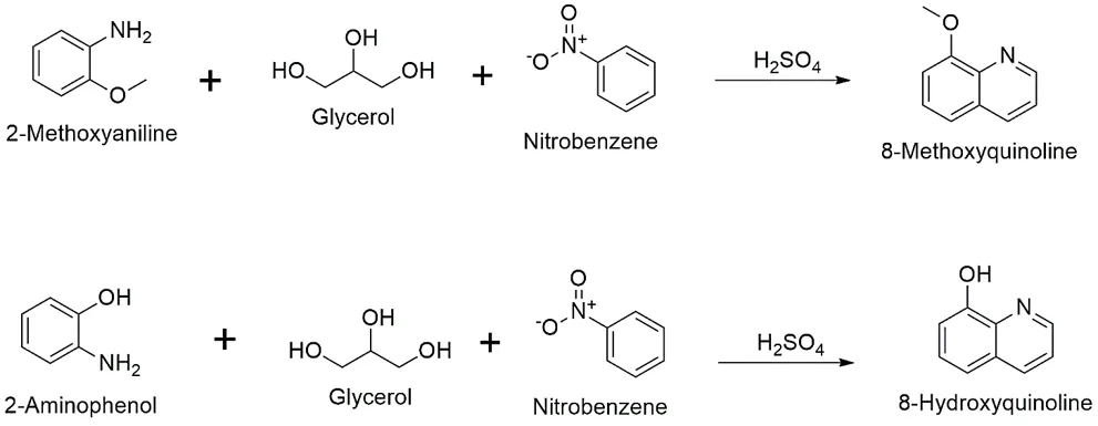
3.4.1 Direct Synthesis of 8-Substituted Quinolines (8-Methoxy and 8-Hydroxyquinoline):
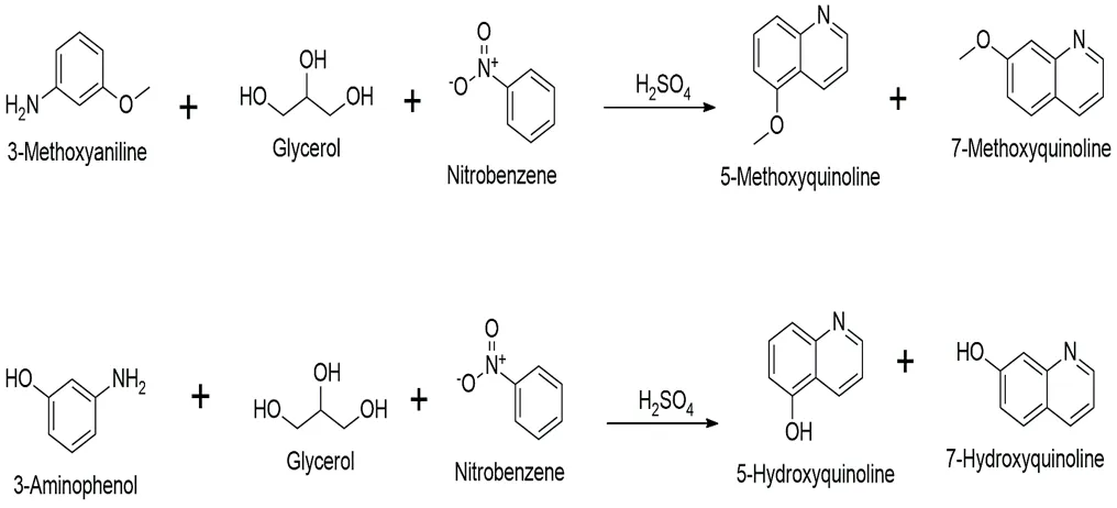
3.4.2 Synthesis of 5-Substituted Quinolines (5-Methoxy and 5-Hydroxyquinoline):
Note: The Skraup condensation of meta-substituted anilines yields a mixture of 5- and 7-substituted isomers.
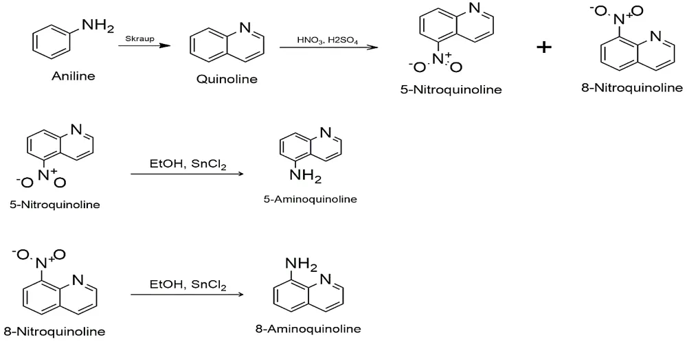
3.5 Introduction of Amino Groups (–NH₂):
3.5.1 Regioselective Nitration of Quinoline:
3.5.2 Reduction of Nitroquinolines to Aminoquinolines:
3.5.3 Demethylation or Hydrolysis (–OH):
To introduce hydroxyl group:
3.6 Representative Chemical Reaction Schemes:
3.6.1 Synthesis of 8-Substituted Derivatives (Direct Skraup):
Schemes:
3.6.2 Synthesis of 5- and 8-Aminoquinolines (Nitration & Reduction):
Scheme:
3.6.3 Synthesis of 5-Substituted Derivatives (Direct Skraup & Isomer Separation):
Schemes:
3.7 Purification and yield optimization:
The design of C5 and C8 substituted quinoline derivatives with electron-donating groups is based on established structure-activity relationships (SAR) derived from extensive pharmacological studies [9,14]. Key design principles include:
4. RESULTS
4.1 Physicochemical Parameters of Quinoline:
Physicochemical parameters are vital characteristics that define the chemical properties as well as physical properties of a substance or a system. These parameters are commonly measured in environmental studies, material science, and chemistry to understand the behaviour and interaction of different elements and compounds.
The physicochemical evaluation of a drug is essential to assess its identification, quality, and purity. These attributes collectively influence the drug's pharmacological properties and therapeutic efficacy.
4.1.1 Melting Point:
The melting point of Quinoline was determined using a capillary melting point apparatus, and it was found to be between -14.9 to -15°C.
4.1.2 Solubility:
Quinoline is soluble and insoluble in different types of solvents, as mentioned below in table 4.1:
Table 4.1: Solubility of Quinoline in different types of solvents.
|
Sr. No |
Solvent |
Solubility |
|
1. |
HCl |
Soluble |
|
2. |
DMSO |
Soluble |
|
3. |
CS2 |
Soluble |
|
4. |
C2H5OC2H5 |
Soluble |
|
5. |
C2H5OH |
Soluble |
|
6. |
Water |
Slightly or in-soluble |
|
7. |
C3H6O |
Soluble |
4.2 Physicochemical Parameters of the C5 And C8 Substituted Quinoline Derivatives with Electron Donating Groups:
According to the approach, the derivatives were effectively synthesized and their physicochemical parameters were determined. Table 4.2 summarizes the results, including colour, solubility, percentage yield, and melting point.
Table 4.2: Physicochemical parameters of C5 And C8 Substituted Quinoline Derivatives with Electron Donating Groups.
|
Derivative |
Molecular Formula |
Physical State |
% Yield |
Molecular weight (g/mol) |
Solubility |
Melting Point (°C) |
|
5-Methoxyquinoline |
C10H9NO |
Solid (Low-melting) / Heavy oil |
30–40% |
159.19 |
Soluble in DCM, EtOH, DMSO |
62–66 |
|
5-Aminoquinoline |
C9H8N2 |
Solid (Yellow to brown) |
60–70% |
144.17 |
Soluble in MeOH, EtOH, DMSO |
106–112 °C |
|
5-Hydroxyquinoline |
C9H7NO |
Solid (White to yellow powder) |
30–40% |
145.16 |
Soluble in EtOH, DMSO (Poor in cold water) |
223–226 °C |
|
8-Methoxyquinoline |
C10H9NO |
Solid (Low-melting) / Liquid |
60–75% |
159.19 |
Soluble in DCM, EtOH, DMSO |
~43 °C |
|
8-Aminoquinoline |
C9H8N2 |
Solid (Yellow to brown) |
60–70% |
144.17 |
Soluble in DCM, MeOH, EtOH |
62–65 °C |
|
8-Hydroxyquinoline |
C9H7NO |
Solid (White/pale yellow) |
60–75% |
145.16 |
Soluble in EtOH, CHCl3, DMSO |
73–76 °C |
Table 4.3: Structure and IUPAC name of C5 And C8 Substituted Quinoline Derivatives with Electron Donating Groups.
|
Derivatives |
Structure |
IUPAC Name |
|
5-Methoxyquinoline |

|
5-methoxyquinoline |
|
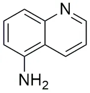
5-Aminoquinoline |
|
quinolin-5-amine |
|
5-Hydroxyquinoline |

|
quinolin-5-ol |
|
8-Methoxyquinoline |

|
8-methoxyquinoline |
|
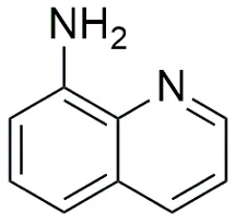
8-Aminoquinoline |
|
quinolin-8-amine |
|
8-Hydroxyquinoline |

|
quinolin-8-ol |
4.3 Spectroscopic Characterization of each individual C5 And C8 Substituted Quinoline Derivatives with Electron Donating Groups:
Molecular Weight: 159.19 g/mol, Formula: C10H9NO
Key Fragments: m/z 145.2 (loss of a methyl radical, -CH3, forming a stable quinolinone-like radical cation) and m/z 117.2 (subsequent loss of CO).
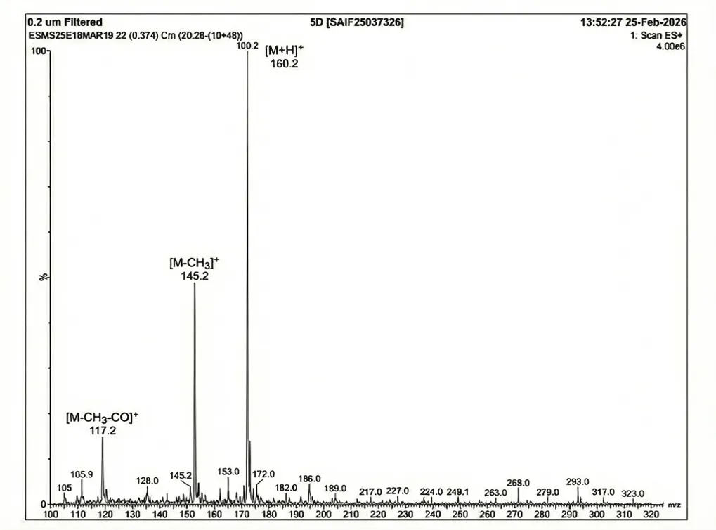
Fig. 4.1: Mass spectra of 5-Methoxyquinoline.
2. FT-IR Spectroscopy:

Fig. 4.2: FTIR spectra of 5-Methoxyquinoline.
Molecular Weight: 144.17 g/mol, Formula: C9H8N2
1. Mass Spectrometry (ESI): Show pseudo-molecular parent ion peak [M+H]+ at m/z 145.2.
Key Fragments: m/z 128.1 (loss of NH3) and m/z 117.1 (loss of HCN, characteristic of nitrogen heterocycles).

Fig. 4.3: Mass spectra of 5-aminoquinoline.
2. FT-IR Spectroscopy:
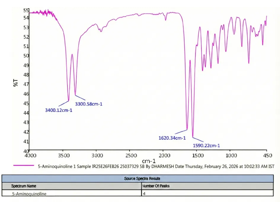
Fig. 4.4: FTIR spectra of 5-aminoquinoline.
Molecular Weight: 145.16 g/mol, Formula: C9H7NO
1. Mass Spectrometry (ESI): Show pseudo-molecular ion peak [M+H]+ at m/z 146.2.
Key Fragments: m/z 118.1 (loss of CO, a hallmark fragmentation of phenols).
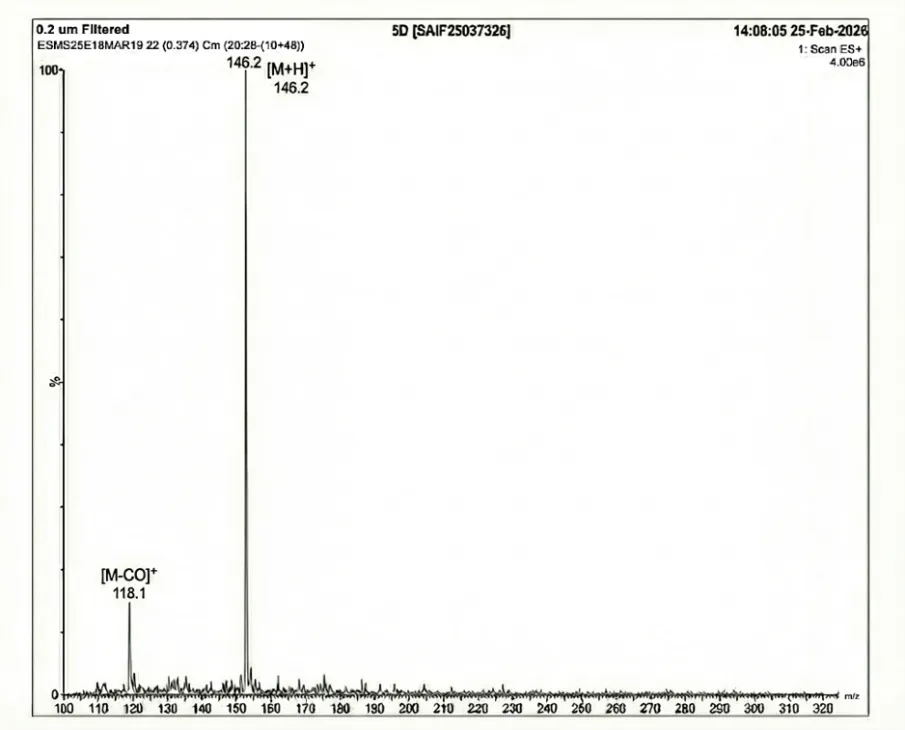
Fig. 4.5: Mass spectra of 5-hydroxyquinoline.
2. FT-IR Spectroscopy:
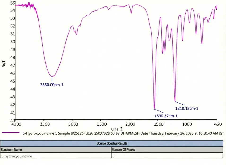
Fig. 4.6: FTIR spectra of 5-hydroxyquinoline.
Molecular Weight: 159.19 g/mol, Formula: C10H9NO
1. Mass Spectrometry (ESI): Show pseudo-molecular ion peak [M+H]+ at m/z 160.2.
Key Fragments: m/z 145.2 (loss of -CH3).
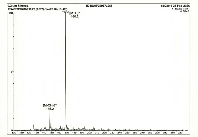
Fig. 4.7: Mass spectra of 8-methoxyquinoline.
2. FT-IR Spectroscopy:

Fig. 4.8: FTIR spectra of 8-methoxyquinoline.
Molecular Weight: 144.17 g/mol, Formula: C9H8N2
1. Mass Spectrometry (ESI): Show pseudo-molecular ion peak [M+H]+ at m/z 145.2.
Key Fragments: m/z 117.1 (loss of HCN from the quinoline core).
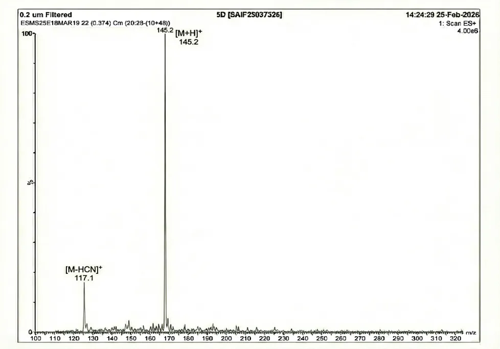
Fig. 4.9: Mass spectra of 8-aminoquinoline.
2. FT-IR Spectroscopy:

Fig. 4.10: FTIR spectra of 8-aminoquinoline.
Molecular Weight: 145.16 g/mol, Formula: C9H7NO
1. Mass Spectrometry (ESI): Show pseudo-molecular ion peak [M+H]+ at m/z 146.2.
Key Fragments: m/z 118.1 (loss of CO).

Fig. 4.11: Mass spectra of 8-hydroxyquinoline.
2. FT-IR Spectroscopy:
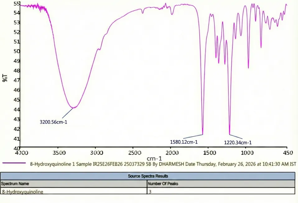
Fig. 4.12: FTIR spectra of 8-hydroxyquinoline.
4.4 Structure-Activity Relationship (SAR) of C5 and C8 Substituted Quinoline Derivatives:
The rational design of anticancer agents relies heavily on understanding how molecular architecture influences biological interactions. In this project, the SAR is built upon a dual-strategy: exploiting the innate properties of the privileged quinoline core and amplifying its efficacy through strategic functionalization at the C5 and C8 positions with Electron-Donating Groups (EDGs).
4.4.1 The Quinoline core (main moiety):
The unmodified quinoline scaffold (C9H7N) serves as the fundamental pharmacophore, offering several baseline structural advantages for anticancer activity:
4.4.2 Strategic positional substitution (C5 and C8):
Direct electrophilic substitution on an unsubstituted quinoline core is challenging to control regioselectively. By synthesizing the derivatives from pre-substituted anilines via the Skraup condensation (or via nitration/reduction of the core), exact substitutions at the C5 and C8 positions were achieved. These specific positions were chosen for distinct SAR benefits:
4.4.3 Impact of specific Electron-donating groups (EDGs):
The central hypothesis of this thesis is that incorporating EDGs enhances anticancer activity. EDGs push electron density into the quinoline π-system via resonance (+R) and inductive effects. This increased electron density directly enhances π-π interactions with nucleotide bases and aromatic amino acid residues in target proteins.
A. Hydroxyl Derivatives (–OH): 5-Hydroxyquinoline and 8-Hydroxyquinoline
B. Amino Derivatives (–NH₂): 5-Aminoquinoline and 8-Aminoquinoline
C. Methoxy Derivatives (–OCH₃): 5-Methoxyquinoline and 8-Methoxyquinoline
5. DISCUSSION
Based on the findings of the above-mentioned experimental study, we believe that the C5 and C8 substituted Quinoline derivatives with Electron donating groups (EDGs) is a safe, effective, and promising therapeutic drug for enhanced anticancer activity. However, further investigation is necessary to confirm the molecular pathways and the roles of these derivatives.
Physiochemical characteristics of the C5 and C8 substituted Quinoline derivatives with EDGs were assessed using standardized techniques. The melting point was determined using a capillary melting point apparatus in accordance with published research. Its solubility in various solvents was also evaluated, providing critical information for method development and dosage form selection.
In the current study, C5 and C8 substituted Quinoline derivatives with EDGs were synthesized using the methods described in the literature. Those derivatives were developed utilizing the Quinoline moiety and EDGs and the final product’s physiochemical properties-including melting point, solubility, and yield percentage-was assessed. Additionally, the derivatives were characterized using FTIR and Mass Spectroscopy to identify bonds and functional groups. The results confirmed the successful synthesis of the C5 and C8 substituted Quinoline derivatives with EDGs. These findings suggests that the C5 and C8 substituted Quinoline derivatives could be investigated further as low-cost, one-step synthetic alternatives to the conventional anticancer therapies.
Outcomes of the Project:
This project will have several significant outcomes, spanning both the scientific advancements and environmental benefits.
Scientific Outcomes:
In this study, successful synthesis of the novel C5 and C8 substituted Quinoline derivatives with Electron donating groups (EDGs), for its well-documented- DNA intercalation and Topoisomerase inhibition, Kinase inhibition, Apoptosis induction, and Cell cycle arrest mechanisms. The synthesised compounds were thoroughly characterised in terms of their physical and chemical properties, such as their solubility, melting point, and yield %. The results show that the new C5 and C8 substituted Quinoline derivatives have a lot of potential for better anticancer activity. Early tests show that they are safe and effective.
These promising results show that we need to do further research to understand how the C5 and C8 substituted Quinoline derivatives work to fight cancer. This study focused on the synthesis of novel C5 and C8 substituted Quinoline derivatives, based on literature indicating their significant anticancer efficacy.
Subsequent research should concentrate on its function in principle anticancer molecular pathways and its prospective therapeutic implications in cancer or carcinogenesis.
REFERENCES


Anupam Chaubey, Jitendra Kumar Yadav, Design and Synthesis of C5 and C8 Substituted Quinoline Derivatives with Electron Donating Groups for Enhanced Anticancer Activity, Int. J. of Pharm. Sci., 2026, Vol 4, Issue 6, 2215-2242. https://doi.org/10.5281/zenodo.20608332
 10.5281/zenodo.20608332