

We use cookies to ensure our website works properly and to personalise your experience. Cookies policy
Department of Pharmaceutical Sciences, JNTUH University College of Pharmaceutical Sciences, Sultanpur, Sangareddy-502001, Telangana, India
The present study was undertaken to develop and evaluate a niosomal film-forming topical lotion incorporating ethanolic extract of Murraya koenigii (curry leaves) for enhanced anti-inflammatory activity. Murraya koenigii is a medicinal plant rich in bioactive phytoconstituents, including alkaloids, flavonoids, phenols, tannins, and other secondary metabolites with reported anti-inflammatory and antioxidant properties. The leaves were extracted using ethanol by Soxhlet extraction following petroleum ether defatting. The extract was incorporated into niosomes using the thin-film hydration method, employing Tween 80 and stearic acid as formulation components. A 2² factorial design was applied to optimize the niosomal formulation based on selected formulation variables. The optimized niosomal dispersion was subsequently incorporated into a film-forming lotion containing hydroxypropyl methylcellulose, ethyl cellulose, glycerine, sodium chloride, and a menthol–camphor eutectic mixture. The prepared formulations were evaluated for physicochemical characteristics, pH, spreadability, viscosity, phase separation, greasiness, stability, film formation, flexibility, drying time, stickiness, and water vapour permeability. The in vitro anti-inflammatory activity was assessed using the heat-induced protein denaturation assay, with diclofenac sodium used as the standard. Phytochemical screening of the ethanolic extract confirmed the presence of carbohydrates, alkaloids, phenols, tannins, flavonoids, saponins, amino acids, sterols, phytosterols, anthocyanins, and reducing sugars. Among the formulations, F3 demonstrated superior overall physicochemical and film-forming characteristics, including a viscosity of 1300–1500 cP, satisfactory spreadability, good film formation, flexibility, and stability. The final formulation demonstrated concentration-dependent inhibition of protein denaturation, increasing from 12.68 ± 0.5% at 100 µg/mL to 85.90 ± 4.8% at 500 µg/mL, compared with 75.20 ± 4.1% for the crude extract and 99.60 ± 5.3% for diclofenac sodium at the same concentration. The findings suggest that incorporation of Murraya koenigii extract into a niosomal film-forming system can enhance its in vitro anti-inflammatory activity and provide a promising approach for topical delivery. Further in vivo, long-term stability, and clinical studies are warranted to establish its therapeutic potential.
Since ancient times, medicinal plants have played a vital role in the treatment and prevention of various diseases. Natural products derived from plants continue to serve as an important source of therapeutic agents due to their wide pharmacological activities and relatively lower side effects compared to synthetic drugs. In recent years, there has been a growing interest in the development of plant-based formulations with improved efficacy, safety, and patient compliance.
Inflammation is a complex biological response of body tissues to harmful stimuli such as pathogens, damaged cells, or irritants. Although it is a protective mechanism, chronic inflammation is associated with several pathological conditions including rheumatoid arthritis, osteoarthritis, psoriasis, and inflammatory bowel diseases. Conventional therapies such as non-steroidal anti-inflammatory drugs (NSAIDs), corticosteroids, and disease-modifying antirheumatic drugs (DMARDs) are widely used for the management of inflammation. However, prolonged use of these agents is often associated with adverse effects such as gastrointestinal irritation, cardiovascular complications, hepatotoxicity, and immunosuppression. These limitations highlight the need for safer and more effective alternatives, particularly from natural sources.[1]
Murraya koenigii (family: Rutaceae), commonly known as curry leaf, is a well-known medicinal plant widely used in traditional Indian medicine. It is rich in bioactive constituents such as carbazole alkaloids, flavonoids, glycosides, and essential oils, which contribute to its diverse pharmacological activities including antioxidant, antimicrobial, antidiabetic, analgesic, and anti-inflammatory effects. [2]
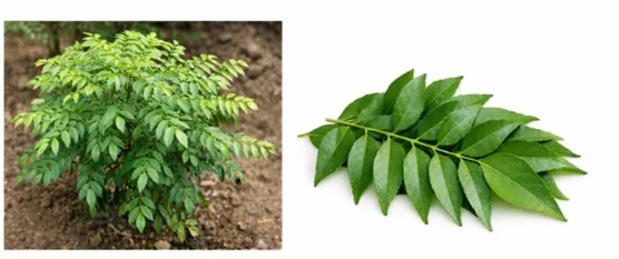
Figure 1: Murraya koenigii (Curry leaf plant and leaves)
Previous studies have reported that the anti-inflammatory potential of Murraya koenigii is primarily attributed to the presence of carbazole alka loids such as mahanimbine and koenigine, which exhibit significant free radical scavenging and nitric oxide inhibitory activities. Despite its promising therapeutic potential, the clinical application of herbal extracts is often limited due to poor solubility, low bioavailability, and instability. To overcome these challenges, novel drug delivery systems such as niosomes have been explored. Niosomes are non-ionic surfactant-based vesicular systems capable of encapsulating both hydrophilic and lipophilic drugs, thereby enhancing drug stability, bioavailability, and targeted delivery. Additionally, niosomes improve transdermal drug permeation and provide controlled drug release, making them suitable carriers for topical drug delivery. [3,4]
In recent years, film-forming systems (FFS) have emerged as an innovative approach for topical and transdermal drug delivery. These systems form a thin, transparent film upon application to the skin, which enhances drug residence time, improves penetration, and provides sustained drug release.
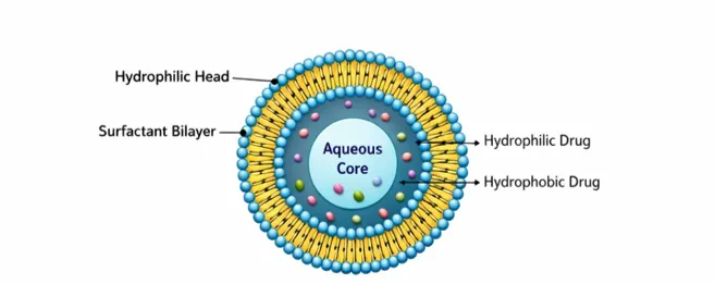
Figure 2: Structure of niosomal vesicle showing hydrophilic and hydrophobic regions
Compared to conventional topical formulations such as creams and ointments, FFS offer advantages including non-greasy nature, better patient compliance, and improved aesthetic acceptability.
Furthermore, the application of factorial design in formulation development allows systematic optimization of formulation variables and their interactions, thereby enhancing the efficiency and reliability of the formulation process. A 2² factorial design is particularly useful in evaluating the influence of formulation parameters on critical quality attributes such as entrapment efficiency, particle size, and drug release behavior.[5]
Therefore, the present study aims to develop and evaluate a niosomal film-forming lotion containing Murraya koenigii extract for enhanced topical anti-inflammatory activity. The formulation is optimized using factorial design and evaluated for its physicochemical properties and in vitro anti-inflammatory activity to establish its potential as an effective and safer alternative to conventional therapies.[6]
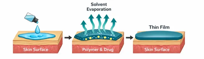
Figure 3: Mechanism of film formation after topical application
2. MATERIALS AND METHODS
2.1 Ethanolic Extraction of Murraya koenigii Using Soxhlet Extraction Method
2.1.1 Plant Material
The leaves of Murraya koenigii (Spreng.) were collected during the summer season (April 2025) from local areas of Sultanpur, Telangana, and were authenticated by the Department of Pharmacognosy, JNTUH. The collected leaves were thoroughly washed 1–2 times with water to remove adhering impurities and subsequently shade dried at room temperature. After complete drying, the leaves were coarsely powdered using a mechanical grinder (mortar and pestle) and passed through sieve No. 40 to obtain a uniform particle size. The powdered material was stored in a sterile, airtight container at room temperature until further use.[7,8]
2.1.2 Preparation of Plant Extract
The powdered plant material (25–30 g) was accurately weighed and loaded into a thimble, which was placed in the Soxhlet apparatus. Initially, the material was subjected to defatting using petroleum ether for 4–6 hours. After completion of defatting, the marc was dried to remove any residual solvent. The dried plant material was then subjected to extraction with ethanol for 6–8 hours. The obtained extract was concentrated using a rotary evaporator or water bath, and the concentrated crude extract was stored at 4°C for further use. [9]
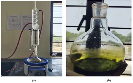
Figure 4: (a) Soxhlet extraction of Murraya koenigii (curry leaves) using ethanol as solvent; (b) Concentrated extract of Murraya koenigii after evaporation showing greenish-black appearance.
2.2 Formulation of Herbal Niosomes Using Thin Film Hydration Method
2.2.1 Thin Film Hydration Technique
Niosomes were prepared using the thin film hydration technique with varying ratios of surfactant (Tween 80) and stearic acid. The surfactant and stearic acid were dissolved in 8 mL of chloroform, while the plant extract was dissolved separately in 2 mL of ethanol. Both solutions were combined in a round-bottom flask and subjected to magnetic stirring. The solvent was evaporated to form a thin lipid film on the inner wall of the flask. This thin film was subsequently hydrated using 10 mL of phosphate-buffered saline (pH 7.4) for 1 hour at a temperature range of 55–65°C with continuous rotation. The resulting hydrated dispersion was further sonicated for 20 minutes to obtain a uniform niosomal dispersion. [10,11]
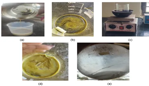
Figure 5: Stepwise preparation of curry leaves–incorporated niosomal formulation using the thin film hydration method: (a) weighing and mixing of curry leaves extract and lipids, (b) dissolution of components in organic solvents, (c) magnetic stirring for thin film formation, (d) thin film appearance after solvent evaporation, and (e) hydration of thin film to form niosomes.
2.2.2 Factorial Design for Niosomal Dispersion
A 2² factorial design was employed to evaluate the effect of formulation variables on the characteristics of niosomal dispersion. The independent variables selected were Factor A: stearic acid concentration (250 mg–500 mg) and Factor B: Tween 80 concentration (300 mg–750 mg). The dependent variables studied included particle size, entrapment efficiency, film-forming capacity, spreadability, and in vitro anti-inflammatory activity. Based on different combinations of these factors, four formulations (F1–F4) were prepared. [12]
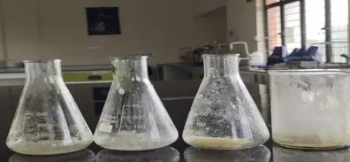
Figure 6: Four different formulations (F1-F4) of curry leaves loaded niosomes.
2.3 Formulation of Film Forming Lotion
The film-forming lotion was prepared using the dispersion method. Ethyl cellulose (1.5 g) was dispersed in 20 mL of ethanol, while hydroxypropyl methylcellulose (HPMC) (3 g) was dispersed separately in 40 mL of water. Both polymeric solutions were allowed to swell for 24 hours to obtain clear solutions. A eutectic mixture was prepared by mixing menthol (0.5 g) and camphor (0.5 g). Subsequently, 10 mL of niosomal dispersion was dissolved in 5 mL of ethanol, followed by the addition of glycerine (5 mL), sodium chloride (0.5 g), and the eutectic mixture (1 g). The prepared polymeric solutions were then incorporated into the mixture and stirred thoroughly to achieve homogeneity. The final volume was adjusted to 100 mL using ethanol, and the formulation was homogenized for 10–15 minutes. The prepared lotion was stored in amber-colored glass bottles at room temperature. [13]
Table 1: Composition of film forming lotion.
|
SR. NO. |
Components |
Activity |
Type of formulation |
|||
|
F1 |
F2 |
F3 |
F4 |
|||
|
1. |
Plant extract loaded Niosomal |
API or herbal extract |
10ml |
10ml |
10ml |
10ml |
|
2. |
Hydroxypropyl methylcellulose (HPMC) |
Polymers |
1g (Low) |
3g (High) |
3g (High) |
1g (Low) |
|
3. |
Ethyl cellulose |
1.5g (High) |
0.5g (Low) |
1.5g (High) |
0.5g (Low) |
|
|
4. |
Ethanol |
Solvent |
q.s. to 100ml |
q.s. to 100ml |
q.s. to 100ml |
q.s. to 100ml |
|
5. |
Glycerine |
Plasticizer, Humectant |
5ml |
5ml |
5ml |
5ml |
|
6. |
Sodium chloride (NaCl) |
Crosslinker |
0.5g |
0.5g |
0.5g |
0.5g |
|
7. |
Menthol and camphor (Eutectic mixture) |
Permeation Enhancer |
1:1 |
1:1 |
1:1 |
1:1 |
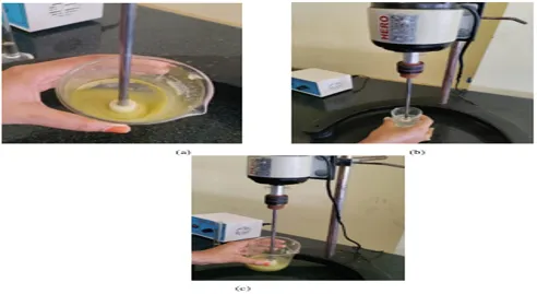
Figure 7: Steps wise preparation of film forming system.(a) Mixing of plant extract, penetration enhancers, cross linker, and plasticizer. (b) Mixing of polymeric solutions and (c) Mixing of both the solutions.
2.3.1 Factorial Design for Film Forming System
A 2² factorial design was applied to evaluate the influence of formulation variables on the film-forming system. The independent variables were Factor A: HPMC concentration (1%–3%) and Factor B: ethyl cellulose concentration (0.5%–1.5%). The dependent variables evaluated included film-forming capacity, viscosity, spreadability, drying time, drug release profile, and anti-inflammatory activity. [15]
2.4 Evaluation of Formulated Film Forming Lotion Incorporated with Ethanolic Extract of Murraya koenigii
The formulated film-forming lotion containing the ethanolic extract of Murraya koenigii was subjected to systematic evaluation to assess its quality, stability, and suitability for topical application. This evaluation included physicochemical characterization, assessment of film properties after application, and evaluation of therapeutic efficacy, particularly anti-inflammatory activity. Two categories of evaluation were performed: physicochemical evaluation and evaluation of film formation and film properties.
2.4.1 Physicochemical Evaluation
The formulation was evaluated for appearance, clarity, colour, odour, and texture through visual and organoleptic examination.[16]
Appearance, clarity, and color evaluation
The pH of the formulation was measured using pH paper or a digital pH meter.
Spreadability was determined using the slide method by placing 1 g of formulation between two glass slides and applying a standard weight. The time required for separation of slides was recorded. Spreadability was calculated using:
S=m×lt
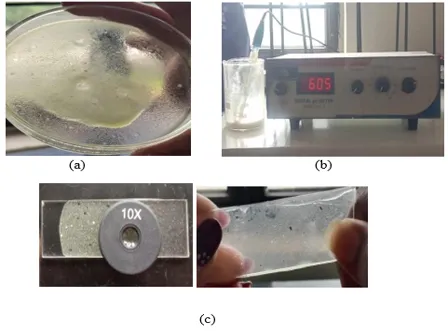
Figure 8: Appearance, clarity and colour. ,Measurement of PH of FFS, Measurement of Spreadability of FFS.
Additional tests included irritancy test (24 h observation), removal test, stability test at 37°C for 72 hours, viscosity measurement using Ostwald viscometer, phase separation analysis, and greasiness evaluation.[17]
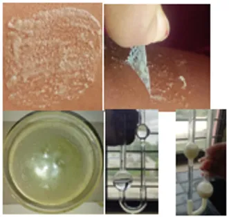
Figure 9: Visual observation of Irritancy test, Visual observation of Removal Test , Visual observation of Stability Test, Visual observation of Viscosity Test.
2.4.2 Evaluation of Film Properties (Post Application)
Film formation was evaluated using a petri dish or pig ear skin and categorized based on uniformity and appearance. Film flexibility was assessed by stretching the film in multiple directions to check cracking. Drying time was evaluated by applying the formulation to the forearm and checking dryness using a glass slide. Stickiness was tested using cotton fibers and graded accordingly. Water vapor permeability was determined using the desiccator method, where weight loss was measured over time and correlated with surface area and duration.[18]
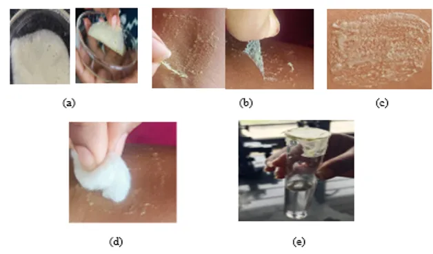
Figure 10: Visual representation of film formation,(b)Visual representation of film flexibility. (c) Visual representation of film drying. (d)Visual representation of Stickiness. (e)Visual representation of Water vapour permeability.
2.5 In Vitro Anti-inflammatory Activity (Protein Denaturation Assay)
The anti-inflammatory activity was evaluated using the protein denaturation assay, based on the inhibition of heat-induced protein denaturation.
Procedure
A reaction mixture containing 0.45 mL of 1% albumin solution, 0.45 mL of test sample, and 0.05 mL of 1N HCl (pH ~6.3) was prepared. The control contained PBS instead of the sample, while the standard contained diclofenac sodium. The mixtures were incubated at 37°C for 20 minutes, followed by heating at 51°C for another 20 minutes. After cooling to room temperature, absorbance was measured at 660 nm using a UV-Visible spectrophotometer.[19]
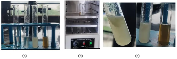
Figure 11: (a)Test Reaction Mixtures. (b)Incubation of the test reaction mixtures,(c) Observation of denaturation in control, No observation of denaturation in final lotion(ffs) and plant extract.
Calculation
The percentage inhibition of protein denaturation was calculated using:
% Inhibition=Ac-AtAc×100

Where:
Ac = Absorbance of control
At = Absorbance of test sample
3. RESULTS AND DISCUSSION
3.1 Phytochemical Screening of Murraya koenigii
The ethanolic extract of Murraya koenigii was evaluated for its physical characteristics and phytochemical constituents. The extract appeared green to dark green in colour with a strong characteristic aromatic odour due to the presence of volatile oils. It exhibited a slightly bitter and pungent taste with a coarse powder texture.[20]
Solubility studies showed that the extract was partially soluble in water, leaving behind fibrous material. It was more soluble in ethanol and methanol, indicating the presence of polar compounds such as flavonoids and phenols. Limited solubility in petroleum ether confirmed the presence of lipophilic substances like oils and waxes.
Phytochemical screening (Table 12) confirmed the presence of carbohydrates, alkaloids, phenols, tannins, flavonoids, saponins, amino acids, sterols, phytosterols, anthocyanins, and reducing sugars. These bioactive compounds are known for their anti-inflammatory and antioxidant properties, supporting the therapeutic potential of Murraya koenigii.[21,22]
Table 12: Phytochemical screening of murraya koenigii.
|
Sr. No. |
Chemical test |
Procedure |
Observation |
Inference |
|
1. |
Molisch’s test |
2mL plant extract + 2 drops of 5% alcoholic α–naphthol + 1mL conc. H2SO4 (along the sides of test tube) |
A violet ring 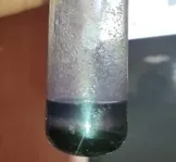
|
Presence of carbohydrates. |
|
2. |
Mayer’s test |
2ml plant+ 1-2 drops of Mayer’s reagent (Along the sides of test tube)
|
A creamy white/yellow precipitate 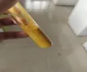
|
Presence of alkaloids. |
|
3. |
Ferric chloride test
|
Extract aqueous solution + few drops 5% ferric chloride sol.
|
Dark green/deep blue 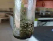
|
Presence of phenols. |
|
4. |
Liebermann- Burchard Test |
2 ml of plant extract+ 12 ml acetic anhydride + 2 drops of concentrated H2SO4 (from the side of the test tube). |
Bluish green colour ppt. 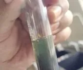
|
Presence of phytosterols. |
|
5. |
Alkaline reagent test
|
Add 2ml of NAOH to 2ml of extract then add dilute HCL drop by drop |
A green or blue ppt 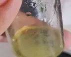
|
Presence of flavonoids. |
|
6. |
Ferric chloride test (for tannins) |
Add 1-2 drops of 5% ferric chloride solution to 2ml of extract |
Blackish blue colour 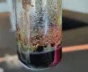
|
Presence of Tannins |
|
7. |
Saponins test |
2ml solution of extract + diluted with distilled water to 20 ml and shaken in a graduated cylinder for 15 minutes |
stable foam is observed 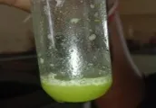
|
Presence of saponins. |
|
8. |
Ninhydrin test
|
2mL plant extract+ 2 drops of Ninhydrin solution(10mg ninhydrin + 200mL acetone).Boil for 510mins. |
A purple Biuret test. 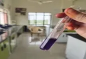
|
Presence of amino acids. |
|
9. |
Biuret test |
Add 1 ml of 40% NAOH and 2 drops of 1% CuSO4 solution to 2ml extract. Mix and observe.
|
Appearance of a violet or purple colour 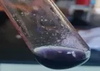
|
Presence of amino acids. |
|
10. |
Salkowski’s Test
|
2–3 mL of the ethanolic curry leaf extract + 2 mL of chloroform + Carefully add 2 mL of concentrated sulfuric acid along the side of the test tube(using a pipette) to form a layer at the bottom. |
Reddish brown colour at the interface. 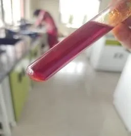
|
Presence of sterols. |
|
11. |
Wagner’s Test |
2–3 mL of the curry leaf ethanolic extract + add 1–2 mL of Wagner’s reagent dropwise. Shake gently and allow the mixture to stand for a few minutes. |
Reddish-brown or brown precipitate 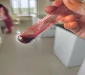
|
Presence of alkaloids. |
|
12. |
Seliwanoff’s Test
|
1mL extract solution + 2ml seliwanoff’s reagent + heated on water bath for 2-5min.
|
Pink/Red{ketoses} 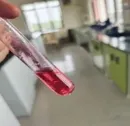
|
Presence of carbohydrates. |
|
13. |
HCl test
|
2mL plant extract + 2mL 2N HCl (+ Few mL ammonia)
|
Pink-red sol. which turns blue-violet after addition of ammonia 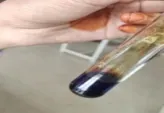
|
Presence of anthocyanins. |
|
14. |
Fehling’s test
|
1mL each of Fehling’s solution A & B + 2mL plant extract &boiled in water bath.
|
Brick red precipitat 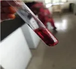
|
Presence of reducing sugars. |
3.2 Physicochemical Evaluation
All formulations (F1–F4) showed a homogenous appearance, indicating proper mixing and uniformity. The colour ranged from light greenish-yellow to faint yellow due to the herbal extract. All formulations had a fresh, mild minty odour because of menthol and camphor.
The pH of the formulations was found between 6.0 and 6.6, which is suitable for skin application and indicates that the formulations are non-irritating.
Spreadability results showed that F4 had the highest spreadability (8–9 mm), indicating easy application, while F2 showed lower spreadability (5–6 mm). All formulations showed no irritation during the irritancy test, confirming their safety.
In the removal test, F2, F3, and F4 were easily removed, whereas F1 showed slight cracking. Stability studies indicated that F3 was the most stable formulation with no changes, while minor changes were observed in other formulations.
Viscosity results showed that F3 had the highest viscosity (1300–1500 cp), which is beneficial for film formation, while F4 had the lowest viscosity (650–750 cp). Phase separation was not observed in F2 and F3, but mild separation was seen in F1 and F4. All formulations were non-greasy, which improves patient acceptability.
Table 13: Evaluations and observations of formulations F1-F4.
|
SR. NO |
TEST |
OBSERVATION |
|||
|
F1 |
F2 |
F3 |
F4 |
||
|
1. |
Appearance/ Homogeneity |
Homogenous Lotion. |
Homogenous Lotion. |
Homogenous Lotion. |
Homogenous Lotion. |
|
2. |
Colour |
Light Greenish yellow |
Faint yellow |
Greenish yellow |
Light Greenish yellow |
|
3. |
Odour |
Fresh, mild minty |
Fresh, mild minty |
Fresh, mild minty |
Fresh, mild minty |
|
4. |
PH |
6.4-6.6 |
6.0-6.2 |
6.05-6.1 |
6.3-6.5 |
|
5. |
Spreadability (mm) |
7-8mm |
5-6mm |
6-7mm |
8-9mm |
|
6. |
Irritancy test |
Nil |
Nil |
Nil |
Nil |
|
7. |
Removal Test |
Cracking is observed while removing from skin by applying water/ peeling |
Easily removed from skin by applying water/ peeling |
Easily removed from skin with slight effort |
Easily removed from skin by applying water/ peeling |
|
8. |
Stability Test (after 3weeks) |
Slight thickening |
Slight colour change |
No changes |
Slight sedimentation. |
|
9. |
Viscosity(cp) |
900-1000cp |
1100-1300cp |
1300-1500cp |
650-750cp |
|
10. |
Phase separation |
Mild separation |
No phase separation |
No phase separation |
Mild separation |
|
11. |
Greasiness |
Non-greasy |
Non-greasy |
Non-greasy |
Non-greasy |
3.2.2 Evaluation of Film Properties
Film formation studies showed that F1 formed a non-uniform film, while F2, F3, and F4 formed uniform films. This indicates better formulation performance in F2–F4.
Film flexibility testing showed that F2 was highly flexible, F3 was flexible and strong, and F4 was flexible, whereas F1 was brittle.
Drying time results indicated that F4 dried faster (3–4 minutes), making it more convenient for use. F2 and F3 required more time (5–7 minutes).
Stickiness evaluation showed that F2 had high stickiness, F3 moderate stickiness, and F1 and F4 low stickiness, making F4 more comfortable for use.
Water vapour permeability results showed that F2 had very high permeability due to higher HPMC content, while F1 had low permeability due to higher ethyl cellulose content. F3 showed moderate permeability, indicating a balanced formulation.[23]
Overall, F3 and F4 showed better performance compared to other formulations.
Table14: Post application observations of FFS.
|
SR. NO. |
Test performed |
Observed results |
|||
|
F1 |
F2 |
F3 |
F4 |
||
|
1. |
Film formation |
Non- Uniform film |
Uniform film |
Uniform film |
Uniform film |
|
2. |
Film flexibility |
Brittle |
Highly Flexible |
Flexible and strong |
Flexible |
|
3. |
Drying time |
Moderate (4-5min) |
Slow (6-7min) |
Slow (5-6min) |
Fast (3-4min) |
|
4. |
Stickiness |
Low |
High |
Moderate |
Low |
|
5. |
Water vapour permeability |
Low (high EC content) |
Very High (More HPMC content) |
Moderate (balanced EC and HPMC) |
High (less EC barrier) |
3.3 In Vitro Anti-inflammatory Activity
The anti-inflammatory activity was evaluated using the protein denaturation method. The results showed that both the curry leaf extract and the formulated lotion exhibited concentration-dependent activity.
At 100 µg/mL, the extract showed 10.52% inhibition, while the lotion showed 12.68%, which is close to the standard drug diclofenac sodium (14.77%). As the concentration increased, the activity also increased.
At 500 µg/mL, the extract showed 75.20% inhibition, while the lotion showed 85.90%, which is very close to the standard (99.60%).
The higher activity of the lotion compared to the extract indicates improved effectiveness due to the niosomal formulation and better drug delivery.
These results confirm that the formulation has significant anti-inflammatory activity and can be effectively used for topical application.
Table 15: % Inhibition of protein denaturation at 660nm.
|
SR. NO. |
Concentration (µg/mL) |
Curry Leaf Extract |
Final Lotion |
Diclofenac Sodium (Std) |
|
1. |
100 |
10.52 ± 0.4 |
12.68 ± 0.5 |
14.77 ± 0.6 |
|
2. |
200 |
25.32 ± 1.8 |
30.15 ± 2.2 |
32.78±2.5 |
|
3. |
400 |
58.62 ± 3.7 |
67.39 ± 3.9 |
75.43±4.3 |
|
4. |
500 |
75.20 ± 4.1 |
85.90 ± 4.8 |
99.60±5.3 |
4. CONCLUSION
The present study successfully developed and optimized a niosomal film-forming topical lotion incorporating the ethanolic extract of Murraya koenigii for enhanced anti-inflammatory activity. The use of niosomal vesicles significantly improved the stability, encapsulation, and potential skin permeation of the bioactive phytoconstituents. In addition, the film-forming system provided better skin adherence and sustained drug release, which are essential for effective topical therapy.
The optimized formulation exhibited suitable physicochemical properties, including appropriate pH, viscosity, and spreadability, confirming its compatibility for dermal application. The in vitro anti-inflammatory activity demonstrated a concentration-dependent inhibition of protein denaturation, with the formulated lotion showing higher activity compared to the crude extract and comparable results with the standard drug.
Overall, the study highlights the potential of this herbal niosomal film-forming system as an effective and safer alternative to conventional topical anti-inflammatory formulations. However, further in vivo studies, stability evaluations, and clinical investigations are required to confirm its therapeutic efficacy and support its future development and commercialization.
REFERENCES

Dr. R. Aparanjitha, Dr. M. Sunitha Reddy, Development and Evaluation of Niosomal Film-Forming Lotion of Murraya Koenigii for Enhanced Topical Anti-Inflammatory Activity, Int. J. of Pharm. Sci., 2026, Vol 4, Issue 9, 2838-2851. https://doi.org/10.5281/zenodo.22913206
 10.5281/zenodo.22913206