
We use cookies to ensure our website works properly and to personalise your experience. Cookies policy
University Department of Chemical Technology, Dr. Babasaheb Ambedkar Marathwada University, Chhatrapati Sambhajinagar 431004.
Picroside II (PK-II), a bioactive compound obtained from Picrorhiza kurroa shows hepatoprotective, antioxidant, and neuroprotective properties but suffers from low oral bioavailability, limiting its therapeutic application. This study aimed to develop and optimize a proliposomes formulation of PK-II using a Quality by Design (QbD) approach to enhance its solubility, stability and ultimately the bioavailability. Proliposomes were prepared by varying phosphatidylcholine-to-cholesterol and phosphatidylcholine-to-carrier ratios, and the formulations were systematically optimized using Central Composite Design (CCD). The optimized formulation, with a phosphatidylcholine to cholesterol ratio of 1:1.7071 and a phosphatidylcholine to carrier ratio of 1:10, exhibited a particle size of 104.4 nm, zeta potential of ±20.2 mV, and entrapment efficiency of 35.24%, confirming the formation of nanosized, stable, and reproducible proliposomes. SEM analysis revealed porous mannitol matrices coated with lipid in the dry powder, and spherical, uniform liposomes upon hydration. FTIR and DSC studies confirmed chemical compatibility and molecular dispersion of PK-II within the lipid matrix. The optimized proliposomes were stable at 4°C and room temperature for one month, with no significant changes in entrapment efficiency or morphology. Overall, the developed proliposomes system demonstrates potential to enhance solubility, stability, and bioavailability of PK-II, suggesting its suitability as an effective oral drug delivery platform
Picrorhiza kurroa Royle ex Benth, commonly known as Kutki, is a perennial, rhizomatous medicinal herb belonging to the family Plantaginaceae. It is predominantly found in the alpine and subalpine regions of the Himalayas, particularly in India, Nepal, and Tibet, at altitudes ranging from 3,000 to 5,000 meters. Traditionally, P. kurroa has been extensively used in Ayurveda, Unani, and Tibetan medicine for the management of liver disorders, fever, respiratory ailments, and inflammatory conditions. The therapeutic potential of the plant is attributed mainly to its iridoid glycosides, of which Picroside II (PK-II) is one of the key bioactive constituents. Picroside II is a hydrophilic iridoid glycoside characterized by limited solubility and susceptibility to hydrolytic degradation, all of which contribute to its inherently low oral bioavailability. Despite these limitations, PK-II is well recognized for its broad pharmacological activities including hepato-protective, antioxidant, anti-inflammatory, neuroprotective, and nephron-protective effects. These diverse therapeutic benefits highlight its potential in the treatment of liver diseases, oxidative stress-mediated disorders, and neurodegenerative conditions. However, the clinical translation of PK-II remains limited, largely due to its unfavourable physicochemical properties and poor pharmacokinetic profile, emphasizing the need for an advanced drug delivery strategy. Various formulations, such as PLA nanoparticles [6], palmitic acid–modified nanoparticles [7], and iridoid-rich fractions [8], have been developed to enhance the bioavailability of PK-II; however, the proliposomes approach for improving its bioavailability has not yet been explored, and therefore PK-II proliposomes formulations were developed and investigated in the present study. In recent years, proliposomes have emerged as a promising nanocarrier system within the domain of novel drug delivery systems (NDDS). Proliposomes are dry, free-flowing formulations that spontaneously form liposomes upon hydration. They offer several advantages over conventional liposomes, including enhanced stability, improved entrapment efficiency, ease of reconstitution, better scalability, and superior handling characteristics. These attributes make proliposomes highly suitable for delivering drugs with solubility and permeability challenges, such as PK-II. By improving membrane permeability, protecting the drug from degradation, and facilitating controlled release, proliposomes can significantly enhance the overall bioavailability of the compounds. To ensure systematic development and robustness of the formulation, the Quality by Design (QbD) approach has become increasingly relevant in pharmaceutical research. QbD enables a comprehensive understanding of formulation variables, identification of critical material attributes (CMAs) and critical process parameters (CPPs), and optimization of the formulation through statistical design tools. Incorporating QbD in proliposomes development of PK-II allows rational selection of lipid composition, cholesterol ratio, and carrier quantity to achieve optimal entrapment efficiency and performance. Considering these aspects, the present study aims to develop and optimize a PK-II-loaded proliposomes formulation using a QbD-based systematic approach to overcome the biopharmaceutical limitations of PK-II and enhance its therapeutic potential.
MATERIAL AND METHODS
Materials
Picroside-II (purity ≥98%) was procured from TCI Chemicals, India. Phosphatidylcholine (lecithin, LR grade), cholesterol, and mannitol were obtained from HiMedia Laboratories, Merck, and Finar Reagents, respectively. Ethanol (AR grade) was sourced from J.T. Baker. In-house AR-grade deionized water was used throughout the study. All other chemicals and reagents employed were of analytical reagent (AR) grade.
Analytical method development for PK-II determination
The UV–Visible spectrophotometric method was developed for the quantitative estimation of Picroside-II using a double-beam UV spectrophotometer (JASCO). Deionized water was used as the solvent for preparing standard and sample solutions. Picroside-II exhibited a maximum absorbance (λmax) at 218 nm, which was selected for all analytical measurements. The method demonstrated good sensitivity, with the limit of detection (LOD) as 10 µg/mL and limit of quantification (LOQ) as 30 µg/mL. A calibration curve was constructed over the selected concentration range, showing excellent linearity with a correlation coefficient (R² = 0.9989). The developed method was validated in accordance with the ICH guidelines for analytical method validation, assessing linearity, accuracy, precision, specificity, and sensitivity. The detailed procedure has been reported in our previous study [9].
Preparation of PK-II proliposomes
Picroside-II proliposomes were formulated using solvent evaporation method, varying ratios of cholesterol and mannitol were taken by keeping the quantities of phospholipid (phosphatidylcholine) and Picroside-II constant. The composition of each formulation is presented in (Table 02). Accurately weighed quantities of phospholipid, cholesterol, mannitol, and Picroside-II were transferred into a clean beaker and dissolved in 5 mL of ethanol with gentle mixing. The resulting clear solution was subjected to solvent evaporation using a rotary evaporator (Heidolph) until a dry proliposomes powder was obtained. The dried mass was then gently scraped and passed through a #45 size mesh sieve to obtain a uniform and free-flowing proliposomes powder. The powder was stored in a desiccator for 24 hours to ensure complete removal of residual moisture and subsequently transferred to airtight containers. All formulations were stored at 4 °C until further evaluation.
QbD-Based Design of Experiments
Formulation optimization was carried out using Response Surface Methodology (RSM) in Minitab™. A Central Composite Design (CCD) with two independent factors—phospholipid-to-cholesterol ratio and phospholipid-to-carrier ratio—was employed, comprising 13 randomized runs (4 factorial, 4 axial, and 5 centre points). Entrapment efficiency was selected as the response variable. The experimental results were analysed using response surface regression, including model fitting, ANOVA, Pareto analysis, and diagnostic plots. Three-dimensional surface plots were generated to visualize factor interactions. Optimal levels of the formulation variables were identified using the software’s Response Optimizer tool. The independent factor ranges and corresponding batch central composite design are represented in (Table 01) and (Table 02) respectively.
Table 01 Factors for QbD
|
Sr. No |
Factors (Ratio) |
Lower Value |
Higher Value |
|
1 |
PL:CL |
0.5 |
1.5 |
|
2 |
PL:C |
5 |
15 |
Table 02 Central Composite Design for two factors
|
Run |
Coded A |
Lipid: Cholesterol |
Coded B |
Lipid: Carrier |
Type |
|
1 |
-1 |
1:0.5 |
-1 |
1:5 |
Factorial |
|
2 |
1 |
1:1.5 |
-1 |
1:5 |
Factorial |
|
3 |
-1 |
1:0.5 |
1 |
1:15 |
Factorial |
|
4 |
1 |
1:1.5 |
1 |
1:15 |
Factorial |
|
5 |
-1.414 |
1:0.3 |
0 |
1:10 |
Axial (A low) |
|
6 |
1.414 |
1:1.7 |
0 |
1:10 |
Axial (A high) |
|
7 |
0 |
1:1 |
-1.414 |
1:2.9 |
Axial (B low) |
|
8 |
0 |
1:1 |
1.414 |
1:17.1 |
Axial (B high) |
|
9 |
0 |
1:1 |
0 |
1:10 |
Center |
|
10 |
0 |
1:1 |
0 |
1:10 |
Center |
|
11 |
0 |
1:1 |
0 |
1:10 |
Center |
|
12 |
0 |
1:1 |
0 |
1:10 |
Center |
|
13 |
0 |
1:1 |
0 |
1:10 |
Center |
Characterization of PK-II proliposomes
Entrapment efficiency
The entrapment efficiency (EE %) of Picroside-II in the proliposomes formulations was determined using the indirect centrifugation method. In this method, the accurately weighed 10 mg of proliposomes formulation was transferred into a microcentrifuge tube, followed by the addition of 2 mL of deionised water. The mixture was vortexed for 10 seconds to ensure complete hydration of the proliposomes. The hydrated dispersion was then centrifuged at 14,000 rpm for 20 minutes at room temperature. After centrifugation, 1 mL of the clear supernatant was carefully collected and suitably diluted with deionised water for UV spectrophotometric analysis at 218 nm to quantify the amount of unentrapped (free) drug. All measurements were performed in triplicate. The entrapment efficiency was calculated based on the difference between the initial amount of absolute Picroside-II present in the formulation and free Picroside-II amount detected in the aqueous supernatant. The following equation (Eq. 01) is used:
% drug entraped=total drug added-free drugtotal drug addedX 100

Particle size and zeta potential
Picroside-II liposomes were generated by reconstituting the proliposomes in deionized water. Proliposomes equivalent to 10 mg/mL of PK-II were dispersed in a small volume of deionized water and vortexed for 10 seconds to facilitate hydration and vesicle formation. The volume was then adjusted to 10 mL with deionized water to obtain a uniform liposomes dispersion. The resulting liposomes suspension was evaluated for particle size distribution, polydispersity index (PDI), and zeta potential using a dynamic light scattering (DLS) instrument (Horiba SZ-100).
SEM analysis
The surface morphology of the prepared Picroside-II proliposomes powder was examined using Scanning Electron Microscopy (SEM). SEM imaging was performed on a JSM-6510 (JEOL, Japan) high-resolution scanning electron microscope. A small quantity of sample (1–2 mg) was carefully placed onto an aluminium stub using double-sided conductive carbon tape. As the proliposomes powder is non-conductive, the mounted sample was sputter-coated with a thin layer of gold to enhance conductivity and improve image quality. Following vacuum stabilization of the instrument, the sample was loaded into the SEM chamber and evacuated to a pressure of 10??–10?? mbar. Imaging was carried out using an accelerating voltage in the range of 5–20 kV, selecting lower voltages to minimise charging effects. The working distance was maintained between 8–12 mm to obtain optimal focus and resolution. Micrographs were captured at 500× and 2000× magnifications to visualize both overall particle structure and fine surface details. Images were obtained from multiple fields to ensure representative observation. The acquired micrographs were evaluated for particle shape, surface texture, and uniformity of the proliposomes powder.
Liquid SEM analysis
The surface morphology of liposomes formed after hydration of the Picroside-II proliposomes powder was examined using a JSM-6510 (JEOL, Japan) SEM. Proliposomes powder equivalent to 10 mg of PK-II was hydrated with 1 mL distilled water and vortexed for 10 seconds to obtain a uniform liposomes dispersion. A 0.5 mL portion of the dispersion was mixed with an equal volume of 2.5% glutaraldehyde and fixed for 30 minutes at 4 °C. After gentle washing, a drop of the fixed sample was placed on an aluminium stub with conductive carbon tape and allowed to air-dry. The dried sample was sputter-coated with gold using a JEOL JFC-1600 coater. The stub was loaded into the SEM chamber and evacuated to 10??–10?? mbar. Images were captured at an accelerating voltage of 5–15 kV and magnifications of 500× and 2000×. The micrographs were analysed for particle shape, surface texture, and size uniformity of the liposomes
DSC analysis
The thermal behaviour of Picroside-II, its proliposomes formulation and blank proliposomes powder was evaluated using a Hitachi EXSTAR DSC7020 instrument. Approximately 3–5 mg of each sample (pure PK-II, proliposomes powder and blank proliposomes powder) was accurately weighed and sealed in aluminium DSC pans, with an empty sealed pan serving as the reference. The instrument was calibrated as per the manufacturer’s guidelines. Samples were scanned from 25°C to 300°C at a heating rate of 10°C/min, under a nitrogen purge of 50 mL/min to prevent oxidation. Thermograms were recorded and analysed to observe endothermic/exothermic transitions and assess any changes in the thermal behaviour of PK-II within the formulation.
FTIR analysis
FTIR spectroscopy was performed to identify functional groups and evaluate possible drug–excipient interactions in the Picroside-II proliposomes formulation. The analysis was carried out using a BRUKER FTIR Alpha spectrometer. Approximately 2–5 mg of proliposomes powder was mixed with 100–200 mg of dry, spectroscopic-grade KBr and compressed into a transparent pellet using a hydraulic press (5–10 tons). A background scan using a blank KBr pellet was recorded prior to sample analysis. The sample pellet was placed in the holder, and spectra were collected over a range of 4000–400 cm?¹ with a resolution of 4 cm?¹, averaging 16–32 scans to improve signal quality. The obtained spectrum was analysed to identify characteristic peaks of PK-II and assess any potential interactions within the formulation.
Stability study
The stability of the Picroside-II proliposomes formulation was assessed following previously reported procedures. Proliposomes powder was stored in tightly closed glass vials wrapped in aluminium foil and kept at room temperature (~25°C) and refrigeration (4°C) for one month. After storage, samples were hydrated with distilled water and examined under an optical microscope to check for drug crystallization or any physical changes. The formulations were also evaluated for colour and entrapment efficiency to determine any stability-related variations.
RESULTS AND DISCUSSION
QbD based optimization
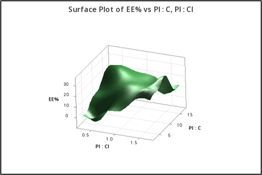
The QbD-based optimization of PK-II proliposomes using a Central Composite Design showed wide variation in entrapment efficiency (–7.29% to 35.24%), indicating a strong influence of the formulation variables. ANOVA confirmed that the quadratic model was significant, with both factors (phospholipid-to-cholesterol and phospholipid-to-carrier ratios), their interaction, and their quadratic terms contributing meaningfully to the response, while the non-significant lack-of-fit indicated good model suitability. Increasing the phospholipid-to-cholesterol ratio consistently improved entrapment, whereas the phospholipid-to-carrier ratio showed an optimum around the mid-level (1:10), beyond which EE% decreased. The response surface (Fig. 01) and contour plots (Fig. 02) showed clear curvature and strong interaction between the two factors, with the highest efficiency (35.24%) observed at a higher phospholipid-to-cholesterol ratio (1.7071) combined with a mid-level phospholipid-to-carrier ratio (10). Overall, the results identified the region around PL: CL ≈ 1.6–1.7 and PL: C ≈ 10 as the optimal design space for maximizing entrapment efficiency (Table 03) represent the runs and their response in the form of entrapment efficiency.
Table 03: Total Runs and their response data
|
PL:CL |
PL:C |
EE% |
|
1 |
10 |
10.6701 |
|
1 |
10 |
19.0792 |
|
1 |
2.9289 |
10.1764 |
|
1 |
17.0710 |
18.6950 |
|
0.5 |
15 |
26.2389 |
|
1.5 |
5 |
31.2107 |
|
1 |
10 |
24.0982 |
|
1 |
10 |
24.4064 |
|
1.7071 |
10 |
35.2419 |
|
0.5 |
5 |
-7.2920 |
|
0.2928 |
10 |
9.3652 |
|
1.5 |
15 |
6.8686 |
|
1 |
10 |
27.1360 |
|
Optimized region |
|
Optimized region |
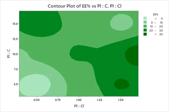
Fig. 02 Contour Plot representing the response of PL: CL and PL: C ratios on EE%
Entrapment Efficiency
The entrapment efficiency of the developed Picroside-II proliposomes was assessed to determine the amount of drug incorporated within the lipid matrix. The optimized formulation, prepared using a phosphatidylcholine: cholesterol ratio of 1:1.7 and a phosphatidylcholine to carrier ratio of 1:10, demonstrated an entrapment efficiency of 35.24% (Table 03). This value reflects satisfactory drug loading in the proliposomes system. The entrapment efficiency was influenced by lipid composition and carrier ratio, where an appropriate balance between phosphatidylcholine and cholesterol facilitated stable vesicle formation and efficient encapsulation of Picroside-II.
Particle size and zeta potential
The particle size and zeta potential of the optimized PK-II proliposomes formulation were evaluated after hydration. The average liposomes size was found to be 104.4 nm, confirming the formation of nanosized vesicles (Fig. 03). Liposomes within the 1–200 nm range are classified as nanosized particles, which is advantageous for enhancing intestinal absorption, facilitating transport across biological membranes, and potentially improving the bioavailability of PK-II. The zeta potential of the optimized formulation was measured as ±20.2 mV, indicating good electrostatic stability (Fig. 04). Typically, particles with a zeta potential within ±30 mV range exhibit sufficient repulsion to prevent aggregation, suggesting that the developed liposomes dispersion is physically stable and less prone to settling over time. Overall, the nanoscale particle size and favourable zeta potential values support the suitability of the optimized PK-II proliposomes formulation for improved absorption and stability.
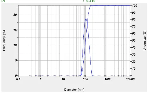
Fig. 03 Particle size of PK-II proliposomes graph
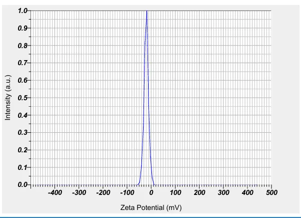
Fig. 04 Zeta potential of PK-II Proliposomes graph
SEM analysis
The surface morphology of the Picroside-II proliposomes powder was evaluated using SEM. The micrographs showed irregular, porous particles characteristic of mannitol, which acts as the carrier matrix (Fig. 05). These particles appeared uniformly coated with a thin lipid layer, confirming successful deposition of phospholipids onto the carrier surface. As expected, liposomes vesicles were not visible in the dry proliposomes powder, since vesicle formation occurs only after hydration. The porous and irregular structure of mannitol is beneficial, as it promotes rapid hydration and efficient lipid dispersion, enabling quick liposome formation in aqueous media. Overall, SEM analysis verified the successful preparation of PK-II proliposomes, demonstrating effective lipid coating over the carrier. This morphology is expected to support improved hydration, stability, and eventual bioavailability of PK-II.
Fig. 05 Dry proliposomes SEM images from different magnification length
Liquid SEM analysis
The surface morphology of the liposomes formed after hydration of the PK-II proliposomes powder was evaluated using SEM. The micrographs showed predominantly spherical vesicles with smooth, well-defined surfaces (Fig. 06), indicating efficient hydration and self-assembly of phospholipids into stable liposomes structures. The spherical shape and uniform surface suggest well-formed lipid bilayers, which are essential for maintaining vesicle integrity and providing an effective encapsulation environment for PK-II. Such morphology supports improved stability, drug protection, and controlled release behaviour. Overall, the SEM analysis confirmed successful transformation of the proliposomes powder into uniform liposomes vesicles upon hydration. The smooth and spherical structures are expected to contribute to enhanced solubility and bioavailability of PK-II.
Fig. 06 Liquid liposomes SEM images from different magnification length
DSC analysis
The thermal behaviour of Picroside-II, blank proliposomes, and the optimized PK-II proliposomes formulation was evaluated using DSC (Fig. 07), (Fig. 08), (Fig. 09). Pure PK-II exhibited a sharp endothermic peak at its characteristic melting point, confirming its crystalline nature. Blank proliposomes showed a broad endothermic peak corresponding to the lipid carrier and excipients. PK-II-loaded proliposomes displayed a thermogram similar to that of the blank, with the characteristic peak of PK-II absent. The absence of the drug’s melting peak in the loaded formulation indicates that PK-II is molecularly dispersed or encapsulated within the lipid matrix rather than existing in crystalline form. The similarity between blank and drug-loaded thermograms also suggests no significant chemical interaction between PK-II and the lipid excipients, confirming the thermal stability of the formulation.
Fig. 07 DSC of PK-II
Fig. 08 DSC of PK-II proliposomes
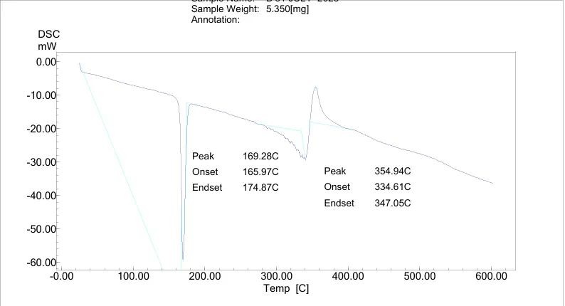
Fig. 09 DSC of PK-II blank proliposomes
FTIR analysis
The FTIR spectrum of pure PK-II (Fig. 10) exhibited characteristic absorption bands at 1015.52 cm?¹, 1265 cm?¹, and 796.93 cm?¹, confirming the presence of its functional groups. The blank proliposomes formulation spectrum (Fig. 12) showed prominent peaks at 1411.62 cm?¹, 1073.12 cm?¹, and 1010.07 cm?¹, corresponding to the phospholipid matrix and carrier components. The PK-II–loaded proliposomes (Fig. 11) demonstrated similar peaks at 1411.74 cm?¹, 1073.77 cm?¹, and 1010.54 cm?¹, with only minimal shifts compared to the blank formulation. The close alignment of peak positions between the blank and drug-loaded proliposomes indicates the absence of any significant chemical interaction between PK-II and the excipients. The slight variations observed are within the acceptable range and suggest physical encapsulation rather than chemical modification. These results confirm that PK-II was successfully incorporated into the phosphatidylcholine-coated mannitol matrix without structural incompatibility.
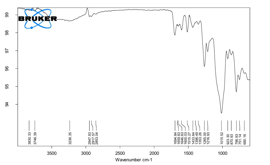
Fig. 10 FTIR of PK-II
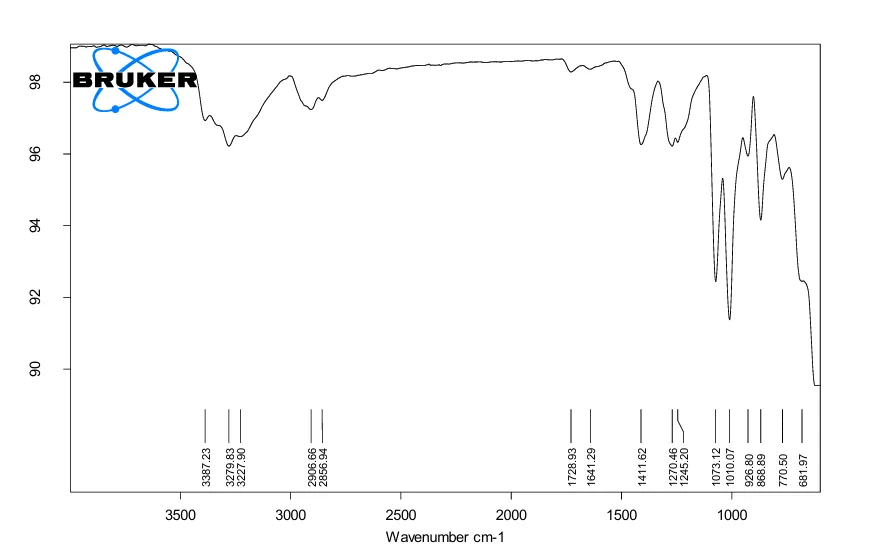
Fig. 11 FTIR of PK-II proliposomes
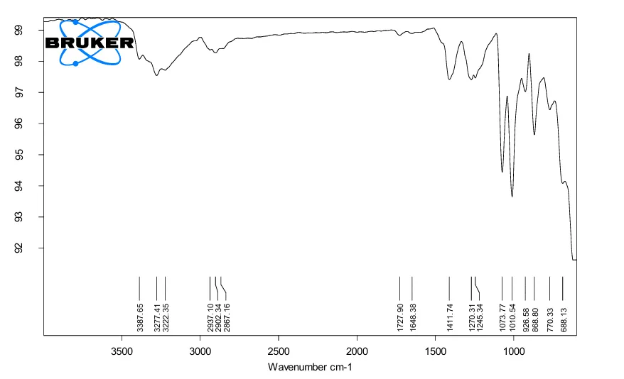
Fig. 12 FTIR of blank Proliposomes
Stability testing
The optimized proliposomes batch was stored at 4°C and room temperature (~25°C) for one month. Samples were evaluated for entrapment efficiency, particle size and zeta potential. No significant changes were observed, and the entrapment efficiency remained largely unchanged, indicating that the formulation was physically stable under the tested conditions. The stability data are summarized in (Table 04).
Table 04 Stability study data
|
Parameter |
Before Stability |
After Stability |
Difference |
SD |
Interpretation |
|
Entrapment Efficiency |
35.24% |
35.55% |
+0.31% |
0.8758 |
Negligible change (stable) |
|
Particle Size |
104.4 nm |
103.9 nm |
-0.5 nm |
0.4800 |
Highly stable |
|
Zeta Potential |
±20.2 mV |
±21.4 mV |
+1.2 mV |
5.7692 |
Minor improvement, stable |
CONCLUSION
A proliposomes formulation of Picroside II (PK-II) was successfully developed and optimized using a Quality by Design (QbD) approach. PK-II, despite its potent therapeutic properties, suffers from poor stability, permeability and low bioavailability, limiting its clinical application. The optimized formulation, prepared with a phosphatidylcholine: cholesterol ratio of 1:1.7071 and a phosphatidylcholine: carrier ratio of 1:10, exhibited a particle size of 104.4 nm, zeta potential of ±20.2 mV, and entrapment efficiency of 35.24%. These characteristics indicate the formation of nano-sized, stable, and reproducible proliposomes capable of efficient drug encapsulation. Overall, the study demonstrates that proliposomes delivery can enhance the stability, solubility and potential bioavailability of PK-II, supporting its use as an effective oral drug delivery system.
REFERENCES

Rushikesh Dasare, Pravin Wakte, Sachin Bhusari, Development, Optimization and Characterization of Picroside-II Loaded Proliposomes Using Quality by Design (Qbd) Approach, Int. J. of Pharm. Sci., 2026, Vol 4, Issue 6, 3639-36, https://doi.org/10.5281/zenodo.20485601
 10.5281/zenodo.20485601