
We use cookies to ensure our website works properly and to personalise your experience. Cookies policy
Department of Pharmaceutics, KMCH College of Pharmacy, Coimbatore-641048.
Chitosan, the most common biopolymer material, is a natural biomass that has been transformed into chitosan by means of the use of alkali to deacetylate chitosan and produce chitosan, which is subsequently used to form nanoparticles. Chitosan is an excellent surface biocompatible, biodegradable, cationic, and mucoadhesive biopolymer material, and has become a leading platform material in nanomedicine due to its superior biocompatibility, biodegradability, cationic surface chemistry, and This narrative review presents a broad synthesis of the existing information on the engineering of chitosan-loaded nanoparticles (CS-NPs), organized around three main themes, namely, formulation strategies, physicochemical characterization methodologies, and therapeutic applications. The techniques of fabrication mentioned are ionic gelation by using sodium tripolyphosphate (TPP), emulsification solvent evaporation, nanoprecipitation, self-assembly of amphiphilic chitosan derivatives, and nanofabrication with the help of microfluidics. Characterization techniques may be Fourier-transform infrared spectroscopy, atomic force microscopy (AFM), scanning electron microscopy (SEM), dynamic light scattering (DLS) or zeta potential. Critical evaluation is done on (FTIR) and in vitro drug release profiles. The therapeutic market is in oncology, antimicrobial treatment, nasal and pulmonary delivery of drugs, ocular delivery, wound healing, and gene therapy. Some of the strategies applied to functionalize surfaces such as PEGylation, folate receptor targeting, hyaluronic acid coating as well as CD44-mediated active targeting are explained. The problems of translation and future research directions are also reported to direct further evolution of chitosan nanomedicine to clinical translation
Nano medicine as a field has radically changed the science of drug delivery by allowing the creation of Nanoparticulate systems that are able to overcome physiological barriers that limit the efficacy of traditional formulations.1-3 Nanoparticles materials of a wide range of different types have been considered to date, but biopolymers are given a special niche due to their natural source, inherent biocompatibility, and ease of structuring into various forms subject to chemical modifications. In this category, chitosan has been found to be one of the most scientifically fruitful and clinically promising platform materials in pharmaceutical nanotechnology over the last thirty years.4-6
Chitin is the second most abundant natural polymer on earth and it is alkaline deacetylated to form chitosan which is a linear polysaccharide consisting of beta -linked D-glucosamine and N-acetyl-D-glucosamine.4 Two major structural variables that dictate chitosan behavior both in solution and at biological interfaces are the degree of deacetylation (DDA) which is usually between 60 and 100 percent and the molecular weight.7,8 By physiological conditions that cause the major amine groups on the backbone of the chitosan to be protonated, giving them a positive charge that allows them to electrostatically interact with negatively charged biological surfaces, anionic crosslinkers, and nucleic acids, which is the basis of the applications of chitosan in nanoparticle engineering.9-11 It promotes contact with negatively charged cell membranes to enhance cellular uptake, spontaneously pulls the chitosan chains with multivalent polyanions into tight nanoparticles and acts as a mucoadhesive agent to epithelial surfaces to increase the contact time of drugs at mucosal delivery sites.10 In addition, chitosan has large amounts of both amine and hydroxyl groups to conjugate targeting ligands, fluorescent probes, PEG chains and stimuli-reactive functional groups, allowing the creation of sophisticated multifunctional nanoplatforms.12-13Chitosan nanoparticles (CS-NPs) enhance drug solubility, prevent enzymatic degradation of sensitive cargo, enhance systemic circulation and passive and active targeted delivery.1,5 With selective formation parameter, physicochemical properties of CS-NPs, including size, zeta potential, encapsulation efficiency and drug release kinetics can be precisely regulated, affording outstanding flexibility in therapeutic indications.14-17
This narrative review combines evidence on a selected set of primary study and quality reviews to present a mechanistically-founded, structured description of the design, characterization, and therapy deployment of chitosan-loaded nanoparticles. The review is an effort to be a comprehensive guide to researchers and formulation scientists advancing the area of chitosan nanomedicine by uniting the information of formulation science, analytical chemistry and biomedical application.
2. Physicochemical Properties of Chitosan Relevant to Nanoparticle Engineering
The logical way to move towards the designing of CS-NPs is to have a good comprehension of the physicochemical characteristics that regulate the action of chitosan in solution and at the biological interfaces.4,8,17 These properties are closely connected with molecular architecture and can be logically used during the fabrication and functionalization of nanoparticles.18
2.1 Molecular Weight and Degree of Deacetylation
Molecular weight assesses the extent to which the amino acid chain has been broken down through removal of amino acids.4 The two main structural parameters that characterize the chitosan functional behavior are the molecular weight and DDA. Increased DDA increases the density of free amino groups, and increases the cationic properties and ionic crosslinking, mucoadhesion and electrostatic drug loading.6,8,10 When molecular weight is increased it makes the solutions more viscous and entangles the chain, which influences the kinetics of nanoparticle formation and size distribution.17 Chitosan with low molecular weight, less than 50 kDa, is usually of interest in the formation of nanoparticles below 100 nm in diameter, since the low molecular weight of chitosan permits higher chain mobility and the crosslinking equilibrium of the process to reach tighter and more uniform nanostructures that can be utilized to deliver drugs into cells.19,20,21
2.2 pH-Dependent Solubility and Surface Charge
At neutral and alkaline pH, chitosan is not soluble but is easily soluble in dilute organic acids with pH lower than 6.5 at which amine protonation is carried out.4 This acid-base solubility is a key factor of nanoparticle preparation procedures, whereby, acidic environments are preserved throughout the crosslinking process to guarantee the sufficient movement of the polymer chains.11,12,16,17 The obtained CS-NPs possess positive zeta potentials, typically ranging between +20 and +50 mV that enable them to react with negatively charged biological membranes and mucus glycoproteins and provide colloidal stability via electrostatic repulsion.22,23
2.3 Mucoadhesion and Epithelial Permeation Enhancement
The protonated amino groups of chitosan are electrostatically reacted with the negatively charged Salic acid and sulfate functional groups of the mucosal glycoproteins to give its mucoadhesive properties.1,9, This long-term adhesion increases the residence time of the drug at the nasal, ocular, gastrointestinal, and pulmonary delivery sites.11,12 Also, chitosan temporarily opens the epithelial tight junctions through its interactions with claudin and occludin proteins, and increases paracellular penetration of hydrophilic macromolecules otherwise excluded by transcellular transport, thereby chitosan is especially useful in mucosal vaccine and peptide delivery systems.24,25
3. Formulation Strategies for Chitosan Nanoparticles

3.1 Ionic Gelation with Sodium Tripolyphosphate
The ionic gelation using Sodium Tripolyphosphate was performed as follows: 0.50g of NaOH was added to the 1.00g of DPP-4 solution, and the mixture was mixed thoroughly before the gel was transferred into a 4.00ml round bottom flask. 1.00g of NaOH was subsequently added to the mixture and the gel was poured into a 4.00ml round bottom flask.4,5,6Ionic gelation is the most common method of CS-NP preparation and the method is valued due to its simplicity, aqueous processing, and the wide range of compatible medicinal cargo. The method takes advantage of the polysaccharide chitosan that is polycationic in nature and, as such, spontaneously gels when in the presence of multivalent polyanions. The most widely used crosslinker is sodium tripolyphosphate (TPP) which is biocompatible, water soluble and capable of forming a good network with chitosan amine groups.14,16,17
The molecular chitosan-TPP gelation has been explained using density functional theory (DFT) calculations that indicated that TPP only forms a conformational network by binding adjacent glucosamine units through its triphosphate chain, and preserves the volume of water-solubility needed to release drugs with the required specificity.18.19 It is thermodynamically spontaneous and produces nanoparticles in seconds of addition of TPP to an acidic chitosan solution that is magnetic stirred with.
The relationship between the chitosan mass ratio and TPP mass ratio, pH of the solution, polymer concentration, temperature, and mixing rate are important parameters that define the size of the nanoparticle, PDI and zeta potential.20,21 The best nano-particles usually have hydrodynamic diameters of between 100 to 400 nm, PDI less than 0.3, and zeta potentials greater than +25 mV, which is characteristic of stable monodisperse dispersions to use in the pharmaceutical industry.23 The high-shear homogenization of the ionic gelation technique has also enhanced the suitability of the technique into sub-100 nm CS-NPs, as dual drug combinations can be simultaneously encapsulated.26 TPP-crosslinked CS-NPs have been optimized as cisplatin and 5-fluorouracil co-loaded nanoparticle by taking advantage of chemotherapeutic synergy and reducing systemic toxicity, although optimization of drug-polymer-crosslinker ratios would be necessary to balance the encapsulation efficiency of each drug and the structural integrity of the nanoparticle.27
3.2 Emulsification-Solvent Evaporation
Encapsulation of hydrophobic drugs that are poorly soluble in water using CS-NPs is most preferably with the method emulsification-solvent evaporation. An aqueous solution of chitosan is mechanically agitated or ultrasonically evaporated to emulsify the drug dissolved in a solvent that is organic and then a surfactant is added.14,15 The next step of the reduction pressure removal of organic solvents precipitates the nanoparticles by solidification of the matrix. Lipophilic drugs can be encapsulated with encapsulation efficiencies of over 80% whilst toxicity of left over organic solvents and biocompatibility of emulsifiers will need to be overcome.17,18 The variants of double emulsion (W/O/W) enable the applicability to hydrophilic drugs and proteins because an aqueous drug solution is encapsulated in the inner water phase.28
3.3 Nanoprecipitation
Nanoprecipitation is a fast precipitation of a chitosan solution with a miscible non-solvent in which the chitosan precipitation is caused by solvent diffusion and results in the formation of nanoparticles that have narrow size distributions.15,17 The procedure is especially open to the on-going production with the use of microfluidic devices and yields highly reproducible batches. Its main weakness is that it is only limited to hydrophilic substances that selectively partition into the aqueous layer instead of the forming polymer network and that cosolvent systems are needed in order to gain sufficient chitosan solubility in partially organic solvents.18,20
3.4 Self-Assembly of Amphiphilic Chitosan Derivatives
The self-assembly of amphiphilic chitosan derivatives is the other technique that has been applied in creating nanopores in the shell of prawns. Hydrophobic moieties including palmitoyl, stearoyl or deoxycholic acid groups introduced chemically result in amphiphilic chitosan derivatives which spontaneously self-assemble to form core-shell nanostructures in aqueous conditions.13,15 The hydrophobic core is used as a drug delivery system of lipophilic therapeutics and the hydrophilic chitosan shell is used to provide colloidal stability and bio-compatibility.29,30 The most investigated derivatives in this respect are n-palmitoyl chitosan and glycol chitosan which are found to encapsulate paclitaxel and curcumin with high efficiency . Such systems also respond to pH swelling in acidic microecosystems allowing directed intracellular drug release in endolysosomal compartments of cancer cells, which has been used strategically in cancer chemotherapy.31,32
3.5 Microfluidics-Assisted Nanofabrication
Microfluidic systems are a new development in the production of CS-NPs, which provides accurate control over mixing dynamics, residence time, and particle size by controlling flow velocities and channel configurations.17,23 The Y-shaped or herringbone-patterning pattern of continuous-flow microreactors generate CS-NPs with mean diameters down to 50 nm and PDI down to 0.15, much better in terms of size uniformity and reproducibility than batch methods.23 Despite being still mainly restricted to research environments, microfluidic nanofabrication has a significant future potential as a GMP-scalable and compatible product manufacturing system of clinical-grade chitosan nanomedicine, especially as regulatory bodies progressively specify tighter quality requirements of nanoparticles regarding critical quality attributes.33
4. Physicochemical Characterization of Chitosan Nanoparticles
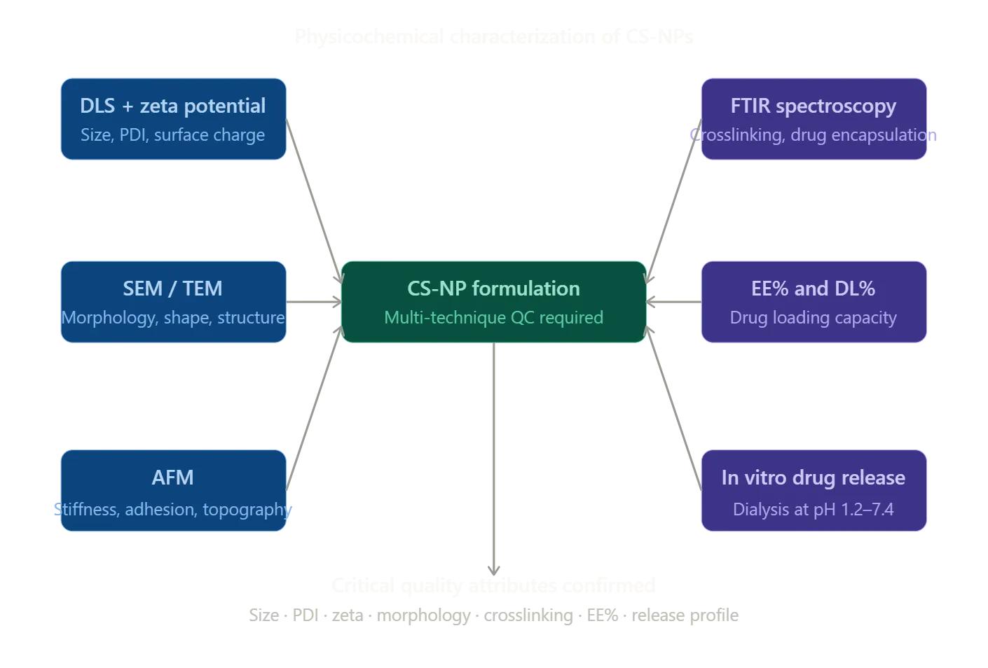
4.1 Dynamic Light Scattering and Zeta Potential
Dynamic light scattering (DLS) is the main technique of measuring the hydrodynamic diameter and polydispersivity index (PDI) of CS-NPs in suspension. Change of laser light with time scattered due to Brownian movement is discussed through the method of autocorrelation to determine the translational diffusion coefficient, using which the hydrodynamic radius is determined based on the Stokes-Einstein equation.16, 23 The values of PDI of less than 0.3 are normally considered to be representative of sufficient monodispersity to be used in pharmaceutical purposes. Using electrophoretic light scattering, zeta potential is used to determine electro kinetic surface charge and forecasts colloidal stability and capability to interact with biological membranes.10,11,24 A typical positive zeta potential of +20 to +50 mV of protonated surface amine groups indicates adhesive properties to negatively charged cell membranes and mucus in CS-NPs. Anionic polymers or targeting ligands cause surface alteration to shift zeta potentials either to neutral or negative values, and special attention should be paid to prevent action that leads to a loss of colloidal stability. Measurements of DLS and zeta potential measured in simulated physiological fluids like phosphate-buffered saline, simulated nasal fluid and simulated tear fluid are critical predictor of nanoparticle stability and behaviour in the biological environment.12, 34
4.2 Electron Microscopy
Transmission electron microscopy (TEM) and scanning electron microscopy (SEM) can be used to obtain direct morphological information on a nanoscale. SEM indicates topography, shape uniformity, and aggregation of dried samples, which usually verifies the morphological appearance of an ionically crosslinked CS-NPs, and which is usually a smooth, round surface.10,13,18 TEM can offer internal structural resolution that has the ability to differentiate between the core-shell structures in functionalized or hybrid nanoparticle systems.23,24The cryo- TEM of nanoparticles in the moist environment removes the dehydration effect and provides a more faithful depiction of the morphology of nanoparticles in physiological conditions and is especially useful in characterizing self-assembled and lipid-chitosan hybrid systems.26
4.3 Atomic Force Microscopy
Using atomic force microscopy (AFM), the mechanical rigidity, adhesion forces and surface topography of single nanoparticles can be measured with sub-nanometer resolution, which has expanded the morphological characterisation of nanoparticles into nanomechanical space.23 With CS-NPs to be used as cellular internalization or mucosal delivery vectors, nanomechanical characterization is especially informative: stiffer, less deformable particles have a higher membrane interaction, efficiency of endosomal escape and cellular uptake.1,8 In a study by Khatib et al. (2025), the force spectroscopy approach through AFM was used to describe the characteristics of chitosan microparticles formed by an ionic gelation process, with their values of the Young modulus ranging between 20 and 80 kPa, which corresponds to the viscoelastic behavior of hydrogel that could be controlled by the density of the crosslinking points and molecular weight of chitosan.21,23 Dry-state AFM dimensions and hydrodynamic DLS diameters complement each other to offer a complete profile of characterisation of dimensions.16
4.4 Fourier-Transform Infrared Spectroscopy
FTIR spectroscopy is necessary to confirm the drug encapsulation condition inside the nanoparticle framework, crosslinking chemistry and the interaction between chitosan and the drug. Aspects of the characteristic chitosan FTIR spectrum are broad O-H/N-H at 3450 cm -1, C-H at 2920 cm -1, amide II at around 1655 cm -1, amide II N-H bending at approximately 1590 cm -1, and P=O phosphate crosslinking at 1220 cm -1.P=O cross linking is confirmed by changes in amide II absorption and the appearance of new phosphate vibrations.18,4 Attenuation or loss of drug-specific absorption bands in the CS-NP spectrum (relative to the free drug and physical mixture) indicates that there is drug encapsulation at the molecular level at the polymer matrix level.21 The ATR-FTIR spectroscopy of the undisturbed nanoparticle pellets in the absence of solvent interference has especially clean spectra that can be compared to others.18
4.5 Encapsulation Efficiency and In Vitro Drug Release
Encapsulation efficiency (EE%), and drug loading capacity (DL%), are a measure of the percentage of drug successfully incorporated in nanoparticles compared to the amount of drug input.5,14The determination of these parameters is done through the separation of nanoparticles and the free drug by using ultracentrifugation or ultrafiltration, and subsequently, quantifying the supernatant by using the HPLC or UV-Vis spectrophotometer. EE% values reported to be with CS-NPs are widely ranged between 30 and more than 95 percent based on the drug physicochemical characteristics, drug affinity to chitosan, formulation pH, and concentration of crosslinker.15,17 In vitro drug release In vitro experiments of dialysis membrane diffusion at physiologically relevant pH values (pH 7.4 for systemic delivery, pH 1.2 for stomach and pH 4.5 for endosomal delivery) mimics the conditions of the desired delivery location. CS-NPs are traditionally characterized by a biphasic release of surface-adsorbed drug to an initial burst release, and a sustained diffusion of the polymeric matrix.32,14 The acidic pH-dependent swelling of chitosan accelerates the release in acidic environments; this is intended to target cancer cells in intracellular delivery of a drug that targets tumors.13 The process of the main release is clarified and the optimization of rational formulation is directed by mathematical modeling on the basis of zero-order, first-order, Higuchi, and Korsmeyer-Peppas models.32,17
5. Surface Functionalization Strategies

5.1 PEGylation for Extended Circulation
The steric stabilization of CS-NPs with polyethylene glycol chains forms an hydrophilic corona that inhibits nonspecific adsorption of proteins and slows down mononuclear phagocytic elimination by the mononuclear phagocyte system.10,1,15 PEGylation is obtained by the first reaction between NHS-activated PEG esters, which react with the primary amines of chitosan to create stable amide bonds.1,16 PEGylation is a conventional method to use in systemic oncological applications as PEGylated CS-NPs have reduced zeta potential and elevated hydrodynamic diameter, however they have greatly improved blood circulation half-lives and tumor accumulation as a result of the augmented permeability and retention effect.1,15
5.2 Folate Receptor Targeting
Folate receptors are also overexpressed in various malignant tumors such as ovarian, breast, cervical and lung carcinomas with the level of expression in normal tissues being relatively limited. The EDC/NHS conjugation of folic acid and chitosan amines forms folate-functionalized nanoparticles, which are receptors and can be endocytosed. The systematic assessment of folate-engineered CS-NPs by Albalawi et al. (2024) revealed that folate-to-chitosan molar ratios, which are crucial to optimally forming folate-receptor-binding affinity, PEG spacer length between chitosan and folate is vital in maintaining colloidal stability, and that 3 to 4 fold higher tumor uptake of targeted nanoparticles in folate receptor-positive tumor-bearing mice than non-targeted counterparts.34-38
5.3 Hyaluronic Acid Coating and CD44-Mediated Targeting
As a biomimetic targeting agent, hyaluronic acid (HA) was selected as it is a natural glycosaminoglycan with high CD44 receptor affinity and has been utilized as a targeting agent against breast, colorectal, and prostate cancer which expresses high levels of CD44 receptors. HA-coated CS-NPs is a method to use a combination of structural and pH-responsive drug release characteristics of the chitosan core and active endocytosis of the CD44-targeted active endocytosis by the HA shell. In preclinical studies, Doxorubicin-loaded HA/CS-NPs had a high tumor penetration, greater cytotoxicity in three-dimensional cancer models, and decreased cardiotoxicity than free drugs due to selective tumor targeting of the targeted nanoparticles. The HA coating simultaneously inverts the positive surface charge of CS-NPs decreasing nonspecific electrostatic interactions with plasma proteins, erythrocytes.34,36,39,10
5.4 Stimuli-Responsive Functionalization
Stimuli-responsive CS-NPs deliver cargo selectively to tumor microenvironmental or intracellular microenvironmental pathological cues (pH of about 6.5 and 4.5 to 5.5, respectively). N-isopropylacrylamide derivatives of chitosan with temperature responsiveness behavior have lower critical solution temperature behavior that allows drug release under mild hyperthermic conditions. Redox-responsive systems with disulfide crosslinks promptly degrade in the higher glutathione concentration in cancerous cells releasing drug inside the cell with high spatial resolution.13,32,33 These stimulus sensitive mechanisms enhance the therapeutic index, as the drugs are released to pathologically important microenvironment and off-target exposure is limited.40
6. Therapeutic Applications of Chitosan Nanoparticles

6.1 Cancer Drug Delivery and Active Targeting
The most developed use of CS-NPs in therapeutic purposes is oncology. They have inspired intensive effort in research to develop their capacity to increase the therapeutic index of cytotoxic agents by increasing their tumor concentration and decreasing systemic toxicity.1,15 HA/CS-NPs, which were loaded with doxorubicin and altered to target CD44, showed that their pH-sensitive intracellular release of drugs, improved cellular uptake via receptor-mediated endocytosis, and better tumor growth inhibition in xenograft models than free drug.39,36, Similarly, paclitaxel-, methotrexate-, or curcumin-loaded CS-NPs targeted folate receptor have shown specific cytotoxicity to folate receptor-expressing cancer cell lines with considerably low cytotoxicity of normal cells.13,1 Cisplatin and 5-fluorouracil co-loaded CS-NPs are one of the most exquisite uses of ionic gelation to dual drug cancer therapy. Concomitant encapsulation of the two drugs at optimal ratios facilitated the concerted release at the tumor location resulting in synergistic cytotoxicity using much lower total drug doses than with free drug-mixtures. In vitro experiments have shown significantly increased cancer cell kill rates using dual drug-loaded CS-NPs than using the same dosage of free drug combinations, which indicate better intracellular bioavailability.34-36
6.2 Antimicrobial Applications
Chitosan has inherent broad-spectrum antimicrobial property consisting of electrostatic disturbance of bacterial membranes and resistance of intracellular enzyme activity.26 When this antimicrobial potency is in the form of nanoparticles, then this potency is multiplied by an increase in the ratio of surface area-to-volume and increase in contact with the membrane.41,42 CS-NPs can also be used to treat multidrug-resistant infections because they have demonstrated high levels of bactericidal activity against methicillin-resistant Staphylococcus aureus, Escherichia coli, Pseudomonas aeruginosa and Candida albicans due to encapsulation of traditional antibiotics that inhibit the enzymes, enhance biofilm penetration and sustain inhibitory levels over extended durations of use.6,43,44
6.3 Nasal Drug Delivery and Brain Targeting
The nasal route bypasses blood-brain barrier and provides direct access to the central nervous system and the systemic circulation via the olfactory pathway because of the mucoadhesive nature and ability to open tight junctions. Mohamed et al. (2024) produced imipramine hydrochloride-impregnated CS-NPs in a thermoresponsive in situ gel, which was used to deliver the particles through the nose. To enhance the elimination of nasal residence and sustained drug delivery, the composite system was a thermoresponsive gelation at body temperature and chitosan mucoadhesion. Experiments on pharmacokinetics showed that brain drug levels were increased with intranasal administration of CS-NP/gel than intravenous injection of chitosan nanoparticles and this confirmed the nose-to-brain delivery potential of chitosan nanoparticle systems.45-48
6.4 Pulmonary Drug Delivery
The local delivery of respiratory diseases and systemic absorption of inhaled biologics can be achieved through the pulmonary delivery of CS-NPs.12 Spray-dried CS-NPs generate inhalable microparticles that deagglomerate into mucus in the airways to deep-ly land on the lung and mucosally permeate.24,25 Mucoadhesive chitosan improves the residence of nanoparticles in the lung epithelium because of prolonged interaction with mucus, which is eliminated by the cilia. Tobramycin or ciprofloxacin-loaded CS-NPs have been shown to have a superior biofilm penetration and bactericidal efficacy in Pseudomonas aeruginosa-infected airway models, which is therapeutically applicable to cystic fibrosis pulmonary infections. The bioavailabilities obtained with inhalation of insulin-loaded CS-NPs are 20 to 40% compared to those of subcutaneous injected insulin, which is a significant improvement towards needle-free insulin delivery.49-52
6.5 Ocular Drug Delivery
CS-NPs tackle several issues of ocular drug delivery at once: being positively charged, they are able to adhere to the cornea; their mucoadhesion allows them to spend extra time in the precorneal space, and their nanoparticulate nature allows them to permeate the cornea.3,53,54 CS-NPs containing pilocarpine, timol or cyclosporine A have shown significantly enhanced ocular bioavailability to conventional eye drops by having sustained intraocular levels of drugs over 24 hours.33,7 Thiolated chitosan nanoparticles take advantage of the formation of disulfide bonds with corneal mucin cysteine residues of exceptionally high affinity in mucoadhesion, and delivered to the posterior segment with, as yet, potential as a non-invasive alternative to intravitreal injection of anti-VEGF biologics in the treatment of age-related macular degeneration.55,32
6.6 Wound Healing
Chitosan enhances wound healing in a multi complementary manner: macrophage activation leading to inflammatory resolution process, fibroblast proliferation and collagen deposition stimulation and hemostatic effects by platelet aggregation stimulation.1,15 To overcome the rapid degradation of the soluble biologics in the wound exudate, CS-NP-loaded hydrogel composites or electrospun nanofiber scaffold can provide extended release of growth factors, antibiotics, and anti-inflammatory medicines in the wound site.20,10 Wound healing on the diabetic wound model with CS-NP-incorporated dressings with silver nanoparticles or curcumin has shown a faster wound healing, less bacterial colonization, and increased angiogenesis due to the multifaceted pathophysiology of chronic non-healing wounds. This is because of the inherent biodegradability of chitosan that makes the scaffolds to be absorbed with wound healing and removes the trauma cused by removing the dressing.35,36
6.7 Gene Delivery and Nucleic Acid Therapeutics
Chitosan is a polycationic material, which allows the nucleic acids with negative charges to form small polyplexes by electrostatic condensation, avoiding degradation by serum nucleases as well as endosomal degradation due to the proton sponge effect. Non-viral transfection methods of plasmid DNA, siRNA, mRNA, and antisense oligonucleotides use chitosan polyplexes, which are safe and non-toxic.12,13 The therapeutic genome editing efficiencies of CRISPR-Cas9 ribonucleoprotein delivery with chitosan nanoparticles have proven with similar efficiencies as lipid nanoparticles at considerably reduced costs and immunogenicity.34,38 The nanoparticle approach of co-delivery of siRNA and chemotherapy drugs to silence gene expression of drug resistance and induce synergistic cancer cell death through co-modulation of pathways and co-delivery of the chemotherapeutic agent is a particularly novel way of overcoming multidrug resistance in cancer.14,26
7. Comparative Overview of Formulation Methods
The table below summarizes the principal formulation strategies for CS-NPs, their mechanistic basis, typical particle size ranges, key advantages, and primary limitations.
|
Method |
Mechanism |
Size Range |
Key Advantages |
Main Limitations |
|
Ionic Gelation (TPP) |
Electrostatic CS-TPP crosslinking |
50-500 nm |
Mild, aqueous, scalable, no organic solvents |
Limited for hydrophobic drugs; TPP residue concerns9,4 |
|
Emulsification-Solvent Evaporation |
Solvent removal from emulsion droplets |
100-1000 nm |
High EE% for hydrophobic drugs; versatile |
Organic solvent residues; surfactant toxicity18 |
|
Nanoprecipitation |
Anti-solvent polymer precipitation |
50-300 nm |
Narrow size distribution; reproducible |
Poor encapsulation of hydrophilic drugs23,8 |
|
Self-Assembly |
Amphiphilic chitosan derivative aggregation |
20-200 nm |
pH-responsive; core-shell structure |
Complex synthesis; derivative characterization burden16 |
|
Microfluidics |
Controlled mixing in microchannels |
20-150 nm |
Precise control; high batch-to-batch reproducibility |
Low throughput; specialist equipment required20 |
8. Challenges and Future Directions
Although the current study has achieved impressive advancements in the study of chitosan nanoparticles, there are a number of obstacles that have to be addressed in order to achieve the clinical potential of the CS-NP-based medicines. The greatest translational barrier is scale-up manufacturing. The majority of CS-NP preparations are done at laboratory scale, and the shift to GMP-compliant production presents complications in ensuring the size of particles is uniform, batches to batches reproducible, and the control of process parameters. The continuous-flow microfluidic manufacturing is an optimistic direction to move but needs additional engineering advancement in order to produce clinically meaningful amounts of production .
Another major challenge is regulatory and safety issues. Toxicological testing needs to be extensively performed on surface-modified CS-NPs (PEGylated, folate-conjugated and HA-coated) in various species before regulatory approval. High doses of immunogenic properties of chitosan nanoparticles, biodistribution and long-term clearance kinetics of polymer fragments in tissue demand a systematic study . Aggregation of nanoparticles during the lyophilization process, which is the preferred storage method over long periods has to be suppressed by optimization of cryoprotectants, trehalose, mannitol, and sucrose have been tested with mixed success and spray-drying is another option in producing stable nanoparticle powders.
The high incidence of failure to translate attractive in vitro findings to in vivo effectiveness is in part due to insufficient preclinical models. Three dimensional tumor spheroids, patient-derived organoids and organ-on-chip models better approximate tumor microenvironment complexity, interstitial pressure and systemic barriers and need to be implemented into the preclinical assessment pipeline. Theranostic CS-NPs will further develop precision nanomedicine by allowing real-time feedback on drug delivery and response to treatment through the confluence of machine learning with formulation science, which will drastically speed up the development cycle of nanoparticles and decrease the empirical cost of formulation development.
CONCLUSION
Chitosan-loaded nanoparticles are a scientifically versatile and therapeutically revolutionary platform which has shown consistent high-quality performance compared to traditional drug delivery systems in a wide range of biomedical uses. Chitosan has a distinct biocompatibility, biodegradability, cationic surface chemistry, mucoadhesion, and versatile functionalizability which places the biopolymer in a distinct position as the most powerful nanoparticle engineering biopolymer. The most commonly used fabrication technique, ionic gelation offers a mechanistically well-defined, scalable and mild platform of encapsulation of a wide range of therapeutics, with other more recent methods such as self-assembly and microfluidics pushing the limits of miniaturization of particles and manufacturing control.
Strict physicochemical characterization with a complementary collection of DLS, zeta potential measurements, SEM, TEM, AFM, and FTIR measurements are necessary to guarantee the quality of formulation and in vivo behavior. CS-NPs therapeutic applications include oncology, antimicrobial therapy, gene delivery, wound healing, and mucosal delivery of drugs and surface functionalization methods such as PEGylation, folate targeting and HA coating further enhance selectivity and therapeutic effect.
Scalable manufacturing, thorough safety assessment, predictive in vitro models, and regulatory science are all the steps to take to ensure that chitosan nanomedicines will go all the way to the bench to the bedside. As these translational fields continue to be invested in, chitosan-based nanoparticles have the transformative potential to re-write the treatment paradigm of cancer, infectious disease, chronic wounds and central nervous system diseases in the next decade.
REFERENCES

Cathrien J , Kamaleshwari B, Sankar C, Jayaprakash K, Engineering Chitosan-Loaded Nanoparticles: Formulation Strategies, Characterization Techniques, and Therapeutic Applications, Int. J. of Pharm. Sci., 2026, Vol 4, Issue 7, 320-335, https://doi.org/10.5281/zenodo.21131989
 10.5281/zenodo.21131989