

We use cookies to ensure our website works properly and to personalise your experience. Cookies policy
Truba Institute of Pharmacy.
The present study was undertaken to develop and evaluate a polyherbal ointment containing methanolic seed extracts of Sterculia Foetida and Psoralea Corylifolia for wound healing. Preliminary phytochemical screening confirmed the presence of important bioactive constituents, while quantitative analysis revealed higher total phenolic (39.26 mg GAE/g) and flavonoid (46.57 mg RE/g) contents in Sterculia Foetida than in Psoralea Corylifolia. Three ointment formulations were prepared and evaluated for physicochemical properties, antimicrobial activity, and wound healing efficacy. All formulations exhibited acceptable pH, viscosity, spreadability, and were non-irritant, indicating their suitability for topical application. The polyherbal ointment showed superior antibacterial activity against Staphylococcus aureus with a zone of inhibition of 18.3 mm and produced 75.50 ± 0.46% wound closure by Day 21, compared with the individual herbal formulations. Although the standard 0.2% Nitrofurazone ointment exhibited the highest wound healing response, the polyherbal formulation demonstrated significantly enhanced healing due to the synergistic action of the combined plant extracts. The findings suggest that the developed polyherbal ointment is a safe and promising natural topical formulation for wound management.
1.1 Medicinal plants The use of medicinal plants in ancient times and initially developed through empirical observation and experience. In the absence of scientific knowledge regarding the causes of diseases and their treatment, early societies relied primarily on instinct and traditional practices to identify plants with therapeutic potential. Over time, systematic observations and accumulated traditional knowledge established associations between specific medicinal plants and particular ailments. Consequently, the use of medicinal plants gradually evolved from an empirical practice toward a more rational and scientifically supported approach based on their therapeutic properties and underlying mechanisms (Jamshidi et al., 2017). Until the advent of iatrochemistry in 16th century, plants had been the source of treatment and prophylaxis (Chen et al., 2016). Nonetheless, the decreasing efficacy of synthetic drugs and the increasing contraindications of their usage make the usage of natural drugs topical again (Pengelly, 2020). Assyrian pharmacotherapy was referred to. Some of them were given the names after mythological characters from these epics; for instance, Elecampane (Inula helenium L. Asteraceae) was named in honor of Elena, who was the centre of the Trojan War (Petrovska, 2012).
Around the world, plant medicines are widely used. While alternative medicine, including the use of dietary supplements, is aggressively marketed in the developed world using the claims of traditional medicine, local traditional medicine, including herbalism, is the only source of healthcare for people in the majority of developing countries, particularly in rural areas. As of 2015, the majority of medical plant-based items had not undergone safety and efficacy testing, and those sold in developed economies and supplied by traditional healers in developing nations were of varying quality and occasionally contained hazardous chemicals (Chen, 2015).
1.2 Herbal medicine One of the most significant scientific foundations for security in many parts of the world has been established by the use of herbal medicine in both traditional and modern medical systems (Khan and Ahmad 2019). Herbal plants have been utilized for a variety of purposes for many years. Generally speaking, herbal plants are defined as one-year-old gramineous herbs without tight context (Msomi and Simelane 2019). Dependent on the type of plants, one part including (flowers, leaves, branch or roots) or whole sectors (aerial parts or roots) could be accomplished in treatment of acute and chronic diseases, food products and nutritional improvement (Falzon and Balabanova 2017).After entering herbs into digestive systems, body starts consuming them and staves off the waste as well as free radicals bounded to the fibres of the herbs and in general purify useful contents into body cells. Because of unending advantages and their considerable benefits, plants with medicinal potentials are anticipated to be applied widely in human nutrition to improve not only the healthiness of human’s body cells but also ensure the psychological health (Aleksic and Knezevic 2014).
The human body's skin acts as a barrier between it and the outside world. It participates in metabolic processes, serves a resorptive and thermoregulatory role, and shields the body from external chemical and physical stimuli being the first line of defence against pathogenic microorganisms, and it partakes in immunological processes. The complex structure of human skin and its physicochemical characteristics turn it into an effective outermost defence line against exogenous factors, and help maintain homeostasis of the human body (Lafrance, 2018). This role is played by the epidermal barrier, in which the corneal layer of epidermis has a particularly important function to perform. It consists of 15–20 layers of fully cornified keratinocytes – corneocytes. In the bottom part of the cornified layer, the cells closely adjoin each other, while in the top part they are arranged loosely and undergo scaling (Agrawal et al., 2023). Construction of the corneal layer resembles a wall in which corneocytes stand for bricks, and a fat-abundant intercellular matrix is the cement. Skin is the largest organ of the body, accounting for about 15% of the total adult body weight (Boer et al., 2016).
The skin is actually made up of interconnected tissues that work together to perform a variety of essential functions, despite the fact that it may not initially appear to be an organ. The integumentary system, which normally shields the body, is made up of the skin and the tissues that support it. The skin's several layers of cells and tissues are joined to their corresponding supporting structures by connective tissue. The dermal layer has a large number of blood vessels. Its sympathetic and autonomic nerve fibers, together with a number of sensory filaments, provide communication between different parts of the brain.
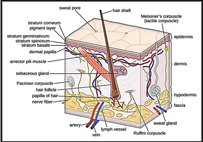
Figure 1: Structure of Skin 1.3.1
Functions of Skin Ø Protection When it comes to weather protection, the skin is the body's outermost layer. The epidermis, which is its outermost layer, serves as a physical barrier that protects internal tissues from harmful substances, bacteria, viruses, and mechanical traumas like cuts or abrasions. It also contains pigments like melanin that protect the body from damaging UV radiation. Infections are also prevented by immune cells and the skin's slightly acidic pH (Harris et al., 2022). Ø Thermal regulation The skin is primarily responsible for maintaining a constant core temperature. When the body's core temperature rises too high, the sweat glands release perspiration. Sweat cools the body when it evaporates from the skin. In order to lower heat, the skin's blood vessels widen at the same time. The body's blood vessels shrink and constrict in cold conditions to maintain body temperature and stop heat loss. The internal environment remains constant despite variations in the outside temperature thanks to this balance (Jung et al., 2023). Ø Sensation A vast array of external stimuli can be detected by the skin's extensive network of sensory nerve endings. These receptors allow us to perceive touch, pressure, pain, heat, and cold. This sensory capability is crucial for protection because it allows the body to swiftly adapt to dangerous situations, such removing your hand from a hot surface or reacting to an accident
Classification of wounds
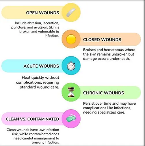
Wounds are fractures in the skin's epithelial integrity, or abnormalities of normal anatomic structure and function brought on by pathological events. Wounds can be classified in many ways, primarily depending on their duration and their depth (Figure 2). Acute wounds usually heal within 3 weeks whereas in the case of chronic wounds the time since injury typically is more than 3 months. Furthermore, wounds may be classified as those that can repair themselves or can be repaired in an orderly and timely process (usually acute wounds) and those that cannot (usually chronic wounds) (Rostami et al., 2021). In superficial-thickness wounds of the skin, damage extends over the epidermis and superficial parts of the dermis, whereas partial thickness wounds involve greater parts of the dermis and full thickness wounds even the subcutaneous tissue layer. Typical superficial-thickness cutaneous wounds are classified by their appearance abrasions, lacerations and incisions as well as superficial thermal wounds
MATERIALS AND METHODS
Procurement of plant material Seeds of Sterculia Foetida (400 g) and Psoralea Corylifolia (340 g) were collected for the study. The plant materials were procured from an authenticated source and collected from the medicinal plant garden of the Pinnacle Biomedical Research Institute, Bhopal (M.P.). Both plant species were authenticated by a qualified botanist before use. The collected seeds were thoroughly cleaned and shade-dried at room temperature until complete drying. After drying, the seeds were separately pulverized into coarse powder using an electronic grinder. The powdered materials were stored in airtight containers and used for further extraction, phytochemical evaluation, formulation development, and pharmacological studies
Extraction of Plant Material by Soxhlation Process The dried and powdered seeds of Sterculia Foetida (400 g) and Psoralea Corylifolia (340 g) were separately subjected to Soxhlet extraction using a suitable solvent. The accurately weighed powdered plant materials were packed into Soxhlet extraction thimbles and placed in the Soxhlet apparatus (Figure 7). The extraction was carried out by continuous hot percolation, where the solvent was heated, evaporated, condensed, and repeatedly passed through the powdered material to extract the soluble bioactive constituents. The extraction process was continued until complete exhaustion of the plant material was achieved, ensuring maximum extraction of phytoconstituents. After completion of extraction, the obtained solvent extracts were filtered to remove any insoluble impurities and concentrated by evaporating the solvent under reduced pressure (Figure 8). The concentrated extracts were collected, weighed, and stored in airtight containers under suitable conditions for further phytochemical evaluation, formulation preparation, and pharmacological studies. The percentage yield of each extract was calculated by comparing the weight of the dried extract with the initial weight of the powdered plant material used Soxhlation Process of Sterculia Foetida and Psoralea Corylifolia
Determination of Percentage Yield of Extract
The percentage yield of the obtained extracts was determined to evaluate the efficiency of the extraction process. After complete removal of the solvent, the dried extracts obtained from Sterculia Foetida (400 g) and Psoralea Corylifolia (340 g) were accurately weighed. The percentage yield was calculated by using the weight of the dried extract obtained and the initial weight of the powdered plant material used for extraction. Finally, the percentage yield of the extract was calculated based on the initial weight of the plant materialPhytochemical Screening of Sterculia Foetida and Psoralea Corylifolia Seed Extracts The extracts were subjected to qualitative phytochemical analysis for the detection of various secondary metabolites using standard procedures.
Phytochemical Screening of Sterculia Foetida and Psoralea Corylifolia Seed Extracts
Quantitative Estimation of Phytoconstituents
1. Estimation of Total Phenolic Content (TPC) The Folin-Ciocalteu colorimetric technique was used to assess the total phenolic content of extracts from Psoralea Corylifolia and Sterculia Foetida. Gallic acid at doses of 20, 40, 60, 80, and 100 µg/mL was used to create a standard calibration curve. One milliliter of the extract solution and five milliliters of diluted Folin-Ciocalteu reagent were combined after the extract solution was made in an appropriate solvent. Following a few minutes of incubation, 4 mL of sodium carbonate solution was added, and the mixture was allowed to develop color for 30 minutes at room temperature. A UV-visible spectrophotometer was used to detect the absorbance at 760 nm. The gallic acid calibration curve was used to determine the total phenolic content, which was then reported as mg gallic acid equivalent (GAE)/g of extract (Al-Owaisi et al., 2014).
2. Estimation of Total Flavonoid Content (TFC)
The aluminum chloride colorimetric method was used to determine the total flavonoid content of extracts from Psoralea Corylifolia and Sterculia Foetida. Rutin at concentrations of 20, 40, 60, 80, and 100 µg/mL was used to create a standard calibration curve. One milliliter of potassium acetate solution and one milliliter of aluminum chloride solution (AlCl₃) were combined with the extract solution, and then distilled water was added. To produce color, the reaction mixture was let to sit at room temperature for half an hour. A UV visible spectrophotometer was used to detect the absorbance at 510 nm. The rutin calibration curve was used to determine the total flavonoid content, which was then reported as mg rutin equivalent (RE)/g of extract. Every measurement was done three times, and mean values were noted (Vongsak et al., 2013). Preparation of polyherbal ointment formulation The ointment formulations were prepared by the fusion method. The required quantities of white soft paraffin, liquid paraffin, beeswax, and cetostearyl alcohol were accurately weighed and melted together in a suitable vessel on a water bath with continuous stirring to form a uniform ointment base. Methyl paraben and propyl paraben were dissolved in distilled water and incorporated into the prepared base with continuous mixing. For Formulation I, 2% of Sterculia Foetida seed extract was gradually incorporated into the ointment base and mixed thoroughly to obtain a uniform dispersion. For Formulation II, 2% of Psoralea Corylifolia seed extract was added to the base using the same procedure. For Formulation III (polyherbal ointment formulation), both Sterculia Foetida seed extract and Psoralea Corylifolia seed extract were incorporated in a 1:1 ratio (1% each) into the ointment base and mixed properly to ensure uniform distribution of herbal extracts (Table 5). The prepared formulations were levigated and triturated until a smooth, homogeneous ointment was obtained. Distilled water was added in sufficient quantity (q.s.) to make the final volume up to 50 mL. The prepared ointments were filled into clean, dry containers and stored under suitable conditions for further evaluation of physicochemical properties and stability parameters (Sultana et al., 2016).
Table 6: Composition of Polyherbal Ointment Formulation (Ojha Et Al., 2024)
| Name of Ingredient | Formulation I (Sterculia foetida) | Formulation II (Psoralea corylifolia) | Formulation III (Polyherbal ointment) |
|---|---|---|---|
| Sterculia foetida seed extract | 2.0% | — | 1.0% |
| Psoralea corylifolia seed extract | — | 2.0% | 1.0% |
| White soft paraffin | 20.0 g | 20.0 g | 20.0 g |
| Liquid paraffin | 5.0 g | 5.0 g | 5.0 g |
| Beeswax | 2.0 g | 2.0 g | 2.0 g |
| Cetostearyl alcohol | 1.0 g | 1.0 g | 1.0 g |
| Methyl paraben | 0.2 g | 0.2 g | 0.2 g |
| Propyl paraben | 0.02 g | 0.02 g | 0.02 g |
| Distilled water | q.s. | q.s. | q.s. |
| Final quantity | q.s. to 50 mL | q.s. to 50 mL | q.s. to 50 mL |
Evaluation of pH, Skin Irritation, Viscosity, and Spreadability of Sterculia Foetida and Psoralea Corylifolia Seed Extract polyherbal ointment formulation 1. Determination of pH: The pH of the prepared Sterculia Foetida and Psoralea Corylifolia seed extract polyherbal ointment formulation was determined using a digital pH meter. A small quantity of ointment was dispersed in distilled water to prepare a uniform dispersion, and the pH was measured after calibration of the instrument using standard buffer solutions. The pH was recorded to assess the compatibility of the formulation with the skin (Islam et al., 2018). 2. Skin Irritation Test: The skin irritation potential of the prepared ointment was evaluated by applying a small amount of formulation on the shaved skin area of experimental animals. The treated area was observed for any visible signs of irritation, such as redness, edema, itching, or inflammation, at regular intervals. The absence of skin irritation indicated the safety and suitability of the formulation for topical application (More et al., 2013).
Determination of Viscosity: The viscosity of the prepared ointment formulation was measured using a Brookfield viscometer. A suitable amount of ointment was placed in the sample container, and the viscosity was measured at a specified spindle speed and temperature. The viscosity value was recorded in centipoise (cP) to evaluate the consistency and flow properties of the ointment (Jadhav et al., 2015). 4. Determination of Spreadability: The spreadability of the ointment was determined by the glass slide method. A fixed amount of ointment was placed between two glass slides, and a known weight was applied on the upper slide to spread the formulation uniformly. The time required for the upper slide to move a specific distance was recorded (Al-Barghouthy et al., 2025).
Pharmacological Study
Animal Protocol
All experimental procedures involving animals were carried out only after obtaining approval from the Institutional Animal Ethics Committee (IAEC) (CCSEA Approval number PBRI/IAEC/ 19012026/023) and were performed in accordance with the guidelines for the care and use of laboratory animals.
Experimental Animals
| Parameter | Information |
|---|---|
| Animal | Wistar albino rats |
| Sex | Male |
| Body Weight | 180 ± 40 g |
| Strain | Wistar |
RESULTS AND DISCUSSION
7.1. Percentage Yield
In phytochemical extraction, percentage yield serves as an important parameter for assessing extraction efficiency across different plant species, plant parts, and solvents. It provides insight into the ability of a particular solvent to extract bioactive constituents effectively. The percentage yield of extracts obtained from the leaves of Sterculia Foetida and Psoralea Corylifolia (Figure 11). The observed variations in yield may be attributed to differences in solvent polarity and the chemical composition of the plant materials. Such data are valuable for selecting suitable extraction conditions for further phytochemical and pharmacological studies (Table 7).
Table 8: Percentage Yield of Crude Extracts of Sterculia Foetida and Psoralea Corylifolia Seed Extract
| S. No. |
Plant Name | Plant Part |
Extraction Solvent |
Initial Weight (g) |
Extract Yield (g) |
Percentage Yield (%) |
|---|---|---|---|---|---|---|
| 1 | Sterculia Foetida | Seeds | Methanol | 400 | 28.3 | 9.43 |
| 2 | Psoralea Corylifolia | Seeds | Methanol | 340 | 19.7 | 5.79 |
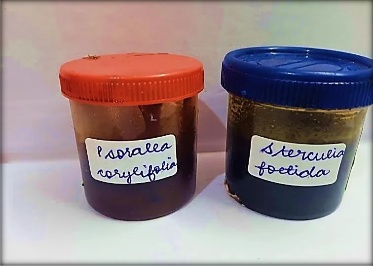
Figure 10: Percentage Yield of crude extracts of Sterculia Foetida and Psoralea Corylifolia seed extract
DISCUSSION:
The percentage yield of crude extract is an important parameter for assessing the efficiency of the extraction process. In the present study, methanolic extraction of Sterculia Foetida and Psoralea Corylifolia seeds produced different extraction yields. Sterculia Foetida showed a higher extract yield of 28.3 g (9.43%) from 300 g of seed powder, whereas Psoralea Corylifolia yielded 19.7 g (5.79%) from 340 g of seed powder. The higher yield obtained from Sterculia Foetida indicates the presence of a greater amount of methanol-
soluble phytoconstituents. The difference in extraction yield may be attributed to variations in the chemical composition of the seeds and the solubility of their bioactive compounds in methanol. These results suggest that methanol is an effective solvent for extracting phytochemicals from both plant species and provided sufficient crude extract for further phytochemical and pharmacological studies.
7.2 Preliminary Phytochemical study
The preliminary phytochemical screening of the methanolic seed extract of Sterculia Foetida revealed presence of several important secondary metabolites, including alkaloids, flavonoids, phenolics and tannins, saponins, glycosides, terpenoids, steroids, and carbohydrates. However, proteins and amino acids were absent in the extract. The presence of these phytoconstituents suggests that methanolic seed extract possesses a rich phytochemical profile, which may contribute to its therapeutic potential. In particular, phenolic compounds and flavonoids are known for their antioxidant activity, while alkaloids, glycosides, terpenoids, and saponins are associated with various pharmacological properties such as antimicrobial, anti-inflammatory, and wound-healing effects (Figure 12). These findings indicate that methanolic seed extract of Sterculia Foetida is a promising source of bioactive compounds for further formulation development and biological evaluation (Table 8).
Table 9: Qualitative Phytochemical Screening of Methanolic Seed Extract of Sterculia Foetida
| S. No. | Phytoconstituents | Test Performed | Result |
|---|---|---|---|
| 1 | Alkaloids | Mayer's Test | Present (+) |
| 2 | Flavonoids | Alkaline Reagent Test | Present (+) |
| 3 | Phenolics & Tannins | Ferric Chloride Test | Present (+) |
| 4 | Saponins | Foam Test | Present (+) |
| 5 | Glycosides | Keller–Killiani Test | Present (+) |
| 6 | Terpenoids | Salkowski Test | Present (+) |
| 7 | Steroids | Liebermann–Burchard Test | Present (+) |
| 8 | Carbohydrates | Molisch's Test | Present (+) |
| 9 | Proteins & Amino Acids | Ninhydrin Test | Absent (−) |
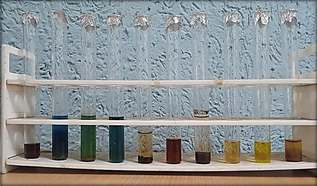
Figure 11: Phytochemical testing of Methanolic extract of Sterculia Foetida
Seed Extract
The preliminary phytochemical screening of the methanolic seed extract of Psoralea Corylifolia demonstrated the presence of several important phytoconstituents, including alkaloids, flavonoids, phenolics and tannins, glycosides, terpenoids, steroids, carbohydrates, and proteins and amino acids. However, saponins were absent in the extract (Figure 13). The presence of these bioactive compounds indicates that the methanolic extract possesses a diverse phytochemical profile, which may be responsible for its pharmacological activities. Phenolics and flavonoids are well known for their antioxidant properties, while alkaloids, glycosides, terpenoids, and steroids contribute to antimicrobial, anti-inflammatory, and wound-healing effects. Overall, the results suggest that the methanolic seed extract of Psoralea Corylifolia is a valuable source of bioactive constituents and is suitable for further formulation development and pharmacological evaluation (Table 9).
Table 10: Qualitative Phytochemical Screening of Methanolic Seed Extract of Psoralea Corylifolia
| S. No. | Phytoconstituents | Test Performed | Result |
|---|---|---|---|
| 1 | Alkaloids | Mayer's Test | Present (+) |
| 2 | Flavonoids | Alkaline Reagent Test | Present (+) |
| 3 | Phenolics & Tannins | Ferric Chloride Test | Present (+) |
| 4 | Saponins | Foam Test | Absent (−) |
| 5 | Glycosides | Keller–Killiani Test | Present (+) |
| 6 | Terpenoids | Salkowski Test | Present (+) |
| 7 | Steroids | Liebermann–Burchard Test | Present (+) |
|---|---|---|---|
| 8 | Carbohydrates | Molisch's Test | Present (+) |
| 9 | Proteins & Amino Acids | Ninhydrin Test | Present (+) |
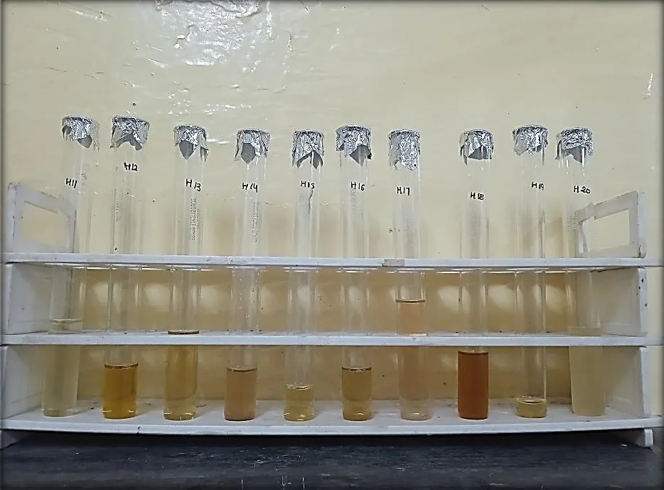
Figure 12: Phytochemical testing of Methanolic extract of Psoralea Corylifolia
Seed Extract
7.3 Quantitative Analysis
Preliminary phytochemical screening of the crude plant extracts confirmed the presence of phenolic compounds and flavonoids. Quantitative assays were subsequently carried out to determine the total phenolic content (TPC) and total flavonoid content (TFC). These analyses provide an estimate of the concentration of major bioactive constituents present in the extracts. The results help in understanding the antioxidant potential and therapeutic relevance of the plant material.
7.3.1 Total Phenolic content (TPC) estimation Table 11: Standard table for Gallic acid
| Standard Solution | Concentration (µg/mL) | Mean Absorbance (AU) |
|---|---|---|
| S1 | 20 | 0.148 |
| S2 | 40 | 0.226 |
| S3 | 60 | 0.311 |
| S4 | 80 | 0.392 |
| S5 | 100 | 0.476 |
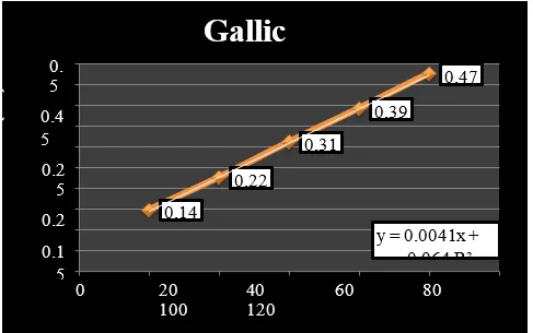
Figure 13: Represent standard curve of Gallic acid
7.3.1.1 Total Phenolic Content
The total phenolic content (TPC) of the methanolic seed extracts was determined using the Gallic acid standard calibration curve. Phenolic compounds are important plant metabolites known for their antioxidant, anti-inflammatory, and antimicrobial properties. The calibration curve of Gallic acid exhibited a linear relationship between concentration and absorbance, indicating the suitability of the method for quantitative estimation of phenolic compounds. The methanolic seed extract of Sterculia Foetida exhibited a total phenolic content of 39.26 mg gallic acid equivalent (GAE)/g of extract (Table 11), whereas Psoralea Corylifolia showed a total phenolic content of 32.19 mg GAE/g of extract (Table 12). The higher phenolic content observed in Sterculia Foetida suggests a greater abundance of phenolic constituents compared to Psoralea Corylifolia. Since phenolic compounds are major contributors to antioxidant activity, the higher TPC of Sterculia Foetida may be associated with enhanced therapeutic potential. Overall, both extracts contained appreciable amounts of phenolic compounds, supporting their use as promising natural sources of bioactive constituents for further pharmacological and formulation studies.
Table 12: Total Phenolic Content in Sterculia Foetida Seed Extract
| S. No. | Absorbance | Mean±SD | TPC in mg/gm equivalent of Gallic acid |
|---|---|---|---|
| 1. | 0.215 | 0.225±0.010536 | 39.26 mg/gm |
| 2. | 0.224 | ||
| 3. | 0.236 |
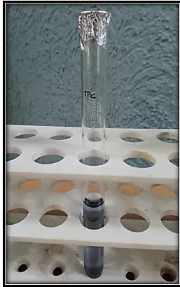
Figure 14: Total Phenolic Content in Sterculia Foetida Seed Extract Table 13: Total Phenolic Content in Psoralea Corylifolia Seed Extract
| S. No. | Absorbance | Mean±SD | TPC in mg/gm equivalent of Gallic acid |
|---|---|---|---|
| 1. | 0.187 | 0.196 ±0.010017 | 32.19 mg/gm |
| 2. | 0.196 | ||
| 3. | 0.207 |
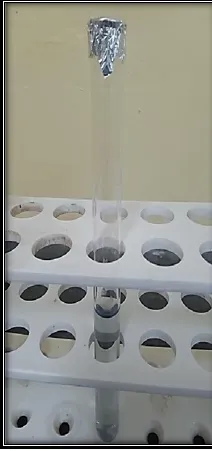
Figure 15: Total Phenolic Content in Psoralea Corylifolia Seed Extract
7.3.2 Total Flavonoids content (TFC) estimation Table 14: Standard table for Rutin
| Standard Solution | Concentration (µg/mL) | Mean Absorbance (AU) |
|---|---|---|
| S1 | 20 | 0.121 |
| S2 | 40 | 0.169 |
| S3 | 60 | 0.221 |
| S4 | 80 | 0.274 |
| S5 | 100 | 0.328 |
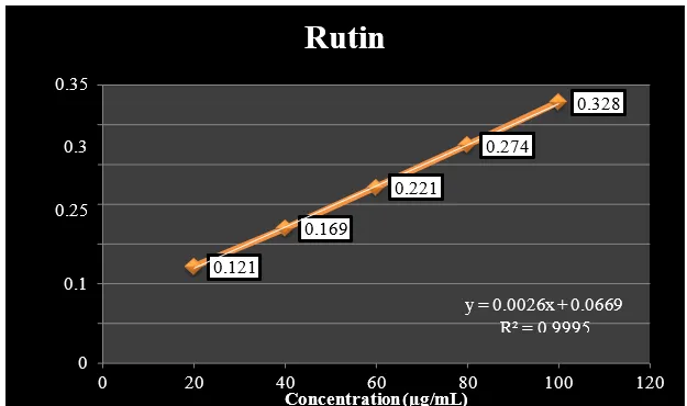
Figure 16: Represent standard curve of Rutin
7.3.2.1 Total Flavonoid Content
The total flavonoid content (TFC) of the methanolic seed extracts of Sterculia Foetida and Psoralea Corylifolia was determined using the Rutin standard calibration curve. The calibration curve exhibited a linear relationship between concentration and absorbance, indicating that the method was suitable for the quantitative estimation of flavonoids in the plant extracts. Flavonoids are important polyphenolic compounds known for their antioxidant, anti-inflammatory, antimicrobial, and wound-healing properties. The methanolic seed extract of Sterculia Foetida showed a total flavonoid content of 46.57 mg rutin equivalent (RE)/g of extract (Table 15), whereas the methanolic seed extract of Psoralea Corylifolia exhibited a total flavonoid content of 34.26 mg RE/g of extract (Table 16). The higher flavonoid content observed in Sterculia Foetida indicates that it contains a greater amount of flavonoid compounds than Psoralea Corylifolia. This difference may be attributed
to variations in the phytochemical composition and biosynthesis of secondary metabolites between the two plant species. Flavonoids are effective free radical scavengers that help protect cells from oxidative damage and contribute to various biological activities, including antimicrobial and tissue repair effects. Therefore, the higher flavonoid content of Sterculia Foetida suggests that it may possess comparatively stronger antioxidant and therapeutic potential. Nevertheless, both plant extracts contained appreciable amounts of flavonoids, supporting their use as rich sources of natural bioactive compounds. These findings further justify the selection of both extracts for the development of polyherbal ointment formulations and their subsequent pharmacological evaluation.
Table 15: Total Flavonoid Content in Sterculia Foetida Seed Extract
| S. No. | Absorbance | Mean±SD | TFC in mg/gm equivalent of Rutin |
|---|---|---|---|
| 1. | 0.179 | 0.188±0.010017 | 46.57 mg/gm |
| 2. | 0.188 | ||
| 3. | 0.199 |
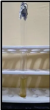
Figure 17: Total Flavonoid Content in Sterculia Foetida Seed Extract Table 16: Total Flavonoid Content in Psoralea Corylifolia Seed Extract
| S. No. | Absorbance | Mean±SD | TFC in mg/gm equivalent of Rutin |
|---|---|---|---|
| 1. | 0.149 | 0.156±0.007506 | 34.26 mg/gm |
| 2. | 0.157 | ||
| 3. | 0.164 |
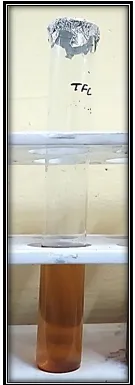
Figure 18: Total Flavonoid Content in Psoralea Corylifolia Seed Extract
7.4 Evaluation parameters of the Prepared Polyherbal Ointment Formulations
The prepared Sterculia Foetida, Psoralea Corylifolia, and polyherbal ointment formulations were evaluated for various physicochemical parameters, including pH, viscosity, spreadability, and skin irritation, to assess their suitability for topical application. The results obtained are presented below.
7.4.1 Measurement of pH, Viscosity, Spreadability, and Skin Irritation
7.4.1.1 Determination of pH
The pH of the prepared ointment formulations was evaluated to determine their suitability for topical application and compatibility with skin. The pH values of all formulations were found to be within the acceptable physiological skin pH range, indicating their safety and non-irritant nature. The pH of Formulation I containing Sterculia Foetida seed extract ointment was found to be 6.55 ± 0.03 (Table 16), while Formulation II containing Psoralea Corylifolia seed extract ointment showed a pH of 6.63 ± 0.05. The polyherbal ointment formulation (Formulation III; 1:1 ratio) exhibited a slightly higher pH value of 6.71 ± 0.04. The minor variation in pH among the formulations may be attributed to the differences in phytoconstituents present in the individual plant extracts. The obtained pH values indicate that the formulations are compatible with the natural pH of the skin and are unlikely to cause irritation or discomfort during topical application. The suitable pH of the prepared ointments supports their potential use as wound healing formulations by maintaining skin integrity and promoting patient acceptability (Figure 20).
Table 17: pH of Prepared Ointment Formulations
| Formulation | pH (Mean ± SD) |
|---|---|
| Formulation I (Sterculia Foetida Ointment) | 6.55 ± 0.03 |
| Formulation II (Psoralea Corylifolia Ointment) | 6.63 ± 0.05 |
| Formulation III (Polyherbal Ointment; 1:1) | 6.71 ± 0.04 |
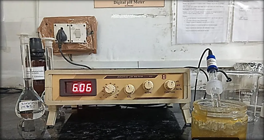
Figure 19: pH of Prepared Ointment Formulations
7.4.1.2 Determination of Viscosity
Viscosity is a critical quality attribute of ointments as it influences consistency, ease of application, and residence time on the skin. The viscosity values of the formulations ranged from 4725 ± 18 cP to 4986 ± 20 cP. Formulation I exhibited a viscosity of 4725 ± 18 cP, whereas Formulation II showed a slightly higher viscosity of 4868 ± 22 cP. The polyherbal ointment (Formulation III) demonstrated the highest viscosity (4986 ± 20 cP) (Table 16), suggesting improved consistency and structural integrity due to the synergistic incorporation of both herbal extracts. The observed viscosity values indicate that all formulations possessed adequate rheological properties for smooth application and prolonged retention at the site of administration (Figure 21).
Table 18: Viscosity of Prepared Ointment Formulations
| Formulation | Viscosity (cP) (Mean ± SD) |
|---|---|
| Formulation I (Sterculia Foetida Ointment) | 4725 ± 18 |
| Formulation II (Psoralea Corylifolia Ointment) | 4868 ± 22 |
| Formulation III (Polyherbal Ointment; 1:1) | 4986 ± 20 |
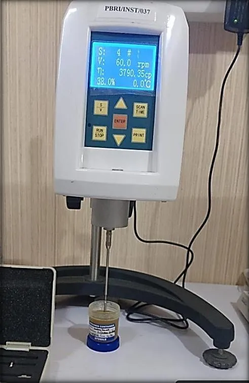
Figure 20: Viscosity of Prepared Ointment Formulations
7.4.1.3 Determination of Spreadability
Spreadability is an important parameter that determines the ease with which an ointment can be uniformly applied over the affected skin surface. The spreadability values ranged from
7.82 ± 0.12 to 8.63 ± 0.11 g·cm/s. Formulation I showed the lowest spreadability (7.82 ± 0.12 g·cm/s), while Formulation II exhibited a moderate value (8.14 ± 0.09 g·cm/s). The polyherbal ointment (Formulation III) displayed the highest spreadability (8.63 ± 0.11 g·cm/s), indicating superior ease of application and better distribution over the skin. The enhanced spreadability of the polyherbal formulation may be associated with the balanced interaction between the ointment base and the combined herbal extracts, resulting in improved patient compliance and effective topical delivery (Table 18).
Table 19: Spreadability of Prepared Ointment Formulations
| Formulation | Spreadability (g·cm/s) (Mean ± SD) |
|---|---|
| Formulation I (Sterculia Foetida Ointment) | 7.82 ± 0.12 |
| Formulation II (Psoralea Corylifolia Ointment) | 8.14 ± 0.09 |
| Formulation III (Polyherbal Ointment; 1:1) | 8.63 ± 0.11 |
7.4.1.4 Skin Irritation Test
The safety of topical formulations was assessed by evaluating the presence of erythema and edema following application. None of the prepared ointment formulations produced visible signs of erythema or edema throughout the observation period. All formulations, including Sterculia Foetida ointment, Psoralea Corylifolia ointment, and the polyherbal ointment, were
found to be non-irritant and well tolerated. These findings indicate that the ointment base and incorporated herbal extracts are dermatologically compatible and safe for topical use. The absence of any adverse skin reactions further supports the suitability of the developed formulations for subsequent pharmacological and wound-healing investigations (Table 19).
Table 20: Skin Irritation Test of Prepared Ointment Formulations
| Formulation | Erythema | Edema | Observation |
|---|---|---|---|
| Formulation I (Sterculia Foetida Ointment) | Nil | Nil | No signs of skin irritation |
| Formulation II (Psoralea Corylifolia Ointment) | Nil | Nil | No signs of skin irritation |
| Formulation III (Polyherbal Ointment; 1:1) | Nil | Nil | No signs of skin irritation |
7.4.1.5 Polyherbal Ointment Formulations
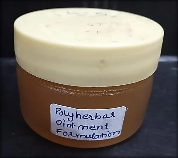
Figure 21: Polyherbal Ointment Formulations
7.5 In-vitro antimicrobial activity
The in-vitro antimicrobial activity of the prepared ointment formulations was evaluated against Staphylococcus aureus using the zone of inhibition method. The results demonstrated that all formulations exhibited antibacterial activity, although the extent of inhibition varied among the formulations. Formulation I containing Sterculia Foetida ointment produced a zone of inhibition of 15.6 mm, indicating good antibacterial activity against Staphylococcus aureus. Formulation II containing Psoralea Corylifolia ointment showed a comparatively lower zone of inhibition of 10.6 mm. In contrast, the polyherbal ointment (Formulation III) exhibited the highest antibacterial activity with a zone of inhibition of 18.3 mm, suggesting enhanced efficacy compared with the individual herbal formulations.
The superior antimicrobial activity of the polyherbal ointment may be attributed to the synergistic effect of the bioactive phytoconstituents present in both Sterculia Foetida and Psoralea Corylifolia. The presence of phenolic compounds, flavonoids, alkaloids, terpenoids, and glycosides in the extracts may contribute to bacterial growth inhibition through disruption of the microbial cell membrane, inhibition of essential enzymes, and interference with cellular metabolism. Overall, the findings indicate that the polyherbal ointment possesses better antibacterial activity than the individual herbal ointments, highlighting its potential as an effective topical formulation for the management of bacterial skin infections and wound healing.
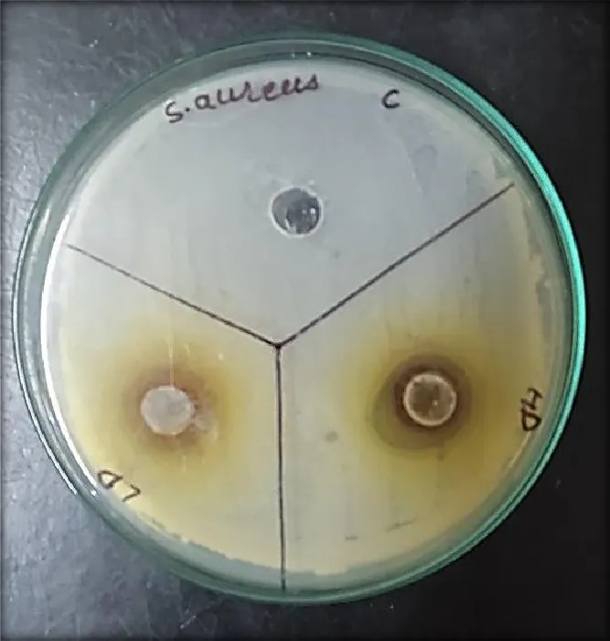
Figure 22: Antimicrobial Activity of Prepared Polyherbal Oinment Formulations Table 21: Zone of Inhibition of the Prepared Polyherbal Ointment Formulations against Staphylococcus Aureus
| S. No. | Sample Name | Zone of Inhibition (mm) |
|---|---|---|
| 1. | Formulation I (Sterculia Foetida Ointment) | 15.6 mm |
| 2. | Formulation II (Psoralea Corylifolia Ointment) | 10.6 mm |
| 3. | Formulation III (Polyherbal Ointment; 1:1) | 18.3 mm |
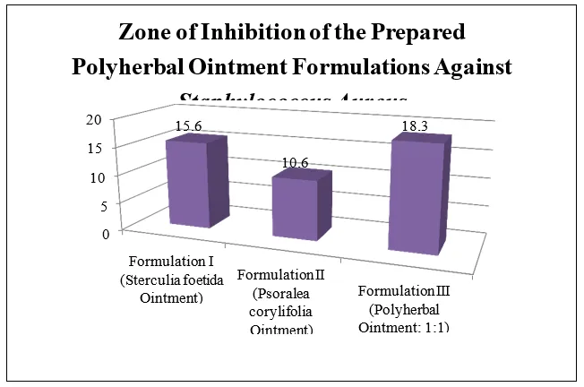
Figure 23: Antimicrobial Activity of the Prepared Polyherbal Ointment Formulations against Staphylococcus aureus (Zone of Inhibition, mm)
7.6 Wound Healing
Table 22: Percentage Wound Closure in Various Treatment Groups
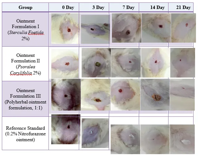
Figure 24: Excision Wound Healing from Polyherbal Ointment Formulations
| Sr. No. |
Formulation | Area of wound during different days of observation (%) |
||||
|---|---|---|---|---|---|---|
| Day 0 | Day 3 | Day 7 | Day 14 | Day 21 | ||
| 1 | Ointment Formulation I (Sterculia Foetida 2 %) |
5.10±0 .4134 |
9.31±0 .3218 |
19.26± 0.456 |
32.26± 0.4892 |
40.36± 0.5127 |
| 2 | Ointment Formulation II (Psoralea Corylifolia 2%) |
9.57±0 .6123 |
19.55± 0.5132 |
38.42± 0.5982 |
53.32± 0.5275 |
65.23± 0.5367 |
| 3 | Ointment Formulation III (Polyherbal ointment formulation, 1:1) |
12.49± 0.5167 |
22.58± 0.7212 |
44.60± 0.5526 |
67.40± 0.5131 |
75.50± 0.4565 |
| 4 | Reference Standard (0.2% Nitrofurazone ointment) |
14.26± 0.6235 |
28.47± 0.6499 |
58.11± 0.5854 |
84.30± 0.4772 |
95.79± 0.5123 |
Area of wound during different days of 120 observation (%) 100 1 Ointment Formulation I (Sterculia foetida 2%) 80 60 40 20 0 Day 0 Day 3 Day 7 Day 14 Day 21 Ointment Formulation II (Psoralea corylifolia 2%) Ointment Formulation III (Polyherbal ointment formulation, 1:1) Reference Standard (0.2% Nitrofurazone ointment)
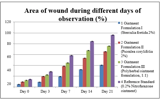
Figure 25: Evaluation of wound healing activity
DISCUSSION:
The wound healing study demonstrated a progressive increase in the percentage of wound closure in all treatment groups throughout the 21-day observation period. However, the rate of healing varied among the formulations. Ointment Formulation I containing 2% Sterculia Foetida showed the lowest wound healing activity, with wound closure increasing from 5.10 ± 0.41% on Day 0 to 40.36 ± 0.51% on Day 21. Although the formulation promoted gradual healing, its effect was comparatively less than the other treatment groups. Ointment Formulation II containing 2% Psoralea Corylifolia exhibited better wound healing
efficacy, achieving 65.23 ± 0.54% wound closure by Day 21. The improved healing may be attributed to the presence of bioactive constituents such as flavonoids, coumarins, and phenolic compounds, which possess antioxidant, antimicrobial, and anti-inflammatory properties that support tissue regeneration.
The polyherbal ointment formulation (Formulation III, 1:1 combination of Sterculia Foetida and Psoralea Corylifolia) demonstrated superior wound healing among the test formulations. The percentage of wound closure increased from 12.49 ± 0.52% on Day 0 to 75.50 ± 0.46% on Day 21, indicating a faster rate of tissue repair compared with the individual herbal formulations. The enhanced activity is likely due to the synergistic effect of phytoconstituents present in both plant extracts, which collectively promote collagen synthesis, fibroblast proliferation, angiogenesis, and epithelialization while reducing inflammation and microbial contamination at the wound site. The reference standard (0.2% Nitrofurazone ointment) produced the highest wound healing response, with 95.79 ± 0.51% wound closure on Day 21, confirming its established efficacy as a topical wound healing agent. Although the polyherbal ointment did not achieve the same level of healing as the standard drug, it exhibited substantially greater wound contraction than either herbal extract alone, highlighting the therapeutic advantage of combining the two plant extracts.
Overall, the results indicate that the polyherbal ointment formulation possesses promising wound healing potential and may serve as an effective natural alternative for promoting wound repair. Its enhanced efficacy compared with the individual formulations suggests that the combination of Sterculia Foetida and Psoralea Corylifolia provides complementary pharmacological actions that accelerate the wound healing process.
REFERENCES


Shraddha Patel, Harpreet Kaur, Evaluation of Wound Healing Potential of a Polyherbal Formulation Prepared from Sterculia Foetida and Psoralea Corylifolia Seeds, Int. J. of Pharm. Sci., 2026, Vol 4, Issue 11, 2596-2616. https://doi.org/10.5281/zenodo.24301142
 10.5281/zenodo.24301142