

We use cookies to ensure our website works properly and to personalise your experience. Cookies policy
1MBBS Third Year, Department of Pharmacology, Fergana Medical Institute of Public Health, Uzbekistan.
2Assistant Professor Department Of Pharmacology, Fergana Medical Institute of Public Health, Uzbekistan.
Ferroptosis is a regulated form of cell death driven by iron-dependent peroxidation of polyunsaturated phospholipids in cellular membranes. Since the term was introduced in 2012, the field has moved quickly from a chemical curiosity observed in RAS-mutant cell lines to a recognised vulnerability across many tumour types. Cancer cells that are mesenchymal, drug-tolerant or metabolically rewired often depend heavily on the glutathione peroxidase 4 (GPX4) axis and on parallel antioxidant systems, and this dependency can be exploited pharmacologically. This review summarises the biochemical core of ferroptosis, including iron handling, phospholipid remodelling and the GPX4, FSP1, GCH1 and DHODH defence systems. We then discuss how oncogenic and tumour-suppressor pathways such as p53, BAP1, NRF2 and Hippo signalling tune sensitivity, and how cancer cells escape ferroptosis through transcriptional, metabolic and microenvironmental adaptation. Pharmacological strategies are surveyed, from system xc? inhibition to covalent GPX4 inhibitors, with emphasis on the limits of the currently available compounds. Combination approaches with chemotherapy, targeted therapy, radiotherapy and immunotherapy are examined, along with the conflicting evidence on how ferroptotic cells shape antitumour immunity. Finally, we consider candidate biomarkers for patient selection, the obstacles to clinical translation, and research priorities. We argue that progress will depend less on discovering further inducers than on defining which tumours are truly ferroptosis-prone and on delivering inducers with acceptable selectivity.
Cell death in cancer therapy has long been discussed almost entirely in terms of apoptosis. Most cytotoxic drugs, and many targeted agents, were assumed to work by triggering caspase-dependent death, and resistance was assumed to arise mainly through defects in that machinery. This picture has been revised over the last decade, as several non-apoptotic regulated cell death programmes have been defined at the molecular level. Among them, ferroptosis has attracted particular interest because its biochemistry is unusually tractable and because it appears to be a genuine metabolic liability of certain cancer cells.
The term ferroptosis was proposed by Dixon and colleagues in 2012 to describe an iron-dependent, non-apoptotic death process triggered by the small molecule erastin [1]. Morphologically, ferroptotic cells show shrunken mitochondria with condensed membranes and little of the nuclear fragmentation seen in apoptosis. Biochemically, death can be blocked by iron chelators and by lipophilic radical-trapping antioxidants, but not by caspase inhibitors [1]. Authoritative overviews have since placed ferroptosis at the interface of metabolism, redox biology and disease [2,3], and the chemistry underlying it has been reviewed in detail [4].
The historical roots of the field lie in chemical genetics. Synthetic lethal screens in engineered cells expressing oncogenic HRAS identified erastin as a compound selectively lethal to RAS-transformed cells [5,6]. Earlier work had already described an oxidative, non-apoptotic death that depended on RAS-RAF-MEK signalling and on voltage-dependent anion channels [7]. A turning point came when GPX4 was identified as the target of the compound RSL3 and as the central enzyme preventing ferroptosis [8]. Genetic deletion of Gpx4 in mice then showed that this protective function matters in whole tissues, with inducible loss causing acute renal failure [9].
For oncology, three observations made ferroptosis attractive. First, many aggressive cancer cells have a high iron requirement and a pro-oxidant metabolism, which should in principle make them closer to the threshold of lipid peroxide toxicity than normal cells. Second, cancer cells that survive targeted therapy or enter a mesenchymal state frequently become sensitive to GPX4 inhibition, a vulnerability that could be used against therapy-resistant disease. Third, ferroptosis is regulated by nodes that can be modulated with small molecules, including the cystine transporter, GPX4 and metabolic enzymes. Broad reviews now describe the role of ferroptosis in tumour suppression and tumour progression, and the opportunities it offers for therapy [10,11].
The enthusiasm needs to be balanced against real difficulties. The first-generation inducers erastin and RSL3 have poor pharmacokinetic properties. Sensitivity differs widely between cell lines in a way that is not easily predicted by a single marker. Ferroptotic death can also have opposing effects on the immune microenvironment, and no ferroptosis-specific agent has yet been approved for cancer treatment. Reviews that treat these limitations explicitly are still comparatively scarce.
This review therefore follows the outline of the problem from mechanism to clinic. Section 2 describes the molecular machinery of ferroptosis. Section 3 examines how cancer genetics and the tumour environment modulate it. Section 4 analyses mechanisms of resistance, and Section 5 reviews pharmacological strategies for induction. Section 6 covers combination therapy, Section 7 biomarkers and patient selection, and Section 8 the state of preclinical and clinical translation. Section 9 outlines future directions, and Section 10 offers conclusions.
2. Molecular Mechanisms of Ferroptosis
Ferroptosis occurs when the capacity of the cell to repair or prevent phospholipid peroxidation is overwhelmed. Conceptually, it can be described as a balance between three inputs: the availability of redox-active iron, the amount of oxidisable polyunsaturated fatty acid (PUFA) in membrane phospholipids, and the strength of the antioxidant systems that neutralise lipid hydroperoxides and lipid radicals [12,13]. When the balance shifts towards peroxidation, membrane integrity is lost and the cell dies.
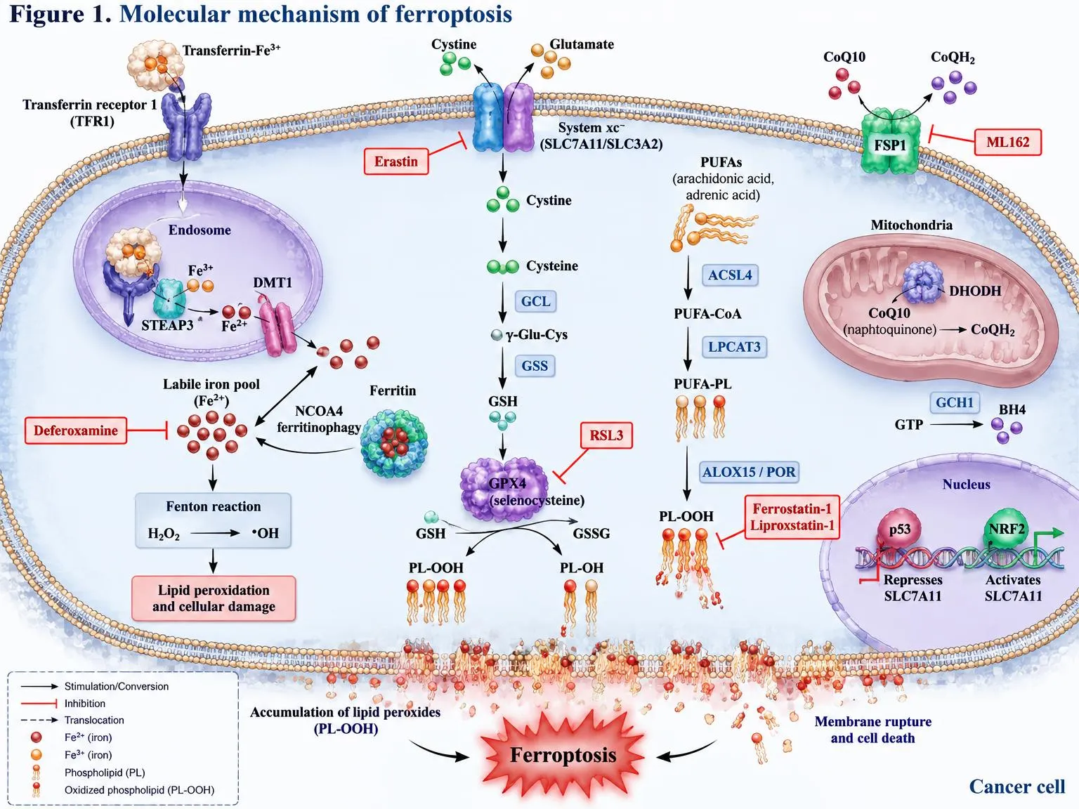
Figure 1. Molecular mechanism of ferroptosis. Iron uptake via TFR1/STEAP3/DMT1 and NCOA4-mediated ferritinophagy expand the labile Fe2+ pool, driving Fenton chemistry. ACSL4 and LPCAT3 incorporate PUFAs into membrane phospholipids that are oxidised by ALOX15/POR to PL-OOH. System xc− supplies cystine for GSH synthesis (GCL, GSS), enabling GPX4 to reduce PL-OOH to PL-OH. FSP1–CoQH2, DHODH and GCH1–BH4 provide GPX4-independent defence; p53 and NRF2 regulate SLC7A11. Inducers (erastin, RSL3) and inhibitors (ferrostatin-1, liproxstatin-1, deferoxamine) are shown in red.
2.1 Iron metabolism
Iron is required because it catalyses reactions that propagate lipid peroxidation. Free ferrous iron can generate radicals through Fenton chemistry, and iron is also a cofactor for enzymes that oxygenate lipids. Cells import iron bound to transferrin through the transferrin receptor, and store it in ferritin. Transferrin and glutamine were shown to be necessary for ferroptosis triggered by serum starvation-type conditions, which linked extracellular nutrient supply to the death programme [14].
Release of iron from ferritin depends on ferritinophagy, a selective autophagy process in which the cargo receptor NCOA4 delivers ferritin to lysosomes [15]. Several groups subsequently showed that autophagy promotes ferroptosis by degrading ferritin, and that genetic inhibition of autophagy components weakens the response to erastin and related compounds [16,17]. Iron homeostasis is therefore not only a passive requirement but an actively regulated determinant of sensitivity, and the labile iron pool is one of the more variable parameters between tumours.
2.2 Lipid metabolism and phospholipid peroxidation
Not all lipids are equally vulnerable. Bis-allylic hydrogens in PUFAs are easily abstracted, so phospholipids containing arachidonic or adrenic acid are the main substrates. Acyl-CoA synthetase long-chain family member 4 (ACSL4) activates these fatty acids, and lysophosphatidylcholine acyltransferase 3 (LPCAT3) incorporates them into phospholipids. Cells lacking ACSL4 resist ferroptosis because they contain fewer peroxidisable species [18]. Lipidomic analysis identified oxidised arachidonoyl and adrenoyl phosphatidylethanolamines as key death signals [19].
The enzymes that generate the first oxidised lipids are still debated. Lipoxygenases can oxidise PUFA-containing phospholipids and contribute to ferroptosis in some systems [20]. Cytochrome P450 oxidoreductase (POR) was found to supply electrons for peroxidation reactions and to be a necessary component in many cell lines [21]. It is likely that both enzymatic and non-enzymatic autoxidation routes operate, with the dominant one depending on cell type. Lipid composition is also shaped by ether lipid synthesis, which was shown to promote ferroptosis susceptibility in some contexts [22]. In contrast, exogenous monounsaturated fatty acids displace PUFAs from membranes and establish a ferroptosis-resistant state [23].
2.3 The GPX4 and glutathione axis
GPX4 is the only mammalian enzyme with a strong ability to reduce phospholipid hydroperoxides within membranes, converting them to the corresponding alcohols. It uses glutathione as a cofactor, and its activity depends on a selenocysteine residue. Work on selenium utilisation showed that incorporation of selenocysteine, rather than cysteine, is needed to prevent hydroperoxide-induced ferroptosis, which explains why selenium availability influences ferroptosis sensitivity [24].
Glutathione is synthesised from glutamate, cysteine and glycine, and cysteine supply is typically rate-limiting. Most cultured cancer cells obtain cysteine by importing cystine through system xc−, a heterodimer of SLC7A11 and SLC3A2, which erastin inhibits [1]. Depletion of glutathione, or direct inhibition of GPX4, therefore leads to the same endpoint: accumulation of lipid peroxides [1,8]. The mechanism of the classical ferroptosis inhibitors ferrostatin-1 and liproxstatin-1 is now understood to be radical trapping within the lipid bilayer, rather than the inhibition of a specific enzyme [25]. This distinction matters when interpreting rescue experiments, since it implies that a rescue by these compounds supports a lipid radical mechanism but does not identify the upstream trigger.
2.4 GPX4-independent defence systems
It became clear that some cell lines survive GPX4 loss. Two groups independently identified ferroptosis suppressor protein 1 (FSP1) as a component of a parallel pathway. FSP1 reduces ubiquinone (coenzyme Q10) to ubiquinol at the plasma membrane, and ubiquinol acts as a lipophilic radical-trapping antioxidant [26,27]. The second known system uses GTP cyclohydrolase 1 (GCH1), which produces tetrahydrobiopterin (BH4), a molecule that acts as a radical-trapping antioxidant and also promotes the synthesis of reduced CoQ10 and the selective retention of certain phospholipids [28]. In the mitochondria, dihydroorotate dehydrogenase (DHODH) also reduces ubiquinone, and its inhibition sensitises GPX4-low cancer cells to ferroptosis [29].
These pathways are of direct therapeutic relevance. A metabolomic and CRISPR-based analysis of cancer cell sensitivity to canonical inducers found that several metabolic features, including lipid and cysteine metabolism, determine the response [30]. It follows that single-agent GPX4 inhibition will only be effective in tumours that depend on GPX4 exclusively, and that rational combinations with FSP1 or DHODH inhibitors may be required elsewhere.
2.5 Organelles, signalling and context
The role of mitochondria in ferroptosis depends on how death is induced. Mitochondrial metabolism and membrane potential are important for death caused by cysteine deprivation, but they appear to be dispensable when GPX4 is inhibited directly [31]. This difference was shown to arise from the contribution of mitochondrial respiration and the tricarboxylic acid cycle to lipid peroxide generation, and the topic has been reviewed in the context of cellular redox regulation [32]. Further, ferroptosis surveillance independent of GPX4 has been reported to be regulated by sex hormones through the membrane-bound O-acyltransferases MBOAT1 and MBOAT2, which remodel phospholipids towards monounsaturated species. This finding has implications for hormone-driven cancers [33].
Broader metabolic influences include amino acid, lipid, iron and energy metabolism, each of which can shift the position of a cell on the continuum between sensitivity and resistance [34]. A global survey of lethal compounds also showed that ferroptosis can be induced by agents with other primary targets, and that sensitivity is regulated by cellular metabolic state [35]. Table 1 summarises the major regulators discussed above.
Table 1: Principal Molecular Regulators of Ferroptosis and Their Relevance to Cancer.
| Pathway | Key components | Effect on ferroptosis | Oncological relevance | Refs |
|---|---|---|---|---|
| Iron handling | TFRC, ferritin, NCOA4 | Higher labile iron promotes ferroptosis | Iron-avid and mesenchymal tumours may be more susceptible | [14–17] |
| PUFA phospholipid synthesis | ACSL4, LPCAT3 | Promotes peroxidisable substrate | ACSL4 level can determine response to inducers | [18,19] |
| Lipid-oxidising enzymes | ALOX family, POR | Generate or propagate lipid peroxides | Context-dependent contribution | [20,21] |
| Ether lipids and MUFAs | Peroxisomal synthesis, SCD1, MBOAT1/2 | Ether lipids sensitise; MUFAs protect | Hormone-linked and lipid-rich tumours | [22,23,33] |
| Glutathione-GPX4 axis | SLC7A11, GCLC, GSS, GPX4 | Suppresses ferroptosis | Primary target of most inducers | [1,8,24] |
| FSP1-CoQ10 axis | FSP1, ubiquinone | Suppresses, parallel to GPX4 | Explains GPX4-inhibitor resistance | [26,27] |
| GCH1-BH4 axis | GCH1, BH4 | Suppresses | Potential co-target | [28] |
| Mitochondrial defence | DHODH | Suppresses in mitochondria | Relevant where GPX4 is low | [29,31] |
3. Ferroptosis in Cancer Biology
Cancer cells are not uniformly sensitive to ferroptosis, and the variation is not random. Oncogenic signalling, tumour-suppressor status, cell state and the local environment all feed into the three determinants described in Section 2. Understanding these links is the basis for any attempt to use ferroptosis therapeutically.
3.1 Tumour suppressors as ferroptosis regulators
The best-studied example is p53. Wild-type p53 represses transcription of SLC7A11, reducing cystine import and sensitising cells to ferroptosis. Importantly, a p53 mutant lacking three acetylation sites, and therefore unable to induce cell-cycle arrest, senescence or apoptosis, retained the ability to repress SLC7A11 and still suppressed tumour growth in xenografts [36]. This result suggested that ferroptosis can contribute to tumour suppression on its own. Later work has complicated the picture, because p53 can also delay ferroptosis in certain metabolic settings, and its net effect seems to depend on cell type and stress.
BAP1, a deubiquitinase that is frequently mutated in uveal melanoma, mesothelioma and renal cancer, offers a second example. BAP1 decreases histone 2A ubiquitination on the SLC7A11 promoter and represses its expression. Cancer-associated BAP1 mutants lose this activity, which allows SLC7A11 to remain high and ferroptosis to be avoided [37]. These findings link a defined genetic lesion to a defined metabolic output, which is the type of association needed for patient stratification.
3.2 Hippo signalling and cell density
Ferroptosis sensitivity also depends on contact between cells. In epithelial cancer cells, high density activates NF2 and the Hippo pathway, which inhibits the transcriptional co-activator YAP. Where YAP is active, for example in sparse cultures or in tumours with NF2 or Hippo defects, expression of ferroptosis-promoting genes including ACSL4 and TFRC rises, and cells become more susceptible [38]. In renal cell carcinoma, the related effector TAZ was shown to regulate ferroptosis through an EMP1-NOX4 route [39]. Since YAP and TAZ are activated in many aggressive and metastatic cancers, these findings fit with the observation that mesenchymal-like cells are often more ferroptosis-prone.
3.3 NRF2 and the antioxidant response
NRF2 is the master transcriptional regulator of the antioxidant response, and it is constitutively activated in many cancers through mutations in KEAP1 or NFE2L2. NRF2 induces genes that protect against ferroptosis, including those for glutathione synthesis, iron storage and quinone detoxification. In hepatocellular carcinoma cells, activation of the p62-Keap1-NRF2 pathway raised expression of NQO1, HO-1 and ferritin heavy chain and decreased the response to erastin and sorafenib [40]. Similar protective effects were seen in glioma, where the NRF2-KEAP1 axis supported proliferation while limiting ferroptotic death [41]. Tumours with hyperactive NRF2 are therefore prime candidates for acquired resistance, and they are also candidates for NRF2 inhibition as a sensitising strategy. They may, though, depend on other pathways that provide opportunities, such as heightened reliance on glutamine.
3.4 Cystine uptake and amino acid dependency
SLC7A11 is overexpressed in many tumour types, where it supports glutathione synthesis under oxidative stress. High expression has a cost, because import of cystine and export of glutamate cause cells to become dependent on glucose and glutamine for NADPH and for glutamate balance, a phenomenon described as nutrient dependency [42,43]. This creates a therapeutic window, since tumours with high SLC7A11 may be sensitive to glucose-transporter or glutaminase inhibition, but also to cystine deprivation.
The latter has been demonstrated experimentally. Depleting cysteine with an engineered enzyme, cyst (e)inase, induced ferroptosis in pancreatic tumours in mice, and genetic deletion of Slc7a11 had similar effects [44]. These data show that dietary or enzymatic manipulation of amino acid availability can be an alternative to small-molecule inhibitors. Beyond transport, mTORC1 was found to link cysteine availability to GPX4 protein synthesis, so that mTORC1 inhibition lowers GPX4 levels and sensitises cells to ferroptosis [45]. This is a notable link because mTOR inhibitors are already used clinically.
3.5 The tumour microenvironment and metastasis
The environment encountered by a cancer cell matters as much as its genotype. Melanoma cells that metastasise through the lymphatics were shown to experience less oxidative stress and less ferroptosis than cells in the blood, because lymph contains more glutathione and oleic acid. Pre-exposure to lymph made cells more resistant to ferroptosis and improved subsequent metastasis in the blood [46]. This indicates that the ability to evade ferroptosis is a selected trait during dissemination. It also suggests that the apparent sensitivity measured in cell culture may not translate to circulating or metastatic cells in vivo. Systematic reviews have emphasised that the clinical picture depends on tissue context and that ferroptosis can promote or suppress tumours depending on circumstances [47].
4. Resistance Mechanisms
Resistance to ferroptosis inducers is likely to limit their use, and it can be intrinsic or adaptive. The mechanisms involved mirror the determinants in Section 2: reduced iron availability, remodelled lipid composition and amplified antioxidant capacity.
4.1 Cell state as a determinant of dependency
One of the most important findings in the field is that cancer cells in a therapy-resistant, mesenchymal-like state become dependent on GPX4. A chemical genetic study showed that cells resistant to multiple drugs, and cells that had undergone epithelial-to-mesenchymal transition, were killed by GPX4 inhibition at concentrations that spared their parental counterparts [48]. A related study found that cells that persist after treatment with targeted therapy, so-called drug-tolerant persisters, are vulnerable to GPX4 inhibition, and that inhibiting GPX4 in these cells prevented the emergence of resistant tumours [49]. In melanoma, dedifferentiated, neural crest-like and mesenchymal states showed greater vulnerability to ferroptosis than the melanocytic state, which suggests that lineage plasticity and ferroptosis sensitivity are connected [50].
These results are conceptually important because they reverse a common assumption. Rather than resistance meaning insensitivity to every form of death, resistance to one therapy can create sensitivity to another. This collateral sensitivity underlies much of the rationale for combining ferroptosis inducers with targeted therapy.
4.2 Upregulation of antioxidant defence
The straightforward route to resistance is increased defence capacity. Elevated NRF2 signalling raises glutathione synthesis, iron sequestration and detoxification (Section 3.3) [40,41]. Increased expression of FSP1 confers resistance to GPX4 inhibition in many cell lines, which is why FSP1 expression has been proposed as a biomarker of intrinsic resistance [26,27]. The GCH1-BH4 and DHODH systems provide further layers of redundancy [28,29]. Notably, a review of acquired drug resistance concluded that these defences are among the most common features of cancers that escape therapy, and that the same adaptations also help cells avoid recognition by the immune system [51].
4.3 Lipid remodelling
Cancer cells can reduce their exposure to peroxidisable lipids. Loss or downregulation of ACSL4 reduces incorporation of PUFAs into phospholipids and confers resistance [18]. Increased uptake or synthesis of monounsaturated fatty acids has a similar effect [23]. Hormone-regulated MBOAT1 and MBOAT2 activity offers a further route in hormone-receptor positive cancers, in which these enzymes can maintain resistance even when GPX4 is inhibited [33]. The ratio of ether and ester lipids is another tunable parameter [22]. A practical consequence is that lipidomic profiles may predict sensitivity better than the expression of individual genes, although such profiling is rarely available in routine practice.
4.4 Microenvironmental and systemic protection
As noted above, lymph-derived and other microenvironmental factors confer protection that is acquired in situ [46]. Cysteine and selenium supply, hypoxia and cell density also change the outcome [24,38,45]. This heterogeneity is likely to produce incomplete responses in vivo even when cell lines appear exquisitely sensitive. It should be taken into account when planning preclinical studies, where cell culture media contain supraphysiological levels of several nutrients.
4.5 Strategies to overcome resistance
The mechanisms above suggest several approaches. Simultaneous inhibition of GPX4 and FSP1 should be effective in tumours that depend on either alone [26,27]. Inhibition of NRF2 or of its downstream effectors could restore susceptibility in KEAP1-mutant cancers [40]. Dietary or enzymatic cysteine restriction can complement pharmacological blockade of cystine uptake [44]. Finally, targeting tumours that are drug-tolerant persisters, rather than bulk cells, may prevent relapse [49]. A recent synthesis of the literature considers ferroptosis induction as a means to reverse drug resistance in several cancer types, but notes that most of the supporting data come from cell lines and xenografts [52]. This is an important caveat, to which we return in Section 8.
5. Therapeutic Strategies to Induce Ferroptosis
Pharmacological induction of ferroptosis can be grouped by the node that is targeted. Each class has distinct strengths, and each has encountered limitations that affect how it can be developed. Table 1 gives the mechanistic background for these nodes.
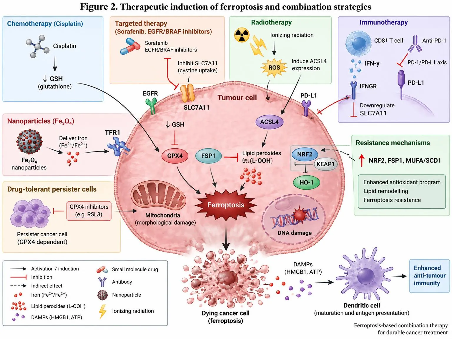
Figure 2. Therapeutic induction of ferroptosis and combination strategies. Chemotherapy (cisplatin) depletes GSH; targeted agents (sorafenib, EGFR/BRAF inhibitors) suppress SLC7A11; radiotherapy generates ROS and induces ACSL4; CD8+ T-cell-derived IFN-γ downregulates SLC7A11, enhanced by anti-PD-1; Fe3O4 nanoparticles deliver iron; drug-tolerant persister cells are GPX4-dependent. Resistance arises through NRF2, FSP1 and MUFA/SCD1 upregulation. Ferroptotic cells release DAMPs (HMGB1, ATP) that promote dendritic cell maturation and antitumour immunity.
5.1 Inhibition of cystine import
System xc− was the first target to be identified, and it remains the most accessible. Erastin inhibits the transporter, and the clinically approved drug sulfasalazine does so with lower potency [1]. In pancreatic cancer models, system xc− supports growth and contributes to resistance to chemotherapy, so its blockade was proposed as a way to sensitise tumours to treatment [53]. Sulfasalazine has also been repurposed in glioma, where it sensitised cells to gamma knife radiosurgery by blocking cystine uptake and depleting glutathione [54]. The repurposing approach is appealing because the safety profile of an approved drug is known. Its drawback is that sulfasalazine is a weak and relatively non-specific inhibitor with poor tissue penetration, and the doses needed for system xc− inhibition are difficult to reach.
Erastin itself is unsuitable for use in animals owing to low solubility and metabolic instability. A derivative, imidazole ketone erastin (IKE), was developed with improved potency and stability, and it slowed tumour growth in a mouse lymphoma model [55]. IKE is the best available tool for in vivo studies of system xc− inhibition, but its pharmacology in larger animals has not been established, and there is no published human experience.
A different approach is to exploit genetic context. In KRAS-mutant lung adenocarcinoma, suppression of the SLC7A11-glutathione axis was synthetically lethal, which indicates that oncogenic KRAS creates a specific dependency on cystine metabolism [56]. Such findings give a stronger rationale for patient selection than broad inhibition would.
5.2 Direct inhibition of GPX4
RSL3 was the first compound shown to inactivate GPX4, and it acts through covalent binding to the active-site selenocysteine [8]. In colorectal cancer cells, RSL3 induced ferroptosis through inactivation of GPX4 and the generation of reactive oxygen species, and reduced cell viability in a dose-dependent manner [57]. As a chloroacetamide, RSL3 is reactive, poorly soluble and unstable in plasma, which limits its use in animals.
Several groups have therefore sought improved GPX4 inhibitors. The compound ML210 was found to be converted inside cells to a reactive nitrile oxide that binds GPX4 covalently, which showed that masked electrophiles may be used to obtain selectivity [58]. A related class, the diacylfuroxans, also release nitrile oxides and inhibit GPX4 covalently [59]. These molecules are valuable because they expand the chemistry available, although none has reached clinical testing. Concerns remain about off-target reactivity, and about whether any GPX4 inhibitor can spare normal tissues, given that conditional Gpx4 loss was lethal to kidney tubules in mice [9].
5.3 Indirect and mechanistically distinct inducers
Not all inducers act on the two main nodes. FINO2, an endoperoxide-containing compound, was found to initiate ferroptosis by oxidising iron and by indirectly reducing GPX4 activity, with no direct inhibition of system xc− [60]. Its mechanism shows that ferroptosis can be induced by compounds that combine iron oxidation with loss of enzymatic function, which could be useful in cells that depend on redundant defences.
Direct targeting of the parallel defence systems is an active area of research. Inhibitors of FSP1 and DHODH are expected to sensitise tumours that rely on these enzymes, and to work in combination with GPX4 inhibition [26,27,29]. Dietary manipulation, in particular cysteine restriction, and enzymatic cysteine depletion offer a metabolic route that avoids the pharmacokinetic problems of small molecules [44]. Targeting the mTORC1 pathway to decrease GPX4 synthesis is a further possibility, although the effect size in tumours has yet to be established [45].
5.4 Practical limitations
Three features limit all of the above. The first is selectivity: ferroptosis machinery is present in normal tissues, and kidney, liver and neurons appear to be sensitive to loss of GPX4 [9]. The second is the lack of validated pharmacodynamic biomarkers. Lipid peroxidation products can be measured, but reliable assays for human tumour tissue are not routinely available. The third is the difficulty of confirming that an observed antitumour effect truly results from ferroptosis, since many compounds have other activities. For this reason, the use of rescue experiments with radical-trapping antioxidants, together with direct lipid peroxidation readouts, is considered the minimum standard of evidence [25]. Reviews that focus on iron-based vulnerabilities in cancer reach the same conclusion and emphasise the need for better delivery and better target validation [47].
6. Combination Therapy Strategies
Because most ferroptosis inducers have modest activity as single agents and because resistance is expected, combinations are likely to determine the clinical value of the approach. Combinations can be based on three rationales: sensitising resistant cells, adding a complementary mechanism of death, or modulating the immune response. Table 2 summarises the principal strategies.
6.1 Chemotherapy
The rationale for combining ferroptosis inducers with cytotoxic drugs rests on collateral sensitivity. Head and neck cancer cells that were resistant to cisplatin were more vulnerable to ferroptosis induced by glutathione depletion, and combining a ferroptosis inducer with cisplatin enhanced the cytotoxic effect [61]. This result is consistent with the broader finding that drug-resistant cell states show an increased dependence on GPX4 [48]. At the same time, it is not clear how general the effect is. Some cytotoxic drugs probably induce oxidative stress that increases dependence on glutathione, while others do not. The sequence and the timing of administration may also matter, and these have been little studied.
6.2 Targeted therapy
Combination with targeted therapy has strong support, mainly because of the findings on persister cells. After exposure to kinase inhibitors, a subpopulation of cancer cells survives in a drug-tolerant state that is dependent on GPX4. Inhibiting GPX4 in this setting reduced the emergence of resistant colonies [49]. Dedifferentiation of melanoma cells during therapy likewise increases vulnerability [50]. In breast cancer cells, the tyrosine kinase inhibitor lapatinib, when combined with the lysosomotropic agent siramesine, induced ferroptosis through increased iron levels that arose from altered transferrin and ferroportin expression [62]. This example shows that targeted drugs can both induce ferroptosis and enhance it in combination, and that the iron handling pathway is a feasible target for combination.
6.3 Radiotherapy
Ionising radiation induces lipid peroxidation, and ferroptosis was shown to contribute to the antitumour effect of radiotherapy. Radiation upregulated ACSL4 and reduced SLC7A11 expression, and ferroptosis inducers acted as radiosensitisers while inhibitors of ferroptosis reduced the effect [63]. The clinical relevance is supported by the earlier glioma study, where blockade of cystine uptake sensitised tumours to radiosurgery [54]. A significant implication is that radioresistant tumours with high SLC7A11 or NRF2 activity could be targeted with this combination, although the effect on surrounding normal tissue will need to be defined with care.
6.4 Immunotherapy
The interaction between ferroptosis and antitumour immunity is the most active and the most contested area. CD8⁺ T cells that are activated by immunotherapy release interferon gamma, which downregulates SLC3A2 and SLC7A11 in tumour cells, reduces cystine uptake and enhances lipid peroxidation and ferroptosis. Ferroptosis then contributes to the efficacy of anti-PD-L1 treatment [64]. Radiotherapy and immune checkpoint blockade were found to act synergistically through repression of SLC7A11, in a manner that depends on ATM and interferon gamma signalling [65]. A further study showed that CD8⁺ T cells and fatty acids act together through ACSL4, since interferon gamma enhances ACSL4-mediated incorporation of arachidonic acid, which then drives ferroptosis in tumour cells [66]. Together, these studies suggest that ferroptosis is an effector arm of T cell immunity, and that inducers might extend the activity of checkpoint inhibitors.
However, the evidence is not one-directional. Ferroptosis of tumour-infiltrating neutrophils, which have immunosuppressive properties, was shown to inhibit antitumour immunity, and blocking ferroptosis in these cells improved the response to radiotherapy in mouse models [67]. This indicates that systemic ferroptosis induction could harm the immune cells that are needed for tumour control. Studies of cell death immunogenicity also point in opposing directions. Cells that are in an early stage of ferroptosis were able to act as a vaccine and induced protective antitumour immunity [68], whereas cells that were dying from ferroptosis impeded dendritic cell-mediated immunity in another model [69]. A likely explanation is that the timing of death, the signals released and the antigen-presenting cells involved all differ between systems. At present, the available data do not allow a reliable prediction of whether a given ferroptosis inducer will promote or suppress immunity in a patient, and prospective immune-monitoring will be needed in clinical studies.
6.5 Metabolic and dual-pathway combinations
Combinations that target two defence systems have a strong theoretical basis. Simultaneous suppression of GPX4 and FSP1, or of GPX4 and DHODH, should be more effective in tumours that use both [26,27,29]. Cysteine depletion could be combined with GPX4 or mTORC1 inhibition [44,45]. These approaches have been tested mainly in cell lines and mouse models, and should be regarded as hypotheses for clinical development.
Table 2: Combination Strategies Involving Ferroptosis Induction in Cancer.
| Partner modality | Rationale | Representative evidence | Principal concern | Refs |
|---|---|---|---|---|
| Cytotoxic chemotherapy | Resistant cells depend on GSH and GPX4 | Cisplatin-resistant head and neck cancer cells are sensitised by ferroptosis inducers | Generality across drugs and tumour types is unclear | [48,61] |
| Kinase inhibitors | Persister and dedifferentiated states are GPX4-dependent | GPX4 inhibition blocks emergence of resistant colonies; lapatinib with siramesine raises iron | Toxicity of long-term GPX4 inhibition | [49,50,62] |
| Radiotherapy | Radiation induces lipid peroxidation and suppresses SLC7A11 | Inducers act as radiosensitisers; sulfasalazine sensitises glioma | Normal tissue damage | [54,63] |
| Immune checkpoint blockade | IFN-γ from CD8⁺ T cells represses SLC7A11 and drives ACSL4-dependent death | Ferroptosis contributes to anti-PD-L1 and radio-immunotherapy effect | Ferroptosis of immune cells may be immunosuppressive | [64–67] |
| Immunogenic cell death | Early ferroptotic cells can act as a vaccine | Protective antitumour immunity in mice | Opposing findings with dendritic cells | [68,69] |
| Dual defence blockade | Parallel pathways provide redundancy | GPX4 loss is compensated by FSP1 and DHODH | Selectivity and delivery of FSP1 or DHODH inhibitors | [26,27,29] |
| Nutrient manipulation | Cysteine or mTORC1 supports GPX4 | Cyst(e)inase induces ferroptosis in pancreatic tumours; mTORC1 inhibition lowers GPX4 | Systemic effects, feasibility of dietary control | [44,45] |
7. Biomarkers and Patient Selection
A ferroptosis-based therapy will only work if the tumours that are treated are those that depend on the targeted pathway. The literature already contains several candidate selectors, but none has been prospectively validated in patients, and this is arguably the largest gap between the laboratory data and clinical use.
7.1 Candidate predictive markers
Genetic markers are the most practical, because tumour sequencing is routine. Loss-of-function mutations in BAP1 remove repression of SLC7A11 and might predict sensitivity to approaches that impair cystine metabolism, although the same mutation could also increase resistance to direct cystine blockade because SLC7A11 is elevated [37]. Activation of KRAS and loss of NF2 were linked to dependency on the cystine-glutathione axis and to YAP-driven sensitivity, respectively [38,56]. In contrast, mutations in KEAP1 or NFE2L2 that activate NRF2 are expected to predict resistance to most inducers [40,41].
Expression-based markers are also under consideration. High ACSL4 expression predicts sensitivity to inducers because it increases the pool of peroxidisable lipids [18], whereas high FSP1 expression predicts resistance to GPX4 inhibition [26,27]. A cell state signature associated with mesenchymal or dedifferentiated features may indicate GPX4 dependency [48–50]. For glutathione-depleting approaches, expression of SLC7A11 itself is informative, although it can point in either direction depending on the intervention, as noted above [42,43].
7.2 Metabolic and lipid readouts
Because the end point of ferroptosis is lipid peroxidation, direct measurement of the lipid pool should in principle be the most informative approach. Mass spectrometry can identify the oxidised phosphatidylethanolamine species that mark ferroptotic death [19], and lipidomic profiles can reflect the balance between PUFA, MUFA and ether lipid content [22,23]. Cysteine and glutathione levels in tissue provide a complementary view of antioxidant capacity [44,45]. At present these methods require specialised equipment and fresh or appropriately preserved tissue, and no standardised assay is available for clinical use.
7.3 Challenges in biomarker development
Three problems recur. First, sensitivity is multifactorial. A single-marker test is unlikely to be adequate, and composite scores that combine genetic, expression and lipid information may be needed [30]. Second, markers measured in cultured cells may not reflect the in vivo state, as the example of lymph-mediated protection demonstrates [46]. Third, pharmacodynamic markers to show target engagement, such as a change in lipid peroxidation in a biopsy or in blood, are lacking. Until these are available, early clinical studies will have to rely on broad enrichment strategies, and will need to include serial tissue sampling to learn which markers matter.
8. Preclinical and Clinical Translation
8.1 Preclinical evidence
The preclinical case rests mainly on cell lines, xenografts and genetically engineered mouse models. Antitumour effects of ferroptosis induction in vivo have been shown with IKE in lymphoma [55], with cyst (e)inase in pancreatic cancer [44], with p53 mutants in xenografts [36], and with GPX4 inhibition against persister cell populations [49]. Radiotherapy and immune checkpoint studies in mice support the role of ferroptosis as an effector of established treatments [63,64]. Together, these data show that the concept is valid in animals.
Several limitations must be acknowledged. Cell culture media contain high levels of cystine, iron-binding proteins and antioxidants that differ from those in tissue, and may bias sensitivity [46]. Many xenograft studies use immunodeficient animals and so cannot capture the immune effects described in Section 6.4 [67,69]. Few studies have measured the toxicity of long-term GPX4 or system xc− inhibition in the organs known to be sensitive, such as the kidney [9]. The reproducibility of some findings across laboratories has also not been systematically assessed. These considerations argue for syngeneic and genetically engineered models, with direct lipid peroxidation readouts, in the next phase of preclinical work.
8.2 Clinical status
There is no ferroptosis-specific drug approved for cancer treatment, and to our knowledge no direct GPX4 inhibitor has entered clinical testing. The clinical experience that exists is indirect. Sulfasalazine, which is approved for inflammatory diseases, inhibits system xc− and has been explored in oncology as a repurposed agent [1,54], and sorafenib has been proposed to induce ferroptosis in some hepatocellular carcinoma models [40]. In both cases, it remains uncertain how much of any clinical effect depends on ferroptosis. Authors who prepare submissions based on this review should check clinical trial registries for current studies, since the field is moving quickly.
The main obstacles to translation are the following. Selective, orally available inhibitors with acceptable pharmacokinetics are not yet available [55,58,59]. The therapeutic window against normal tissue is uncertain [9]. Biomarkers are not validated (Section 7), and the immune consequences of ferroptosis are not predictable (Section 6.4). Reviews of drug resistance conclude that the biology is convincing but that clinical proof of concept is still missing [51,52].
9. Future Perspectives
Several lines of work are likely to determine whether ferroptosis becomes a useful therapeutic principle.
The first is improved chemistry. Masked electrophiles and other strategies that give selective GPX4 inhibition are promising [58,59], as are inhibitors of the parallel systems FSP1 and DHODH [26,27,29]. Compounds that act through iron oxidation, such as FINO2, extend the chemical space [60]. Pharmacokinetic optimisation will be as important as target potency, as the history of IKE shows [55].
The second is better definition of context. Hormone-dependent regulation by MBOAT1 and MBOAT2 [33], cell density and Hippo signalling [38,39], mTORC1 activity [45] and tissue-specific nutrient availability [46] all influence the response. Mapping these factors in patient-derived models and in clinical samples would allow more precise selection of patients.
The third is the immune dimension. The contradictory results on how ferroptotic cells affect immunity [64–69] need to be resolved with models that preserve an intact immune system and with attention to timing, dose and cell type. If the conditions that favour immunogenic ferroptosis can be defined, combination with checkpoint blockade could be made rational. If not, the use of ferroptosis inducers might need to be restricted to settings in which the immune system plays a minor role.
The fourth is nutritional and metabolic intervention. Cysteine restriction and other dietary approaches have shown efficacy in animals [44] and could be combined with drugs, but the safety and feasibility of such approaches in patients with cancer have to be established.
Finally, the field needs common standards. Consistent definitions of ferroptosis, requirement for lipid peroxidation measurement, use of rescue controls and reporting of negative results would improve comparability between studies [4,25].
CONCLUSION
Ferroptosis has moved in little more than a decade from a descriptive observation to a mechanistically defined form of regulated cell death with clear relevance to cancer. The core machinery, which combines iron, peroxidisable phospholipids and the GPX4, FSP1, GCH1 and DHODH defence systems, is now well characterised [1–4,26–29]. Cancer genetics and cell state modulate the response, and resistant or dedifferentiated states are often more vulnerable [36–38,48–50]. These findings suggest that ferroptosis induction could be used to attack tumours that escape conventional therapy, and that combination with chemotherapy, targeted drugs and radiotherapy may be the most credible route [49,61,63].
Open problems remain. Available inducers lack the properties needed for clinical use, resistance mechanisms are numerous, biomarkers are not validated, and the effect of ferroptosis on antitumour immunity is still disputed. Resolving these issues will require improved compounds, careful models and early clinical studies designed to measure target engagement. If these requirements are met, ferroptosis-based approaches could add a new mechanism of cell death to the oncologist's options, particularly for patients whose tumours have become resistant to other treatments.
Declarations
Funding, conflicts of interest, author contributions and data availability statements to be completed by the authors, as required by the target journal.
REFERENCES


Samiksha Anandache, Shivprasad Dhage, Ferroptosis-Based Cancer Therapy: Molecular Regulation, Resistance Mechanisms, and Combination Strategies, Int. J. of Pharm. Sci., 2026, Vol 4, Issue 11, 846-861. https://doi.org/10.5281/zenodo.23971108
 10.5281/zenodo.23971108