

We use cookies to ensure our website works properly and to personalise your experience. Cookies policy
G. H. Raisoni Institute of Life Sciences, Nagpur.
Wound healing continues to be a major concern in healthcare, creating a need for safe and effective treatment approaches. Neem, a well-known medicinal plant, has been widely studied for its ability to support tissue repair and accelerate the healing process. In this study, a transdermal patch containing Neem and marigold extracts was developed and evaluated for its wound healing potential. The findings indicate that the herbal transdermal patch enhances the wound healing process and may serve as a promising alternative or supportive therapy for wound management. Although the results are encouraging, further studies are required to better understand its mechanism of action and to optimize the formulation for future clinical use. This research highlights the potential of combining herbal extracts in transdermal drug delivery systems as an effective strategy for improving wound healing.
Transdermal drug delivery systems (TDDS) are an advanced method of delivering therapeutic agents through the skin in a controlled and sustained manner. These systems provide several advantages over conventional dosage forms, such as improved patient compliance, prolonged drug release, reduced dosing frequency, avoidance of first-pass metabolism, and fewer gastrointestinal side effects. Due to these benefits, transdermal patches have gained significant attention in the treatment of various skin disorders, including wound healing.[1,2]
Wound healing is a natural and highly coordinated biological process that restores the structure and function of damaged skin. It occurs through four overlapping phases: hemostasis, inflammation, proliferation, and tissue remodeling.[3,4] Successful wound healing requires protection from microbial infection, control of inflammation, adequate collagen synthesis, angiogenesis, and regeneration of healthy skin tissue. Delayed healing caused by infection, oxidative stress, or chronic inflammation can lead to serious complications, highlighting the need for effective wound-care therapies.[5,6]
Herbal medicines have become increasingly popular in wound management because they are generally safe, economical, and rich in bioactive compounds with therapeutic potential.[7] Neem (Azadirachta indica) is widely recognized for its antimicrobial, anti-inflammatory, antioxidant, and wound-healing properties. It contains phytochemicals such as azadirachtin, nimbidin, quercetin, and flavonoids that help reduce microbial growth, minimize inflammation, and promote tissue repair.[8,9] Marigold (Tagetes erecta) is another valuable medicinal plant known for its ability to accelerate wound healing. It contains triterpenoids, flavonoids, carotenoids, and essential oils that stimulate collagen production, enhance epithelial regeneration, improve blood circulation, and reduce oxidative damage at the wound site.[10]
The combination of Neem and Marigold extracts in a transdermal patch offers a synergistic approach to wound healing by providing antimicrobial protection, reducing inflammation, and promoting faster tissue regeneration.[11] A transdermal patch ensures continuous and controlled release of the herbal constituents directly at the affected site, maintains a moist environment favorable for healing, minimizes frequent dressing changes, and improves patient comfort. The use of suitable polymers, plasticizers, and permeation enhancers further enhances the mechanical properties and drug-release profile of the patch.[12]
The present study aims to formulate and evaluate a herbal transdermal patch containing Neem (Azadirachta indica) and Marigold (Tagetes erecta) extracts for wound-healing applications. The prepared patches will be evaluated for various physicochemical properties, including thickness, weight variation, folding endurance, moisture content, surface pH, skin irritation, and stability. The developed formulation is expected to provide a safe, effective, and sustained herbal therapy for enhanced wound healing.
MATERIAL AND METHODS:-
Herbal extract powders of Neem and Marigold are obtained from the process of extraction (Soxhlet extraction) of dry leaves and flowers respectively. Petroleum Ether, Chloroform, Ethyl Acetate, Ethanol were used for the extraction of active constituents from the herb. Hydroxypropyl Methylcellulose (HPMC), Chloroform, Methanol, Polyethylene Glycol-400, Glycerol were used in the formulation of the hebal patch. The other equipment used in the formulation of herbal patches were petri dish, electronic balance, hot water bath, hot air oven, magnetic stirrer, vernier caliper, etc.
Plant Authentication:-
Fresh leaves of neem (Azadirachta indica) belonging to family Meliaceae and flowers of Marigold (Tagetes erecta) belonging to family Asteraceae were collected from Shree Sai Herbal shop of Nagpur, Maharashtra, India. The collected plant materials were washed, shade dried, and powdered. The plants were authenticated by a qualified Botanist from the Department of Botany, Rashtra Sant Tukadoji Maharaj University (RTMNU), Nagpur. Voucher specimens were submitted and preserved in the departmental herbarium for future reference.
Experimental Methodolgy:-
The fresh marigold flowers (Tagetes erecta, family Asteraceae) and neem leaves (Azadirachta indica, family Meliaceae) were collected and shade-dried at room temperature for 4-5 days and coarsely powdered and stored in airtight containers.
Process of Extraction: -
Step 1:- Maceration method (Deffating)
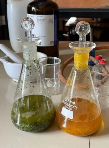
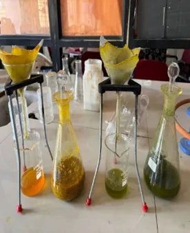
Fig: Maceration process of Powdered Neem leaves and Marigold flowers
Step 2:- Soxhlet Extraction (Successive Extraction Method)
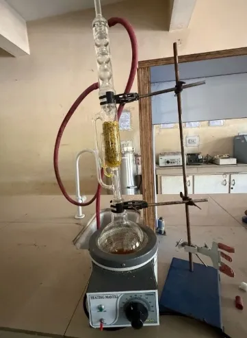
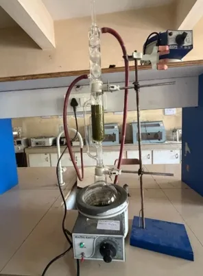
Fig: Soxhlet Extraction Assembly
Phytochemical Screening of Plant Extracts: - [17-20]
| Sr. No. | Identification Test | Observation | Inference |
|---|---|---|---|
| 1. | Alkaloids a. Dragendroffs’s Test: Add 1-2 ml Dragendroff reagent to filtrate |
Yellow coloured precipitate | Presence of Alkaloids |
| 2. | Glycosides a. Legal test: To 2 ml test solution, pyridine and alkaline sodium nitroprusside is added |
Blood red or pink colour | Presence of Glycosides |
| 3. | Flavonoids a. Shinoda Test: 2 ml test solution is added with few fragments of Magnesium ribbon, drop wise conc. H2SO4 is added |
Pink scarlet or Crimson red colour | Presence of Flavonoids |
| 4. | Tannins a. Ferric Chloride Test: The extract solution mixed with drops of ferric chloride solution |
Blue/green, black colour precipitate | Presence of Tannins |
| 5. | Proteins & Amino acids a. Millon’s Test 2 ml test solution is added with Millon’s reagent and heat |
White precipitate turns to red colour precipitate | Presence of Proteins |
| 6. | Carbohydrates a. Molish Test: To 2 ml test solution add Molish reagent, shake and add conc. H2SO4 from sides of test tube |
Formation of violet ring at junction | Presence of Carbohydrates |
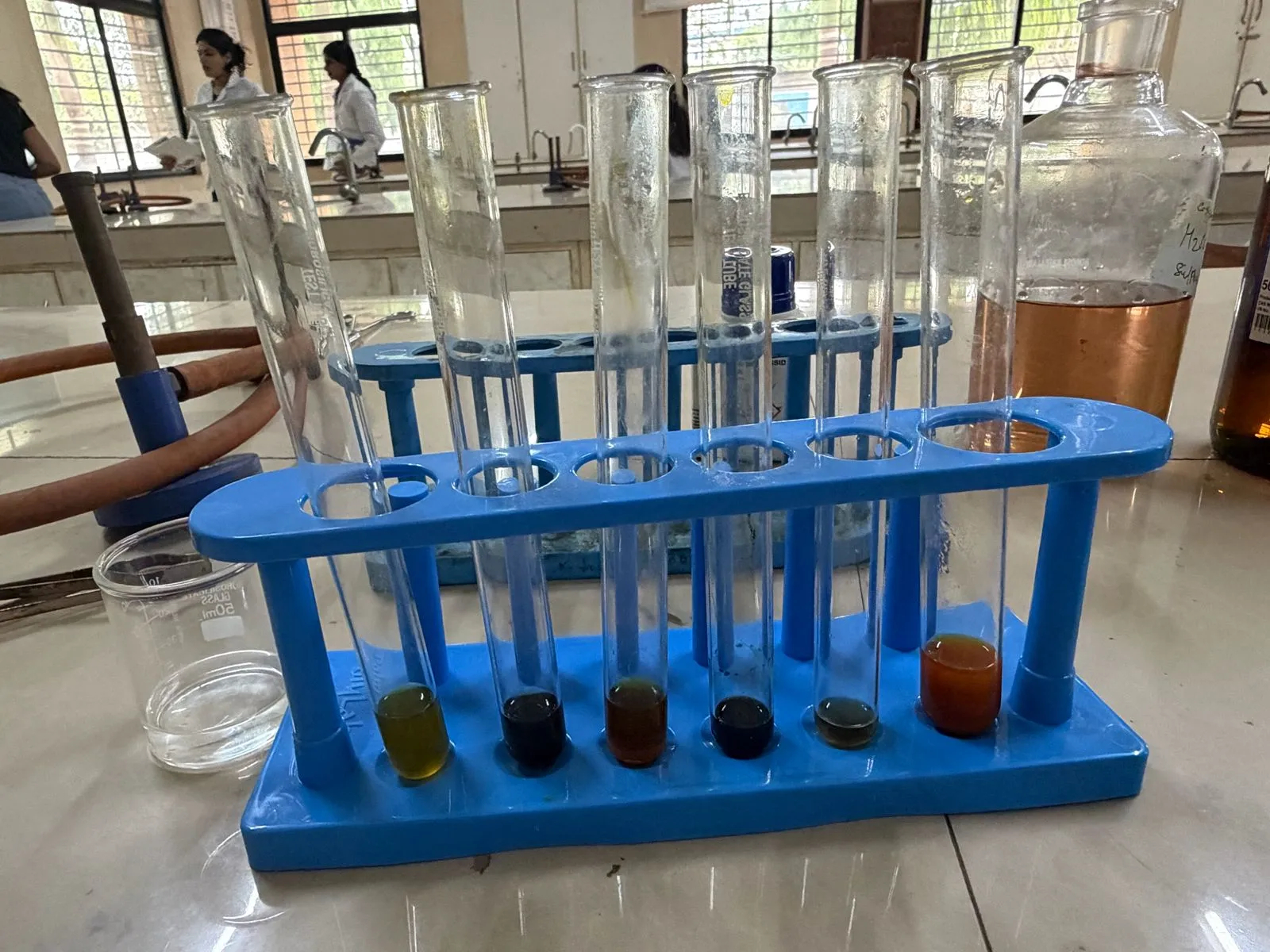
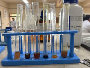
Fig: Phytochemical Screening of Neem & Marigold Extract
Preparation of Transdermal Patch:-[21]
Patch preparation was done through solvent-casting technique because it is a simple, cost-effective and compatible technique with thermosensitive herbal actives. A procedure as follows was adopted:
➢ Add 8 ml of chloroform and 4 ml of methanol to a beaker and stir them properly using the magnetic stirrer.
➢ Then add 2.5 ml glycerol and 2.5 ml polyethylene glycol-400 and again stir it continuously using an magnetic stirrer.
➢ Each medicinal plant extract, i.e., 5ml neem leaves extract and 5ml marigold flower extract, was added with constant stirring for 10-15 minutes. 20
➢ Now, then add HPMC with constant stirring using a magnetic stirrer. But add 1 gm of HPMC at the time interval minutes.
➢ After a time period of 10-15 minutes, when the formulation became viscous, it was added to the glass petri plates, which were coated using the aluminium foil.
➢ Then the petri plate was placed in the Hot Air Oven at 50 degrees for a certain time period for the evaporation of solvent.
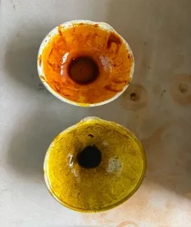
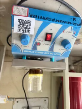
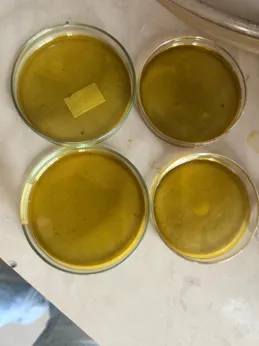
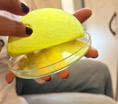
Fig: Formulation of herbal transdermal patch
Evaluation of Transdermal Patch :-[22-24]
I. Organoleptic Characteristics :- The physical appearance of developed patch was evaluated by using a naked-eye examination for its appearance, colour, clarity, flexibility, and smoothness.
II. Physico-Chemical Evaluation
A. Thickness of Patch:- A Vernier caliper was used to measure patch thickness uniformity at six different places. The mean thickness of all six places was then determined.
B. Determination of Surface pH:- The pH of the patch is evaluated by swelling it with 1 ml of distilled water for two hours at room temperature before use. Then, place the pH electrode on the patch's surface to record the pH value and make them to adjust itself for 1 minute.
C. Percent moisture content :- The percent moisture content was studied using Desiccator. Initially the individual patches were weighed and then kept in the desiccator containing activated silica at the Room temperature for time period of 24 hours. Then afterwards the Patches were reweighed. The percent moisture content can be calculated using the following formula:
%Moisture content = (Initial weight - Final weight) / Initial weight × 100
D. Folding endurance:- Folding endurance was evaluated by folding the patches repeatedly in the same area even after it broke. Folding endurance is the number of times the patches can be folded in the same area without breaking.
E. Uniformity of weight :- Each of the three patches were weighed for each batch, and the mean weight were determined.
F. Moisture Uptake:- At room temperature the previously Weighed patches were placed in desiccators for 24 hours in a saturated potassium chloride solution to keep 84% relative humidity. After 24 hours, the patches were reweighed and the % moisture uptake was calculated using the following formula.
%Moisture uptake = (Final weight - Initial weight) / (Initial weight × 100)
G. Skin irritation test [25]
Before applying the patch, the dorsal skin of a volunteer was washed with 70% ethanol. The patches were applied on right forearm for 24hrs. After 24 hours, the patches were removed and the forearms were cleansed with saline. The cutaneous responses were assessed by observing erythema, edema, pruritus and urticaria, skin allergy and irritation at 15 minutes, 1 hour and 24 hours after the test patch was removed.
H. Anti–Microbial Assay Procedure:-
The antimicrobial activity of the extracts was evaluated using the disc diffusion method. In this method, nutrient agar was prepared, sterilized, and poured into sterile Petri dishes. After solidification, the agar surface was inoculated with the test bacterial culture. Sterile filter paper discs impregnated with different plant extracts were placed on the inoculated agar. The plates were then incubated at 37°C for 24 hours. After incubation, the zone of inhibition around each disc was measured in millimeters. A larger zone of inhibition indicated stronger antimicrobial activity of the extract. All procedures were carried out under aseptic conditions in a laminar airflow cabinet using sterilized glassware to prevent contamination.[26]
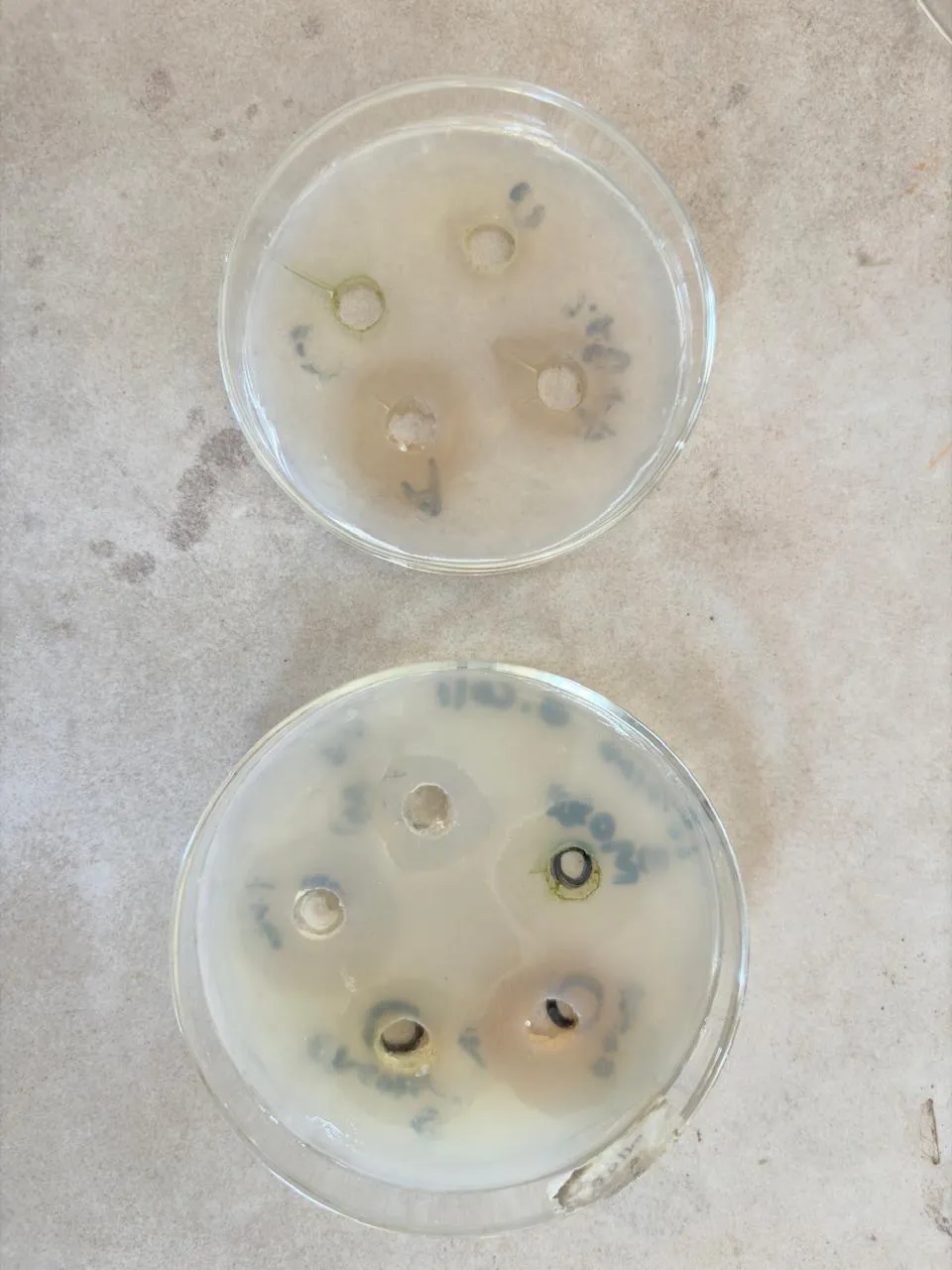
Fig: Anti-microbial activity of Patch against E.coli and P. aeruginosa
RESULTS & DISCUSSION
Phytochemical screening
Phytochemical screening was performed to identify the bioactive compounds present in neem and marigold extracts. The ethanolic extracts showed the highest presence of phytochemicals such as alkaloids, flavonoids, glycosides, tannins, carbohydrates, and proteins. These compounds are responsible for the antimicrobial, antioxidant, and wound-healing properties of the plants, making them suitable for herbal transdermal patch formulation. Results are represented in Table 1.1 and Table 1.2
Table 1.1: Phytochemical Screening of Azadirachta Indica (Neem) Extract
| Phytochemical | Test Applied | Chloroform extract | Ethyl Acetate extract | Ethanolic extract |
|---|---|---|---|---|
| Alkaloids | Dragendroff’s Test | - | + | + |
| Glycosides | Legal Test | - | + | + |
| Flavonoids | Shinoda Test | - | + | + |
| Carbohydrates | Molish Test | - | - | + |
| Proteins /Amino Acids | Millon’s Test | - | - | + |
| Tannins | Ferric Chloride Test | - | + | + |
Table 1.2: Phytochemical Screening of Tagetes Erecta (Marigold) Extract
| Phytochemical Test | Test Applied | Chloroform extract | Ethyl Acetate extract | Ethanolic extract |
|---|---|---|---|---|
| Alkaloids | Dragendroff’s Test | - | - | + |
| Glycosides | Legal Test | - | + | + |
| Flavonoids | Shinoda Test | - | + | + |
| Carbohydrates | Molish Test | - | - | + |
| Proteins /Amino Acids | Millon’s Test | - | - | + |
| Tannins | Ferric Chloride Test | - | + | + |
Physicochemical and Mechanical Evaluation of Herbal Transdermal Patches
The prepared herbal transdermal patches (F1, F2, and F3) were evaluated to determine their physicochemical and mechanical properties. These tests help to ensure that the patches are suitable for topical application and provide effective wound healing. The evaluation included thickness, weight variation, surface pH, folding endurance, moisture content, moisture uptake, and visual appearance. These parameters are important for assessing the quality, stability, flexibility, and patient acceptability of the patches. The results of all formulations are presented in Table 2.1.
Table 2.1: Physicochemical and Mechanical Evaluation Parameters of Patch Formulations F1, F2, and F3
| Evaluation Parameter | F1 | F2 | F3 |
|---|---|---|---|
| Thickness (mm) | 0.28 | 0.35 | 0.42 |
| Weight (mg/patch) | 312 | 387 | 465 |
| Surface pH | 6.2 | 6.0 | 5.8 |
| Folding Endurance (folds) | 145 | 152 | 155 |
| Moisture Content (%) | 3.3 | 2.9 | 2.6 |
| Moisture Uptake (%) | 6.8 | 6.0 | 5.2 |
| Visual Appearance | Smooth, slight opacity | Smooth, uniform, slight translucent | Smooth, uniform, translucent |
5.5 Antimicrobial Activity
The agar well-diffusion method was used to evaluate antimicrobial activity of the optimised formulations against the two principle bacterial strains: Escheriachia coli and Pseudomonas aeruginosa. Table 5.5 summarised the results of the zones of inhibition (ZOI) measured after 24-48 h incubation at 37 oC.
Table 2.2: Antimicrobial Activity – Zone of Inhibition (mm) Against E. Coli and P. Aeruginosa
| Test Sample | E. coli ZOI (mm) | P. aeruginosa ZOI (mm) | Interpretation |
|---|---|---|---|
| F1 | 13.0 | 11.5 | Moderate activity |
| F2 | 16.5 | 14.2 | Good activity |
| F3 | 18.8 | 16.8 | Good activity |
| Amoxicillin (+ve ctrl) | 21.0 | 22.5 | Excellent activity |
| Placebo (-ve ctrl) | 0 (No inhibition) | 0 (No inhibition) | No activity |
CONCLUSION
The present study successfully developed and evaluated a herbal transdermal patch containing Azadirachta indica (Neem) and Tagetes erecta (Pot Marigold) extracts for wound healing. The formulated patches exhibited satisfactory physicochemical, mechanical, and antimicrobial properties, making them suitable for topical application. The combination of both herbal extracts showed promising wound-healing potential due to their antimicrobial, anti-inflammatory, and antioxidant activities. Overall, the developed herbal patch represents a safe, effective, and cost-effective alternative for wound management. Further in vivo and clinical studies are recommended to validate its therapeutic efficacy.
REFERENCES

Pradhnya Kakde, Prachi Chavhan, Sarika Meshram, Formulation and Evaluation of Herbal Transdermal Patch Used in Wound Healing, Int. J. of Pharm. Sci., 2026, Vol 4, Issue 10, 2106-2114. https://doi.org/10.5281/zenodo.23360394
 10.5281/zenodo.23360394