
We use cookies to ensure our website works properly and to personalise your experience. Cookies policy
Vidyaniketan College of Pharmacy, Takarkheda more road, Anjangaon Surji.
Because of their high moisture retention, biocompatibility, and capacity to distribute therapeutic agents directly to the afflicted area, hydrogel-based topical formulations have drawn a lot of attention for the treatment chronic wound infections. In order to improve wound healing and stop microbial growth, the current work focuses on the development and assessment of an antimicrobial hydrogel system. To enable continuous release at the wound site, the hydrogel was made using appropriate polymers and filled with an antibacterial medication. The efficacy of the formulation was evaluated using the variety of physicochemical and the biological tests, including the drug absorption, swelling behavior, antibacterial activity, and rheological analysis. The produced hydrogel shown excellent antibacterial activity and good Spreadability, stability, suggesting that it could be used to treat the infected wounds. Furthermore, the hydrogel's wet environment may promote the quicker tissue regeneration and lessen patient discomfort. Overall, the findings imply that the hydrogel is loaded with antimicrobial drugs that may be used in topically to treat the microbial infections as a safe and the efficient wound dressing.
The largest organ in the body, human skin, acts as a remarkable protective barrier that divides the internal environment from the external surrounding. The skin, owing to its multifunctionality, is vital for moisturization, thermoregulation, sensory perception, maintenance of humoral equilibrium, and protection against pathogens. Nonetheless, when this organ is subjected to external influences over an extended period, it can incur considerable strain. As a result, it may become vulnerable to numerous types of stimuli. When the skin's integrity is compromised, for example through wound formation, it can serve as a predisposing factor for various diseases.[1] Physical injuries that cause the skin to break or open are called wounds. To restore altered anatomical continuity and compromised functional status, wounds must heal properly. The epidermis. Restoring the integrity and functionality of injured tissues is the goal of the intricate process known as healing. Continuous interactions between cells and the matrix enable wound healing to occur in three overlapping phases: inflammation (3days), cellular proliferation (3–12 days), and remodeling (3–6 months). To restore the anatomical continuity and function of the afflicted part, the fundamental idea behind optimum wound healing is to limit tissue damage and provide sufficient tissue perfusion and oxygenation, appropriate nutrition, and a moist wound healing environment.[2]
Hydrogels are polymeric networks arranged in three dimensions that can take up water without disintegrating. Hydrogels have been investigated for important uses in wound healing, drug delivery, water purification, tissue engineering, scaffolding, and 3D printing. Hydrogels are characterized by a high surface-area-to-volume ratio, which enables rapid response and optimal interaction with the surrounding tissue. This is their main advantages.[3]
Hydrogels are classified into two categories based on the nature of bonding within the network: physical and chemical. When a crosslinker is added, it leads to the formation of covalent bonds between the polymer chains, resulting in the creation of a chemical hydrogel. On the other hand, a physical hydrogel arises when the polymer chains bond through hydrogen interactions, ionic forces, or other physical mechanisms. Hydrogels that are physically crosslinked do not necessitate a chemical crosslinker and are simpler to synthesize. However, they are easier to disturb than chemically crosslinked hydrogels.[4] Current methods for treating wounds encompass covering patches, gels, natural remedies like turmeric and aloe vera, sutures, and medications. Nonetheless, each of these approaches has some limitations. Covering Patches serve solely to shield the wound site without offering any treatment. Natural remedies and medicines, such as gels, aid the healing process, but their effect is slow. Sutures are quite painful because they are direct and invasive. [5]
Physiology of Wound Healing
Depending on how long they take to heal, wounds are divided into two groups: acute and chronic. In general, a wound can be classified as chronic if it takes at least twelve weeks to heal. It can be classified as a wound that shows no evidence of healing, though, if the healing process takes longer than 30 days. Numerous internal and external factors influence the rate of healing. Reduced healing rates can be attributed to a number of reasons, including a shortage of extracellular matrix (ECM) proteins, increased production of reactive oxygen species (ROS), worsened swelling, hypoxia, decreased collagen formation, and attenuation of angiogenic growth factors. Chronic wounds include diabetic foot ulcers, pressure ulcers, and vascular ulcers. The wound's underlying protective epithelial layer has been damaged, making it useless. There is an ordered sequence of steps in the wound healing process. Slowdowns in this flow result in a cycle of inflammation, which makes the wound chronic. Increased ROS, chemokines, cytokines, and other chemicals correlated with inflammation are typically responsible for an increase in M1 macrophages and neutrophils at the chronic wound site. These cells also secrete matrix metalloproteinase (MMPs) molecules and reactive oxygen species (ROS), all of which contribute to inflammation. Scar tissues are the outcome.[6]
Wound Healing Phases:
The natural wound repair process, an essential physiological mechanism, requires the coordinated interactions of multiple cellular strains with their corresponding products. The skin's integrity needs to be maintained. To properly steer the wound into complete healing succession, a complex biological healing process comprising hemostasis, inflammation, proliferation, and remodeling must be carried out.[7]
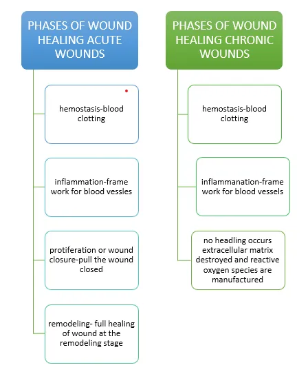
Figure 1: The four phases of wound healing
Molecular Mechanism Of Healing Process:
Coordinated cellular, humoral, and molecular mechanisms are involved in the intricate and dynamic process of wound healing, which starts as soon as an injury occurs and can last for months or years. Open or closed wounds can cause tissues to become impaired structurally and functionally. Regeneration or repair—the latter leading to fibrosis and scar formation—are the two ways that healing happens. This process resembles a coordinated interaction between growth factors, cytokines, and cells . Compensatory mechanisms permit healing to proceed even in the absence of specific cells or mediators. Restoring skin integrity and avoiding infection and dehydration depend on effective wound healing. Hemostasis, inflammation, proliferation, and remodeling are the four overlapping phases of adult wound healing . While lymphocytes build up by day four and begin to decrease after two weeks, macrophages and monocytes peak during the proliferative phase. The second week is when remodeling starts, and it might last for several months. In general, immune cell interactions and the wound microenvironment play a crucial role in controlling tissue repair.[8]
Pathophysiology of Antimicrobial Hydrogels:
compromise the skin’s protective barrier, exposing underlying tissues to microbial invasion. The wound bed becomes a moist, nutrient-rich environment, which:
2. Hydrogel Integration with Wound Physiology:
Hydrogels are 3D polymeric networks that resemble the extracellular matrix (ECM), offering:
3. Release of Antimicrobial Agents in hydrogels
Hydrogels' high-water content, adjustable structure, biocompatibility, and capacity for both prolonged and localized release make them ideal antimicrobial agent delivery vehicles. The hydrogel's polymer network, crosslink density, stimuli-responsiveness, and the kind of antimicrobial agent used all affect the release behavior.
Mechanisms of Drug Release from Hydrogels:
Most antimicrobial release occurs by diffusion, where drugs migrate through the water-filled pores of the hydrogel network. Diffusion depends on mesh size, swelling behavior, and drug molecular weight. Nanoparticle-loaded hydrogels (e.g., AgNPs, ZnO) especially rely on diffusion and slow ionic release for antibacterial effects.[11]
b. Swelling-Controlled Release:
Hydrogels that swell upon absorbing wound exudate increase pore size, enhancing antimicrobial release. Hydrophilic polymers such as alginate, gelatin, or chitosan exhibit significant swelling, making them suitable for topical wound applications.[12]
c. Degradation-Controlled Release:
Biodegradable hydrogels release antimicrobial agents as the polymer matrix undergoes enzymatic or hydrolytic degradation. In infected wounds, enzyme-responsive hydrogels degrade faster due to elevated protease levels, enhancing release.[13]
4. bioflim disruption:[14]
Table 1 : Biofilm Disruption Mechanisms of Antimicrobial Hydrogels.[14]
|
Mechanism |
Description |
How Hydrogels Facilitate This Mechanism |
|
1.Enzymatic Degradation of Biofilm Matrix |
Enzymes (e.g.,DNase, proteases, dispersin B) break down extracellular polymeric substances (EPS) of biofilms. |
Hydrogels load enzymes and release them locally at the infection site to degrade EPS and weaken biofilm structure. |
|
2. Reactive Oxygen Species (ROS) Generation |
ROS such as •OH, H₂O₂, and O₂⁻ cause oxidative damage to bacteria and EPS. |
ROS-generating hydrogels (e.g., metal oxide-based) continuously or stimuli-responsively release ROS at the wound site. |
|
3. pH-Responsive Disruption |
Many biofilms acidify their environment; acidic pH destabilizes certain hydrogel matrices to release antimicrobials. |
pH-responsive hydrogels swell or degrade in low pH to release antibiotics, AMPs, or nanoparticles that destroy biofilm bacteria. |
|
4. Chelation of Divalent Ions (Ca²⁺/Mg²⁺) |
Removing Ca²⁺ and Mg²⁺ ions disrupt cross-linking stability within EPS. |
Hydrogels incorporating chelators (e.g., EDTA) release them to destabilize biofilm ionic bridges. |
|
5. Mechanical Disruption (Swelling Pressure) |
Physical expansion exerts force on biofilm clusters, breaking them apart. |
Super-swelling hydrogels absorb exudates and swell, causing mechanical detachment of biofilm layers from tissue. |
|
6. Nanoparticle-Mediated Disruption |
Metal nanoparticles (Ag, ZnO, CuO) penetrate biofilms, generating ROS and damaging membranes. |
Hydrogels provide a controlled-release platform for sustained delivery and deep penetration of nanoparticles into biofilms. |
|
7. Antimicrobial Peptide (AMP) Action |
AMPs disrupt bacterial cell membranes and destabilize biofilm structure. |
AMPs are embedded in hydrogels and released gradually to inhibit bacterial adhesion, aggregation, and biofilm development. |
|
8. Quorum Sensing Inhibition |
Interferes with bacterial communication pathways, preventing biofilm maturation. |
Hydrogels deliver quorum-quenching molecules (e.g., furanones, RNA analogs) directly to infection sites. |
|
9. Localized Heat/Photothermal Disruption |
Heat damages EPS and bacterial membranes, enhancing drug penetration. |
Photothermal hydrogels containing nanoparticles (e.g., gold nanorods) generate heat upon light exposure to destroy biofilms. |
|
10. Targeted Drug Release Through Biofilm Stimuli |
Biofilms produce enzymes (e.g., gelatinases), lowering O₂ and changing redox potential. |
Stimuli-responsive hydrogels release drugs only in the presence of biofilm-specific signals, enhancing penetration and killing efficiency. |
5. Biofilm Disruption
Biofilms are structured bacterial communities encased in extracellular polymeric substances (EPS), highly resistant to antibiotics. Antimicrobial hydrogels disrupt biofilms through:
6. Host Response and Healing Enhancement
By reducing microbial burden and inflammation, antimicrobial hydrogels positively influence the wound healing cascade:
Factors affecting wound healing process. [17]
Antimicrobial hydrogels
Hydrogels with antimicrobial properties are highly promising materials for application such as wound dressings and fillers. Gels offer a moist and heavily hydrated environment for the wound area, thanks to their high-water content. This environment promotes cellular immunological activity that is crucial for the wound healing process. Nonetheless, the same hydrated environment can also promote microbial infection. Therefore, it is desirable for gels to have antimicrobial properties in addition to serving their primary functions (such as wound healing, drug delivery, etc.).[18]
It has been reported that metallic nanoparticles, such as those made of silver, gold, or copper, can eliminate various kinds of bacteria. Every one of these nano-metals possesses unique characteristics and operates through a distinct mechanism. It is suggested that the primary antimicrobial action of nanometals consists of interfering with metabolic processes in the cell membrane. Microbial mortality can occur when metallic nanoparticles permeate the cell membrane and disrupt related enzymes. Additionally, metal nanoparticles may produce reactive oxygen species (ROS), which can cause DNA damage and bacterial mortality. Bacterial replication is additionally impeded by nanoparticles attaching to the cell membrane. The size, shape, and concentration of antibacterial nanoparticles are important features. [19]
pH-Responsive hydrogel
It is known that pH can vary at multiple locations in the body, including the gastrointestinal tract vagina, and blood vessels. These differences may provide a suitable basis for pH-responsive medication release. Additionally, local pH fluctuations in respect to specific substrates can be created and used to modify the release of drugs. The goal of pH-responsive drug delivery systems is to release pharmaceuticals intravascularly when blood pH is increased in certain cardiovascular abnormalities, mask the taste of bitter therapies, and provide peroral controlled administration of drugs.[20]

Figure 2: Swelling of (a) anionic and (b) cationic hydrogels in responses to pH
Thermosensitive hydrogels
Hydrogels that show temperature-sensitive behavior are referred to as thermosensitive hydrogels. Under low temperatures, these hydrogels stay in a sol form; however, when exposed to body temperature, they convert into a gel state. They can be used as an alternative to traditional wound dressings because of their special quality. Certain substances including β-glycerol phosphate, HPMC, and poloxamer can be added to chitosan to create a temperature-sensitive hydrogel. Odinokov. used Ter phthaloyl diazide to cross-link chitosan to create pH- and thermosensitive hydrogels. Bhattarai et al. created a hydrogel by neutralizing a chitosan solution with a polyol counter-ionic single-head salt. At low temperatures, this hydrogel stays liquid, but at body temperature, it solidifies into a gel. As therapeutic drug delivery methods for encouraging skin tissue regeneration and repair, these thermosensitive chitosan-based hydrogels show promise. They accomplish this by releasing loaded medications in a controlled and prolonged manner.[21]
Ideal properties of hydrogels to promote wound healing. [22]
Natural and Synthetic Hydrogel
Hydrogel wound dressings are developed using diverse natural and synthetic polymers. Natural polymers include chitosan, gelatin, hyaluronic acid, and alginate. Synthetic polymers include polyethylene glycol, polyvinyl pyrrolidone, polyethylene ox ide, and polyvinyl alcohol. Hydrogels can be highly elastic, and this reduces mechanical power; therefore, multipolymeric hydrogels have introduced for improved mechanical Hydrogel wound dressings are developed using diverse natural and synthetic polymers. Natural polymers include chitosan, gelatin, hyaluronic acid, and alginate. Synthetic polymers include polyethylene glycol, polyvinyl pyrrolidone, polyethylene ox ide, and polyvinyl alcohol. Hydrogels can be highly elastic, and this reduces mechanical power; therefore, multi polymeric hydrogels have been introduced for improved mechanical power and absorption. Combining a naturally occurring polymer with a synthetic polymer promises to be a viable approach for generating materials with the desired thermal and mechanical attributes. Advancements in the field have been achieved by harnessing the inherent features of polymers, leading to the development of novel technologies such as sprayable hydrogels, “smart hydrogels”, nanogels, aerogels, and cryogels.[23]
Natural hydrogels are mainly made up of proteins and ECM components, making them inherently biocompatible and bioactive. Their capacity to boost various cellular functions suggests they could be a good fit for a range of biomedical uses. The composition and properties of these materials are similar to the intrinsic qualities of tissue layers. Nevertheless, they are subject to some limitations, mainly due to the challenges associated with their manipulation arising from variations observed between different batches. Hydrogel variants display distinct characteristics that make them more suitable for their intended use. A brief discussion of the different natural hydrogels will follow.[24]
Table 2: Natural Polymers. [25,26,27]
|
Polymer |
Origin / Structure |
Key Properties |
Limitations |
Applications / Notes |
|
Chitosan. |
Derived from chitin; polysaccharide of D‑glucosamine & N‑acetyl‑D‑glucosamine |
Cationic, antibacterial, biocompatible; properties depend on molecular weight & deacetylation |
Low mechanical strength; requires cross‑linking |
CMCS hydrogel: 132% swelling; accelerates diabetic wound healing |
|
Hyaluronic Acid. |
Glycosaminoglycan; N‑acetyl glucosamine + glucuronic acid |
Biocompatible, hydrophilic, biodegradable, non‑immunogenic |
Rapid degradation unless modified |
Maintains moisture; widely used in wound healing |
|
Gelatin. |
Derived from collagen; rich in glycine, proline, hydroxyproline |
Excellent cell adhesion, biocompatible, ECM‑mimicking |
Low mechanical strength; needs cross‑linking |
Used for tissue regeneration; crosslinked via EDC or glutaraldehyde |
B. Synthetic Hydrogels
Due to their mechanical properties, ease of shaping into different configurations, and cost-effectiveness in manufacturing, synthetic polymers have shown significant efficacy in biomedical applications.[28] These polymers are stable and easy to use, but their biocompatibility is limited. Unlike their naturally occurring counterparts, synthetic polymers have the advantage of being able to be produced conveniently on an industrial scale. Moreover, due to their built-in adaptability, they can be used in different configurations that foster optimal tissue development. Furthermore, the capability to accurately control both the hydrophilic and hydrophobic areas of synthetic polymers allows for the creation of frameworks that are more uniform as well as an enhanced ability to retain water. By combining them with biopolymers, hybrid polymers (blended) can be created that demonstrate advantageous physicochemical properties. Bioactive substances obtained from natural sources enhance the positive attributes of blended hydrogels made from synthetic polymers. Blended hydrogels offer a promising option for future wound treatment by integrating their beneficial properties. Examples of synthetic polymers are polyvinyl alcohol, polyethylene glycol, and polyvinyl pyrrolidone.[29]
Table 3: Synthetic Polymers. [30,32,33,]
|
Polymer |
Structure / Nature |
Key Properties |
Limitations |
Applications / Notes |
|
Polyethylene Glycol (PEG)
|
Hydrophilic, ether-based polymer |
Biocompatible, prevents protein adsorption, tunable (PEGDA/PEGDM) |
Not bioactive unless functionalized |
Used in diabetic wound dressings; reduces scarring |
|
Polyvinyl Alcohol (PVA)\
|
Hydrophilic, semi-crystalline polymer |
Biocompatible, stable, moisture-permeable |
Requires antibacterial enhancement |
Double-crosslinked PVA/chitosan hydrogels improve rigidity & healing time |
|
Polyvinyl Pyrrolidone (PVP) |
Synthetic, lactam ring |
Highly hydrophilic, good film-forming ability |
Low mechanical strength alone |
Used in blends with PEG/PVA |
|
Polyethylene Oxide (PEO)
|
High molecular weight polyether |
Highly soluble, flexible |
Weak without crosslinking |
Improves swelling and flexibility in blends |
Table 4: Comparison Between Hydrogel Types in Terms of Biocompatibility and Effectiveness [34]
|
Hydrogel Type |
Biocompatibility |
Effectiveness |
|
Natural Hydrogels (Alginate, Chitosan, Collagen, Gelatin) |
• Highly biocompatible |
• Excellent wound healing |
|
Synthetic Hydrogels (PVA, PEG, Pluronic, PMMA) |
• Good biocompatibility |
• Strong mechanical strength |
|
Semi-Synthetic Hydrogels (GelMA, PEG-chitosan, Carbopol blends) |
• Improved biocompatibility due to combination |
• Balanced strength and biodegradation |
|
Smart/Stimuli-Responsive Hydrogels (pH-, temperature-, enzyme-responsive) |
• Depends on polymer used |
• Targeted drug release |
|
Nanocomposite Hydrogels (AgNPs, ZnO, graphene) |
• Biocompatibility varies |
• Strong antimicrobial activity |
|
Ionic-Crosslinked Hydrogels (Calcium alginate) |
• Excellent biocompatibility |
• Good for rapid healing |
|
Covalently Crosslinked Hydrogels |
• Good if non‑toxic crosslinkers used |
• High stability |
Advantages.[35]
Applications
Table 5:Applications of hydrogel[36-38]
|
Description |
|
|
Wound Healing |
Hydrogels have a cross-linked structure that allows them to hold large amounts of water and drugs. Their high water-holding capacity helps in absorbing and retaining wound exudates, thereby maintaining a moist environment favorable for healing. Polyvinyl pyrrolidone and polyacrylamide hydrogels typically contain 70–95% water. |
|
Colon-Specific Hydrogels |
Polysaccharide-based hydrogels are designed for colon-specific drug delivery due to the high concentration of polysaccharide-degrading enzymes present in the colon. Dextran hydrogels are commonly used for this purpose. |
|
Drug Delivery in the Gastrointestinal Tract |
Hydrogels enable site-specific drug delivery in the GIT. In the presence of intestinal microflora, colon-specific hydrogels exhibit tissue specificity. Variations in pH and enzymatic activity cause hydrogel degradation and controlled drug release. |
|
Transdermal Drug Delivery |
Hydrogel-based drug delivery systems are developed for transdermal administration. These formulations are also explored in iontophoresis to enhance the permeation of drugs such as hormones and nicotine through the skin. |
|
Gene Delivery |
Changes in hydrogel composition allow effective targeting and delivery of nucleic acids to specific cells for gene therapy. Hydrogels show strong potential in treating various genetic and acquired diseases. |
Table 6: Methods of preparation of hydrogel.[39-43]
|
Method |
Description |
|
Free Radical Polymerization |
Uses initiators (APS, UV light) to form polymer networks. |
|
Ionic Crosslinking |
Uses ions like Ca²⁺ to crosslink polymers (e.g., alginate). |
|
Chemical Crosslinking |
Uses covalent crosslinkers such as GA, EDC/NHS, Genipin. |
|
Physical Crosslinking |
Gelation via hydrogen bonding, crystallization, hydrophobic interactions. |
|
Thermo-Responsive Gelation |
Polymers gel upon heating/cooling (e.g., PNIPAM, Pluronic). |
|
pH-Responsive Gelation |
Gel formation triggered by specific pH levels. |
|
Enzymatic Crosslinking |
Uses enzymes like HRP or transglutaminase for gelation. |
|
Radiation Crosslinking |
Gamma/e-beam radiation induces polymer crosslinking. |
|
Click Chemistry |
Fast covalent bonding (Thiol–Ene, etc.). |
|
Freeze–Thaw Method |
Repeated freezing and thawing cycles (commonly for PVA). |
|
Self-Assembly Method |
Peptides or block copolymers self-assemble into gels. |
Evaluation OF Hydrogel. [44-46]
Visual observations were used to assess the produced gels' homogeneity and physical appearance. The commercial formulation served as the standard.
Spread ability can be assessed by spreading the gel on a level surface and looking for any grit in the hydrogel. A 1% hydrogel formulation in deionized water was made, and the pH was measured.
Diclofenac sodium was extracted from 1 g of each gel formulation using 20 mL of phosphate buffer pH7.4 for 30 minutes in order to test the drug in gels. A membrane filter with a pore size of 0.45μm was used to filter the final combination. Following the proper dilution with phosphate buffer pH 7.4, the absorbance of the sample was measured spectrophotometrically at 276 nm using an Elico SL150 UV-VIS spectrophotometer. The calibration curve was used to quantify the diclofenac sodium concentration.
The Brookfield viscometer with spindle number 7 was used to measure the viscosity of the gel compositions at 250C and 100 rpm.
The International Conference on Harmonization (ICH) requirements were followed when conducting stability studies on improved formulations. According to ICH guidelines, the formulation in an aluminum tube was put through three months of accelerated stability testing at a temperature of 40 ± 2oC and a relative humidity of 75 ± 5%. Over the course of three months, samples were collected at regular intervals of one month and examined for changes in pH, spreadability, drug content, and in-vitro drug release using the previously described method. If there were any modifications in the evaluation criteria, these were recorded. Tests were conducted in duplicate, and the standard deviation and mean of the observed data were recorded.
FUTURE PROSPECTS
CONCLUSION
Antimicrobial hydrogels have proven to be advanced wound dressing materials that not only protect the wound but also actively promote the healing process. Their three-dimensional structure maintains optimal moisture, supports cell migration, and enhances tissue regeneration while simultaneously delivering antimicrobial agents to prevent infection and biofilm formation. Stimuli-responsive hydrogels further improve treatment effectiveness by releasing drugs in response to changes in pH, temperature, or enzymatic activity within the wound environment. By combining the biocompatibility of natural polymers with the durability of synthetic materials, modern hydrogel systems offer improved mechanical strength, controlled drug release, and faster wound closure. These properties make antimicrobial hydrogels a promising and efficient alternative to conventional wound care approaches, especially for chronic and infected wounds.
REFERENCES


Saquib Ullah Khan, Yuvraj Tade, Nikhil Kharate, Vaishnavi Gole, Rupeshri Netkar, Prashil Dhumale, Hydrogel-Based Topical Applications in Microbial Infection Control: A Comprehensive Review, Int. J. of Pharm. Sci., 2026, Vol 4, Issue 6, 6063-6077, https://doi.org/10.5281/zenodo.20827557
 10.5281/zenodo.20827557