
We use cookies to ensure our website works properly and to personalise your experience. Cookies policy
1,2,4 Department of Pharmaceutics, Karnataka College of Pharmacy, Bengaluru
3 Department of Pharmacy Practice, Faculty of Pharmacy, M S Ramaiah University of Applied Sciences, Bangalore.
Diabetic foot ulcers (DFUs) represent a significant complication for individuals with diabetes, characterized by infection, ulceration, and tissue damage, which can lead to severe health issues and potentially amputation. Conventional treatment modalities are frequently insufficient, necessitating advancement in therapeutic strategies. Nanofibers have become a game-changing technology in a variety of industries, most notably advanced manufacturing, biomedicine, and environmental management. Nanofibers, which are distinguished by their high surface area-to-volume ratio, porosity, and biomimetic structural characteristics, present unmatched potential for uses in drug delivery, tissue engineering, biosensing, and filtration. For treating Diabetic Foot Ulcer Nanofiber based system found to be an effective method which providing controlled and targeted release of drugs, improved tissue regeneration, and antimicrobial action. By creating the ideal healing environment and delivering bioactive agents straight to target sites, their ability to efficiently load drugs and maintain sustained release makes them especially effective in wound healing, including difficult cases like diabetic foot ulcers. Additionally, combining nanofiber technology with Platelet-rich plasma and bioactive molecules results in a synergistic approach that improves therapeutic efficacy while addressing problems like tissue regeneration and infection control. This current review on PRP and nanofiber in DFU management is compiled , which highlights how they can improve healing processes and get around the drawbacks of conventional treatments. The combination of biotherapy and nanotechnology has the potential to completely transform diabetic wound care by providing individualised, effective, and regenerative solutions that enhance patient outcomes.This edge of medicine holds the promise of not only quicker healing but also a future of efficacious, and transformative DFU treatments.
Diabetic foot (DF) refers to an infection, ulcer, or damage to the tissue in the foot of an individual with diabetes. Prolonged tissue oxygen deprivation, combined with nerve damage, creates a high-risk environment for traumatic events, leading to diabetic foot ulcers (DFU)1. Diabetes mellitus is a significant and prevalent metabolic disorder impacting populations around the globe. When trauma occurs at the affected site the patient will be unable to identify the injury that happened at the lower extremities. As a result, numerous injuries remain undetected and gradually worsen due to ongoing pressure and shear forces from activities like walking and bearing weight2. Approximately more than half of these conditions progress into infection that may result in amputation, disability, prolonged hospitalization, and death4. DFU, especially when infected, is a significant source of illness and can result in serious consequences such as limb amputation. Successful management of these usually requires a multidisciplinary strategy, which typically includes wound debridement, off-loading pressure, management of blood sugar levels, surgical procedures, and occasionally other interventions3.
Platelet-rich plasma (PRP) is abundant in growth factors, including epidermal, platelet-derived, transforming, vascular endothelial, fibroblast, insulin-like, and keratinocyte growth factors. The healing capabilities of PRP stem from its higher levels of growth factors when compared to regular plasma. Recently, PRP has been widely applied across numerous clinical fields, notably in regenerative medicine5. Platelets are essential for maintaining homeostasis and facilitating thrombosis, significantly contributing to different stages of wound healing by encouraging and reinforcing the clot. Platelet-rich plasma contains a greater concentration of platelets compared to standard plasma and is derived from the individual’s own body, which minimizes the risk of any immune-related adverse reactions6.
Gram-positive bacteria, including both methicillin-sensitive and methicillin-resistant Staphylococcus aureus (MRSA), are primarily found in diabetic foot infections. These infections create major difficulties in managing diabetic foot ulcers due to the existence of antibiotic-resistant strains. Effective management requires careful identification and targeted treatment of these pathogens to prevent further complications. By understanding the prevalence and resistance patterns of these organisms, clinicians can enhance their therapeutic approach to improve patient outcomes7.
Antibiotics are crucial in treating DFUs by targeting and eliminating bacteria also preventing cellulitis and osteomyelitis. An antibiotic that is especially effective for managing diabetic foot ulcers caused by gram-positive bacteria, including methicillin-resistant Staphylococcus aureus (MRSA). Broad-spectrum antibiotics effectively treats diabetic foot ulcers by targeting gram-positive bacteria, including MRSA8.
One-dimensional (1D) nanomaterials known as nanofibers have gained significant industrial applications. These nanofibers have diameters that are many times smaller than a single strand of human hair, and they demonstrate enhanced mechanical characteristics, including stiffness and tensile strength, relative to standard base materials. Additionally, nanofibers offer a large surface area with variable 3D topography, porosity, and adaptable surface functionality9. Nanofiber integrated drug delivery system showing a promising approach for treating DFU. These systems can provide targeted and controlled release of therapeutic agents, enhancing wound healing and reducing infections. The increased surface area of nanofiber allows for better interaction with the wound site promoting faster healing.
PATHOGENESIS
Diabetic foot ulcers arise from two key pathophysiological mechanisms: Neuropathy and Ischemia.
Neuropathy and diabetic foot ulcer: Peripheral neuropathy is a major contributor to DFUs, resulting from chronic hyperglycemia that damages peripheral nerves. Sensory neuropathy leads to loss of protective sensation, making an individual unable to detect minor injuries, pressure, or trauma to the foot. Motor neuropathy results in muscle weakness and imbalances, which can lead to deformities in the feet. These deformities create abnormal pressure points, particularly over bony prominences, which are more prone to skin breakdown under repetitive stress. Additionally, autonomic neuropathy impairs the regulation of sweat glands, leading to dry, cracked skin that is more susceptible to cracks and infection. Together these neuropathic changes predispose the foot to unnoticed injuries that can progress to ulcers 8,2.
Ischemia and diabetic foot ulcer: Peripheral arterial disease (PAD) is another critical factor in DFU development, as diabetes accelerates atherosclerosis, leading to narrowing and occlusion of blood vessels in the lower extremities. Reduced blood flow results in poor tissue perfusion, limiting oxygen and nutrient delivery necessary for tissue maintenance and repair. Ischemia impairs the wound healing process and weakens the local immune response, increasing susceptibility to infections. In severe cases, critical limb ischemia can cause tissue necrosis, further exacerbating ulcer formation 8,2. Neuropathy and ischemia can function independently or together to establish a heightened risk for diabetic foot ulcers. The combination of ischemia and neuropathy significantly increases the likelihood of developing diabetic foot ulcers and associated complications such as gangrene.
CHOICE OF WOUND CARE FOR DIABETIC FOOT ULCER
Given the complexities of wound healing and overall health in diabetic patients, choosing a treatment strategy suited to wound type and sensitivity is essential. Effective management of diabetic ulcers involves not just systemic antibiotics and surgical interventions but also precise wound care.
Management techniques
(i) Debridement: - Indicating the removal of dead or damaged tissues leads to enhances the healthy tissues10. Based on type of wound the debridement technique are of following types.
(a) Surgical or Sharp debridement: This is advised for wounds that are necrotic and infected.
(b) Autolytic debridement: This is a careful procedure in which the dead tissue is converted into a liquid form. This technique is not used for infected pressure ulcer11.
(c) Mechanical debridement: It involves removing unhealthy tissue with dressing and wound irrigation, but it can cause bleeding and pain. This technique is utilized for surgical incisions and venous leg ulcers. It is both time-intensive and expensive 12.
(d) Enzymatic debridement: This uses topical enzymes like collagenase, fibrinolysin, or papain to remove devitalized tissue. It is advised for wounds that are sloughy, infected, or necrotic when surgical debridement is not an option. This method targets dead tissue without harming healthy tissues 13.
(e) Maggot debridement: This method uses sterile maggots, typically Lucilia sericata, to treat wounds when conventional methods fail. The maggots are applied to the wound and covered with a dressing, where they consume necrotic tissue and bacteria while secreting antimicrobial enzymes, aiding the healthy process 14.
(ii) Offloading: It is an important aspect of diabetic wound healing, as it involves complete or partial pressure from the weight-bearing area of the foot. This is achieved by providing mechanical support, allowing the wound area to rest and heal.
(iii) Wound care: - This is critical in managing diabetic foot ulcers and involves cleaning with normal saline using an aseptic technique and employing a modern method that ensures a moist healing environment. While topical treatment is important, it is not as relevant as systemic and surgical treatments. The options that can be employed include hydrocolloids, hydrogels, alginates, foam dressings, silver-impregnated dressings, growth factors, silicone-based atraumatic dressings, vacuum-assisted devices, and hyperbaric oxygen therapy. Selection criteria should be based on the patient's health, wound characteristics, dressing properties, and accessibility 16.
An ideal wound dressing should be6,17 : (a)Sterile, simple, and inexpensive to use, (b) Maintain moisture and absorb excess exudate. (c) Be non-toxic, non-allergic, and non-adherent. (d) Protect against microorganisms and avoid contamination. (e) Facilitate the exchange of gases, regulate odors, and offer thermal insulation as well as mechanical protection
(iv) Antibiotic Selection: - The choice of antibiotics for treating diabetic foot infections should be informed by the results of bacterial cultures and sensitivity testing to avoid resistance and negative side effects. Oral antibiotics are provided to individuals with mild soft tissue infections, while those experiencing moderate to severe infections receive parenteral antibiotics. Evidence-based guidelines for infection management in diabetic foot patients must be followed rigorously. There is a need to assess the proper dosing, ideal treatment duration, detection and removal of the infection source, and any possible side effects in both outpatients and inpatients with diabetic foot infections18,19,20.
Antibiotic prescribing is divided into three levels:
Table 1: Antibiotic levels and usage criteria
|
Levels |
Usage criteria |
|
First line |
General infection, Standard treatment |
|
Restricted antibiotics |
Resistant pathogens, polymicrobial infections, special conditions, or costly antibiotics |
|
Reserve antibiotics |
Life-threatening infections |
(v) Revascularization: - With the progress of vascular and orthopedic reconstructive surgeries, most cases which would have been amputation otherwise can now be saved as limbs today. With peripheral arterial disease, patients suffering from diabetes are at a more significant risk of developing ischemic ulcers. There is more than a 95% success rate with the following endovascular treatments: PTA, balloon-expandable stents, self-expanding stents, and covered stents are utilized for stenting the iliac arteries. Revascularization plays a vital role in managing ischemic lower extremity wounds and should be performed prior to drainage or debridement. Emerging endovascular techniques, such as cryoplasty, drug-eluting stents, and plaque-debulking lasers, show potential as beneficial adjuncts to PTA. Subintimal angioplasty performed below the ankle boasts a remarkable limb salvage rate of 94.6%, and revascularization greatly enhances the outcomes of trans-matarsal amputations21.
(vi) Wound dressing: - Dressing of a diabetic foot ulcer is one of the critical wound care interventions, as it encourages healing and prevents infection. The process includes rinsing the wound with normal saline while adhering to aseptic methods to eliminate debris and bacteria. Contemporary wound care strategies focus on preserving a moist environment, which is crucial for the healing process. Different dressings, such as hydrogels, hydrocolloids, alginates, and foam, are used to achieve this. These dressings prevent the entry of external contaminants, absorb excess exudate, and create a barrier against infection. Non-adherent dressings prevent trauma during dressing changes and are more comfortable for patients. The wound is monitored, and proper healing is ensured by frequent dressing changes. Overall, selection of the right dressing material, according to the condition of the wound and the patient's needs, is essential for effective management.
(vii) Novel approaches: -
(a) Tibial Periosteal Distraction: Tibial Periosteal Distraction is a surgical intervention for diabetic foot ulcers. The intervention aims at increasing blood supply and thus improving healing of the ulcer. The technique stretches the periosteum, the membrane covering the bone, to improve microcirculation. Increased blood supply accelerates the healing process while minimizing complications. The procedure is quite simple and not invasive, thus a possible intervention for chronic, non-healing ulcers22.
(b) 3D printing: Customized solutions for wound management in diabetic foot ulcers are offered by 3D Printing technology. Patient-specific data can be used to create 3D-printed dressings and scaffolds that fit the wound perfectly. These custom dressings offer better support, optimal healing conditions, and a reduced risk of infection. Moreover, 3D printing allows the incorporation of bioactive compounds and growth factors into the dressings, enhancing their therapeutic efficacy 23.
(c) Topical Oxygen Therapy (TOT): Topical Oxygen Therapy is application of oxygen on the wound and has shown promising results as it helps with healing and inhibits infection; oxygen is vital to cellular functions participating in wound repair, such as collagen synthesis and fibroblast proliferation. These TOT delivery devices concentrate the oxygen on the wound, making the healing process efficient and better for patients with diabetic foot ulcers24.
(d) Stem Cell Therapy: Stem cell therapy represents a promising and innovative approach for enhancing wound healing in diabetic foot ulcers (DFUs). Autologous adult stem cells, particularly bone marrow-derived mesenchymal stem cells (BM-MSCs), are the most commonly utilized due to their safety profile, ease of harvest, and ability to promote angiogenesis, modulate inflammation, and differentiate into essential skin cell types. These cells can be delivered locally through injections, often in combination with growth factors like G-CSF, to improve healing outcomes. Combining stem cell therapy with revascularization procedures such as angioplasty further amplifies their effectiveness, especially in ischemic wounds. While variability exists in protocols, current evidence supports stem cells as a viable, safe, and potentially superior option for wound care in DFU patients, with ongoing research aiming to standardize treatments and optimize results.
In order to promote stem cell mobilization from the bone marrow, delivery methods usually involve local injections straight into the wound bed, occasionally mixed with systemic administration of granulocyte-colony stimulating factor (G-CSF). Additionally, the application of G-CSF boosts stem cells' capacity to adhere to the wound site. Stem cell therapy frequently combines with other methods such as platelet-rich plasma (PRP), which is abundant in growth factors like VEGF, PDGF, and TGF-β, to increase treatment efficacy. PRP can be applied topically or incorporated into scaffolds to foster angiogenesis, cell adhesion, and proliferation while establishing a favourable microenvironment for stem cell function.
(e) Nanodrug Delivery Systems: Nanodrug Delivery Systems represent a cutting-edge approach for treating diabetic foot ulcers. These systems use nanoparticles to deliver therapeutic agents directly to the wound site, enhancing the effectiveness of treatment. Examples include:
Difficulty in diagnosing infection in diabetic foot infection
Correctly diagnosing infections in diabetic foot ulcers (DFUs) is crucial since almost 50% of these wounds are not infected and therefore do not need antibiotic treatment31. Even though finding microorganisms in aseptically collected samples from typically sterile areas suggests an infection, positive cultures alone are insufficient for diagnosing all open wounds because of the potential for microbial colonization. Therefore, wound management guidelines advocate using clinical signs as a basis for diagnosing infection. However, the diagnosis of infections in diabetic foot wounds is particularly challenging because peripheral neuropathy or foot ischemia obscures or simulates signs of inflammation, reducing their diagnostic value.
Patients with diabetic foot diseases ordinarily show with a later disturbance in the defensive skin obstruction, which is taken after by continuously spreading irritation that may happen over hours, days, or indeed weeks32. These wounds can result from mechanical, chemical, or warm injury but are most commonly caused by weight. DFIs are characterized by an assortment of clinical signs that are reliable with a localized irresistible disorder: redness (erythema), heat (calor), swelling (tumor), pain or tenderness (dolor), and pus discharge. Systemic signs of infection, including fever, chills, an increase in white blood cells (leukocytosis), low blood pressure (hypotension), rapid heartbeat (tachycardia), and quickened breathing (tachypnea), are rare and indicate a more severe condition33. Soft tissue infections in diabetic foot wounds can extend to the underlying bone causing osteomyelitis. Suspect key signs include a "sausage toe" or bone fragments from sinus tracts. The probe-to-bone test aids diagnosis but must be performed technique appropriately. Osteomyelitis changes on x-rays take weeks, and inflammatory markers may be absent in chronic cases.
Nanofiber92
Structure
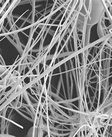
Nanofibers, which are very thin fibers, have a diameter that is usually between 10 nm and several micrometers, with a high surface-area-to-volume ratio as well as porosity. Because of their structural resemblance to the extracellular matrix (ECM), their main applications are in biomedical fields, such as wound healing and drug delivery. Depending on the manufacturing method employed, nanofibers may be oriented randomly or aligned in a specific direction.The orientation gives different mechanical properties and biological interactions. Advanced nanofiber structures, including core-shell and multilayered fibers, provide controlled drug release and enhanced functionality. Their interconnected porous structure promotes oxygen exchange and nutrient transport, facilitating tissue regeneration.
Nanofiber for the treatment of diabetic foot ulcer
Nanofibers are unique and useful in treating diabetic foot ulcers because of their excellent properties. The large surface-area-to-volume ratio enabled effective drug loading and allowed for the sustained release of therapeutic agents including antibiotics, growth factors, and anti-inflammatory chemicals directly to the wound area. The porous structure of nanofibers provides excellent gas exchange and maintains moisture, avoiding desiccation or excessive moisture, resulting in an optimal healing environment. Their ability to mimic the ECM provides structural support for cell adhesion, proliferation, and migration, encouraging tissue regeneration. Nanofibers can be functionalized with bioactive molecules or antimicrobial agents to prevent infections, a critical factor in managing DFUs. Combining these properties makes nanofibers highly effective in accelerating wound healing and reducing complications associated with DFUs.
Characteristic features of Nanofiber 26, 27, 28, 29
Table 2. Description of characteristic features of nanofibers
|
Characteristic |
Description |
|
Size |
Typically ranges from 1-100nm in diameter, providing a large ratio of surface area to volume |
|
Surface area
|
The extensive surface area facilitates effective engagement with the surrounding environment, enhancing mechanical, chemical, and biological properties. |
|
Mechanical properties |
High tensile strength and elasticity compared to other materials of similar size, making them suitable for reinforcement in composites. |
|
Porosity |
High porosity, allows for increased fluid transport and high absorption capacity, which is beneficial for tissue engineering and drug delivery. |
|
Flexibility |
Can be highly flexible and adaptable for various applications, particularly in medical and textile industries. |
|
Electrical conductivity |
Can be engineered to exhibit conductive properties, especially in carbon-based nanofibers, used in sensors or flexible electronics. |
|
Biocompatibility |
Many types of nanofibers are biocompatible, making them suitable for biomedical applications like wound dressing and tissue scaffolding |
|
Surface functionalization |
Ability to modify surface properties through chemical or physical processes, allowing for targeted drug delivery or controlled interaction with cells. |
|
Porous structure |
Enables high diffusion rates for nutrients, drugs, and other molecules, ideal for use in medical and filtration applications. |
|
Thermal stability |
Some nanofibers can withstand high temperatures and exhibit thermal stability. Useful in heat-resistant materials. |
Mechanism treating diabetic foot ulcer by nanofiber27,29
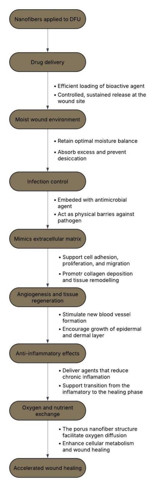
Picture 1: Mechanism of treating DFU
Platelet-rich plasma (PRP) for the treatment of diabetic foot ulcer99
The rates of occurrence of diabetes mellitus on a global scale is rising fast. Based on the International Diabetes Federation (IDF), the rate of prevalence of diabetes mellitus (DM) was 10.5% in 2021 and will rise to 12.2% by 2045. DFU is most prevalent and severe complication in the patients with diabetes and DFU also carries the important mortality and morbidity and is becoming economical burden to the society.So treatment for DFU is now an emergent issue. At present in India the first line treatment of DFU involves blood glucose level control and some traditional method treatment such as infection management wound discharge and dressing and angioplasty for ischemic peripheral artery disease (PAD) but the treatment of DFU is still not effective.
The average healing time for diabetic foot ulcers (DFU) without operation is about 12 weeks. However, around 20% of patients have not experienced healing for over a year. Additionally, the recurrence rate within the same year is about 40%. So the creation efficient and cost-effective treatment for DFU is quite necessary in the modern world. In recent researches have discovered that the application of stem cell or growth factor may be an efficient treatment which would be capable of restoring body's healing process sooner. PRP is the choice in that due to the fact that platelets have a mix of growth factors, which are vital for tissue repair and tissue regeneration. The major function of platelet-rich plasma (PRP) during wound healing is by releasing different bioactive molecules contained in the platelets. These factors are growth factors, cytokines, and other proteins involved in signaling which stimulate tissue repair, inhibit inflammation, and accelerate the body's natural recovery processes.
Over the past few years, many studies have compared the effectiveness of platelet-rich plasma (PRP) in the treatment of diabetic foot ulcers (DFU), but these studies have considered only a few indicators, and hence the conclusions have been different. For example, PRP facilitated wound healing, decreased ulcer volume, reduced the time to full healing, and lowered the rate of adverse events, without influencing the likelihood of wound complications
Clinical trials investigating the use of Platelet-Rich Plasma (PRP)
Table 3. Clinical trials to understand the use of PRP
|
Study |
Study design |
Subjects Included (Treatment vs Control) |
Duration of study |
Aetiology of wound |
Findings of study (Treatment vs Control) |
|
Elasid A et al. 2020 |
Randomized controlled trial comparing PRP gel vs regular saline dressing for clean non-healing DFU |
24 patients (12 PRP gel vs 12 saline) |
20 weeks |
Diabetic foot ulcers (DFU) |
Complete healing: 25% (PRP) vs 0% (saline) |
|
Amir Yarahmadia et al. 2020 |
Randomized controlled trial |
Comparison of PRP-FG dressing + oral vitamins E and C vs PRP-FG dressing + placebo for non-healing diabetic foot ulcers (non-healing DFU) |
8 weeks |
Non- healing diabetic foot ulcers |
Complete wound closure: 46.2% (intervention) vs 16.7% (control) |
|
Ajay guptal et al. 2018 |
Randomized controlled trial |
60 noninfected DFU patients; PRP dressing (Study Group) vs normal saline dressing (Control Group) |
6 weeks or until complete ulcer healing
|
Diabetic Foot Ulcers (plantar surface ≤ 20 cm², Meggitt-Wagner grades 1 and 2) |
No significant difference in healing rate or ulcer area reduction between PRP and normal saline dressing (P > 0.05) |
|
SP Singh et al. 2018 |
Prospective study
|
55 patients; PRP treatment (Study Group, n = 29) vs standard therapy (Control Group, n = 26)
|
28 days
|
Diabetic Foot Ulcers (DFUs)
|
PRP treatment resulted in significant improvement in wound score and faster healing (36.7 ± 3 days for PRP vs 60.6 ± 3.7 days for control, P < 0.0001). No side effects in PRP group. |
|
Hossam EM et al. 2022
|
Prospective randomized controlled study
|
80 patients; PRP injection (Group A, n = 40) vs standard care (Group B, n = 40)
|
12 weeks
|
Diabetic Foot Ulcers (DFUs)
|
PRP treatment resulted in faster healing (complete healing at 6 weeks vs 9 weeks in control, P < 0.001), lower infection rate, and fewer amputations compared to standard care. PRP treatment was also more cost-effective ($247.50 vs $437.50) |
|
Asad ullah et.al, 2022
|
Prospective observational study
|
160 patients; PRP injection (Study Group, n = 80) vs conventional dressing (Control Group, n = 80
|
180 days
|
Diabetic Foot Ulcers (DFUs)
|
PRP injection was significantly more effective in wound reduction (80% in PRP group vs 46.25% in control, P < 0.0001). PRP also showed better healing in females and patients aged >55 years. |
|
Meamar R et.al 2021
|
Randomized controlled trial
|
28 patients with DFUs; Group A (human placenta-derived mesenchymal stem cells hPDMSCs), Group B (human placenta-derived mesenchymal stem cells + PRP), Group C (Standard care). |
12 weeks
|
Diabetic Foot Ulcers (DFUs)
|
Group B (human placenta-derived mesenchymal stem cells + PRP) showed the highest wound size reduction (71%), followed by Group A (66%), compared to 36% in the control. Differences were significant for wound closure and pain-free walking distance between Groups A/B and Control (P < 0.05). |
Multiple clinical trials have demonstrated that the administration of platelet-rich plasma (PRP) to treat diabetic foot ulcers (DFUs) has a generally positive therapeutic effect. Six among the seven evaluated studies indicated that PRP delivered significantly more effective outcomes than conventional therapies. The results included reduced infection and amputation rates, increased ulcer size reduction, rapid wound healing, and improved rates of total wound closure. Some studies also highlighted additional advantages, such as reduced inflammation markers, cost-effectiveness, and improved results in females and older patients. For instance, PRP treatment resulted in wound healing in a shorter time frame (as low as 6 weeks) and demonstrated significant effectiveness when used with adjuvant treatments such as mesenchymal stem cells or oral antioxidants. Only one study trail (Ajay Gupta et al., 2018) found no statistically significant difference between PRP and normal saline dressings. Despite this, the overall evidence supports PRP as an effective and safe treatment modality for promoting wound healing in patients with diabetic foot ulcers100.101.102.103.104.105.106.
Drug-loaded nanofiber for diabetic foot ulcer infection
Diabetic foot ulcer is defined as, which is fibrin-covered, discharges serous fluid, demonstrates undermining of the wound margin, or presents with discolored or friable granulation tissue and has a foul odor. Properly diagnosing a diabetic foot ulcer infection is critical, as half of these ulcers are not clinically infected and therefore do not need to be treated with antibiotics. Diabetic foot ulcers (DFUs) and infections (DFIs) are the complications of diabetes that arise as a result of hyperglycemia, neuropathy, and poor blood circulation. Most DFUs, unless infected, typically heal with wound healing like debridement, appropriate dressings, off-loading pressure, glycemic control in situ, but possible need for antibiotics to avoid more serious complications like amputation when the wound is infected. Recent studies show that some diabetic foot osteomyelitis cases can be treated successfully with antibiotics only, thus, avoiding surgical procedures. However, the unnecessary consumption of antibiotics creates adverse effects of drug toxicity and interactions, thereby promoting antibiotic resistance. Hence, it is paramount to assess how necessary antibiotics will be for patients, especially mild infections, in a way that could minimize the occurrence of risks and optimize benefits. For clinically infected diabetic foot wounds, antimicrobial therapy is essential and can be administered through parenteral, oral, or topical agents. No single treatment regimen is universally optimal. The course of therapy usually lasts 1-2 weeks for minor soft tissue infections and 4-6 weeks for osteomyelitis if bone resection is not needed. Treatment should be tailored to target likely pathogens, taking into account local epidemiology, patient-specific factors, and culture results. Obtaining deep tissue samples rather than superficial swabs provides more accurate microbiological data. To minimize adverse effects and antibiotic resistance, clinicians should restrict antibiotic use to necessary cases, choosing the safest, most cost-effective drugs for the shortest effective duration.
Staphylococcus aureus and other Gram-positive bacteria, including methicillin-resistant S. aureus, continue to be the leading pathogens implicated in diabetic foot infections. Drug-loaded nanofibers have been a promising approach to treating diabetic foot ulcers (DFUs) through the delivery of antibiotics, anti-inflammatory drugs, or other therapeutic agents directly at the wound site. Nanofibers are often prepared using the electrospinning technique, allowing for controlled drug release and improved wound management. However, the method of fabrication is complex for these nanofibers, requiring some control over their parameters to achieve fiber morphology and the desired drug loading. Moreover, biocompatibility might be a bit challenging, and regulatory clearances can pose a problem in some cases (59). Despite these limitations, drug-loaded nanofibers have great prospects in wound healing, with lowered infection risk, enhanced tissue regeneration, and prevention of antibiotic resistance. Currently, ongoing studies aim to combine various therapeutic agent-delivering nanofibers designed to provide overall improvement of the treatment being conducted.
Comparison of DFU treating antibiotics 38,40,41,42,43,44,45,46,47,48,49
Table 4. Comparison of antibiotics used in treating DFU
|
Antibiotic |
Gram-Positive Coverage (MRSA/VRE) |
Tissue Penetration |
Renal Dosing Needed? |
Biofilm Activity |
|
Linezolid |
MRSA, VRE |
Excellent |
No |
Yes |
|
Vancomycin |
MRSA, No VRE |
Poor |
Yes |
No |
|
Daptomycin |
MRSA, VRE |
Good (but not in lungs) |
Yes |
Yes |
|
Clindamycin |
MRSA (some resistance), No VRE |
Good |
No |
No |
|
Fluoroquinolones (Levofloxacin, Ciprofloxacin) |
Weak MRSA, No VRE |
Good |
Yes |
No |
|
Beta-lactams (Piperacillin-Tazobactam, Cefepime) |
No MRSA, No VRE |
Moderate |
Yes |
No |
Method of preparation of drug-loaded nanofiber
1. Electrospinning technique50,51
Electrospinning is a method in which a thin fiber is drawn from a polymer solution with the drug using an electric field. The solution is loaded in a syringe and then exposed to a high-voltage electric field, leading to the formation of fibers that are accumulated on a substrate. Nanofibers resulting from this process are usually dried to remove the solvent. Electrospinning is particularly beneficial in the formation of drug-loaded nanofibers with high surface area, especially for wound dressings and controlled drug release. The technique is versatile and can be used to create materials with fine structures that improve the healing process. However, the use of solvents can negatively impact drug stability, and ecialized equipment is required for electrospining.
2. Solvent evaporation method52
This technique requires the drug (for instance, an antibiotic) and a polymer to be dissolved in separate solvents. The drug and polymer solution are then mixed under controlled conditions. Afterward, the solvent is evaporated, leaving behind drug-loaded particles or fibers. This method is widely used for preparing nanoparticles or nanofibers due to its simplicity and effectiveness in encapsulating drugs with high efficiency. It, however, involves organic solvents and thus may face challenges in toxicity and environmental safety. The volatility of the solvent can also influence the stability of the drug if not well regulated. The technique applies to antibiotics that are soluble in organic solvents but is limited by the characteristics of solubility of the drug.
3. Template synthesis53
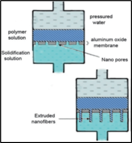
The principle of template synthesis for preparing nanofibers is based on utilizing a nonporous membrane with cylindrical pores to guide the formation of nanofibers. In this method, a polymer solution undergoes a chemical or electrochemical oxidative polymerization process. When this solution is forced through the nanoscale pores of the template under applied pressure, the polymer extrudes from the pores, allowing for the controlled formation of nanofibers. As the solution exits the template, it solidifies upon contact with a solidifying agent, resulting in the formation of structured nanofibers. The dimensions and properties of the resulting fibers are affected by the diameter of the template's pores, which makes this technique efficient for generating consistent nanofibers with designated diameters.
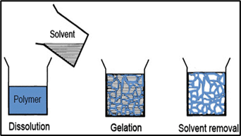
4. Phase separation53,54
The phase separation method for preparing nanofibers involves several systematic steps that utilize the physical incompatibility of components within a polymer solution. It begins with dissolving a polymer in a solvent, which is often carried out at room temperature or elevated temperatures to produce a homogeneous solution.The next critical phase is gelation, whereby the solution is left to undergo phase separation, and this will affect the morphology and porosity of the derived nanofibers; the timeframe of this process depends on the polymer concentration and temperature. Gelation is followed by the removal of the solvent in the gel, typically through the use of water, to allow the generation of fibrous structures. The final step is freezing and subsequent freeze-drying under vacuum. Such a procedure easily results in the formation of nanofibers. This technique of producing fibrous materials has advantages over other methods: it does not allow the forming of long fiber chains, but it is bound by certain restrictions toward the type of polymers to be applied, as one needs the gelling ability for this method to work.
5. Self-assembly53
The self-assembly method is the bottom-up fabrication process of nanofibers: molecules self-assemble into ordered patterns or structures by themselves through non-covalent forces like hydrogen bonding, hydrophobic interactions, and electrostatic interactions.In this approach, the self-assembling molecules, typically small active agents, come together to produce supramolecular architectures, resulting in nanofibers with nanoscale diameters typically between below 100 nm to a few nanometers, whereas the length can be several micrometers. The main driving force for this process is the intermolecular forces that attract and bind the smaller molecular units. However, the self-assembly method has challenges, such as complexity and time consumption, resulting in low productivity and less precise control over fiber dimensions. It is also largely limited to materials that can self-assemble, which is not very versatile compared to other fabrication techniques.
6. Freeze drying method (FD)53, 55
The freeze drying (FD) technique, also referred to as ice segregation-induced self-assembly or solid-liquid phase separation, consists of cooling a polymer solution to low temperatures (-70 to -80 °C) to create ice crystals. The ice is then removed by direct sublimation under reduced pressure, and any residual unfrozen water is eliminated to maintain the porous nanofiber structure. This method allows the direct creation of porous structures from polymers without the need for additives, working at lower temperatures to avoid the thermal degradation of sensitive materials. However, the preparation of complex hierarchical structures is challenging. The resulting nanofiber mats have applications in drug delivery systems and as precursors for highly porous carbon nanofibers.
7. Drawing53
In the setting of nanofiber creation, "drawing" is a prepare utilized to upgrade the introduction and properties of nanofibers after their beginning arrangement. Drawing as a general rule involves stretching the filaments to modify the polymer chains, which may advance the mechanical properties of the nanofibers, e.g., tensile strength and elasticity. This strategy can be adjusted to numerous sorts of nanofibers. The nanofibers may encounter mechanical pressure whereas being drawn, extending and arranging the strands. As a result, their atomic course of action would be more organized, hence making their execution characteristics higher. The applications for drawn nanofibers would be great for tissue building, filtration, and composite materials.
8. Interfacial polymerization53,57
An aqueous and organic phase in general, usually consist of a dissolving phase as well, hence producing nanofibers by exploiting interfacial polymerization is realized by employing a different pair of two monomers separately dissolved in these two immiscible phases. It can be exemplified in an aqueous monomer with dissolved diamine where the other can be represented in diacid chloride being dissolved in a suitable organic solvent. When these two solutions meet, polymerization occurs at the interface of the two phases, and a polymer forms the walls of nanofibers. This technique is characterized by its rapid reaction rate, which enables the production of nanofibers with distinct properties based on the choice of monomers. Although it allows for versatile fiber fabrication, the method is often associated with the production of polyamide membranes. Interfacial polymerization presents a very viable route to preparing lightweight yet robust nanofibers for many applications, including filtration and drug delivery.
Characterization of nanofibers
I. Morphological Characterization60
(i) Scanning Electron Microscopy (SEM): SEM provides a good resolution image of the nanofiber surface to assess fiber diameter, uniformity, and surface texture. The technique is popular because of the high resolution obtained and the simplicity of sample preparation.
(ii) Transmission Electron Microscopy (TEM): TEM offers sub-nanometric resolution to the investigation of the interior structure of nanofibers. It is especially suitable for studying crystallinity and phase distribution within the fibers.
(iii) Atomic Force Microscopy (AFM): AFM measures surface roughness and gives three-dimensional topographical data. It is helpful in the measurement of mechanical properties at the nanoscale and understanding surface interactions.
II. Mechanical Properties61,62,63
(i)Tensile testing: This method measures the force to break a nanofiber and gives data on tensile strength and elongation. Specialized microtensile testing apparatus are often employed because of the very small dimensions of the nanofibers.
(ii)Nanoindentation: This technique measures hardness and Young's modulus by indenting the fiber with a known force and measuring the resulting deformation. It is especially beneficial for assessing the mechanical characteristics of single fibers.
(iii)Dynamic Mechanical Analysis (DMA): It offers a dynamic mechanical examination of the viscoelastic characteristics of nanofibers through controlled oscillations in a mechanical setting to evaluate parameters such as storage modulus, loss modulus, and tan δ, in relation to viscoelastic behavior and temperature sensitivity.
(iv)Rheological testing: Evaluates the reaction to distortions under various conditions. Investigates viscoelastic characteristics. Examines performance with variations in frequency, temperature, and humidity.
III. Physicochemical Properties63,64
(i)Surface area and Porosity: Techniques of Brunauer-Emmett-Teller orBET analysis determines the surface areas and porosities of mats of nanofibers-a critical parameter where filtration and catalysis are employed.
(ii)Wettability: Contact angle measurements evaluate the hydrophilicity or hydrophobicity of nanofiber surfaces, which influences its use in fields like tissue engineering and drug delivery.
(iii)Surface Charge (Zeta potential): The zeta potential measurements give insights into the surface charge of nanofibers, which affects their stability in suspension and how they interact with biological entities.
(iv)X-ray Diffraction (XRD): XRD analysis provides information on the crystalline structure of nanofibers. The degree of crystallinity can influence the mechanical strength and degradation rate of the fibers, which are critical factors in wound healing applications.
IV. Thermal Properties63,64
(i)Differential Scanning Calorimetry (DSC): DSC quantifies the heat flow related to thermal transition that is taking place in nanofibers, such as melting and crystallization points
(ii)Thermogravimetric Analysis (TGA): TGA gives the thermal stability by measuring the weight loss against temperature, so it gives some information about the decomposition temperatures as well as gives compositional analysis.
V. Chemical Characterization63,65
(i)Fourier-transform infrared spectroscopy: FTIR detects functional groups and chemical bonds in nanofibers by analyzing infrared absorption across various wavelengths. This technique serves as a way to detect molecular changes resulting from processing or environmental exposure, which makes it crucial in studying surface modifications and interactions of materials.
(ii)X-ray photoelectron spectroscopy (XPS): XPS offers information on the elemental makeup and oxidation states of surface atoms in nanofibers. It has widely been applied to the studies involving the surface functionalization, interfacial interactions, and the incorporation of additives such as graphene oxide.
VI. Biological Properties63,66
(i)In-vitro cell culture: Evaluating cytocompatibility is achieved by seeding relevant cell types, such as fibroblasts or keratinocytes, onto the dressing material. Assessed parameters are cell viability, proliferation, and morphology, and thus give insights into the suitability of the material for wound healing. For example, Nanofibers that are electrospun and designed to mimic biological structures, which contain bioactive substances, have demonstrated the ability to enhance cell growth and speed up the healing process in models of diabetes.
(ii)Antibacterial Testing: Given the sensitiveness of DFUs to diseases, it's fundamental to evaluate the antibacterial properties of nanofibrous dressings. This can be accomplished through tests that assess the restraint of bacterial development in the nearness of the dressing fabric. Consolidating antimicrobial specialists into the nanofibers can give a multifunctional capability in mending wounds by giving fast retention and anticipating diseases
(iii)In-vivo animal studies: To comprehensively evaluate the performance of nanofibrous dressings, in vivo studies using diabetic animal models are conducted. These studies assess parameters such as wound closure rate, re-epithelialization, collagen deposition, and angiogenesis, providing a holistic view of the dressing's efficacy in promoting wound healing diseases.
Application of nanofibers
The progress in electrospinning technology and the ability to design customizable nanofibers have propelled the creation of innovative applications in numerous areas, such as healthcare, energy devices, the textile sector, and environmental solutions.
(i) Health care67,68
Being a developing and critical field, healthcare with biomedical applications remains an important center of research activity. Nanofiber technology would be a leading solution to answer many of the challenges in the field of biomedicine through exceptional properties capable of enhancing any medical treatment approach. Its utility ranges from repair of organs and real-time vital monitoring to application in burn dressings and the treatment of numerous diseases through wound dressing and the purification of blood.
(ii) Biosensors67,69
As a developing and crucial field, healthcare has seen significant advancements with the incorporation of biomedical biosensors. These analytical devices detect, measure, and monitor various analytes in the human body, including enzymes, bacteria, cells, nucleic acids, and antibodies. With the advent of nanofiber technology, the selectivity and sensitivity of these sensors have been greatly enhanced. Consequently, highly miniaturized sensing platforms capable of operating at micro, nano, and macro scales have been developed. These sensing platforms are categorized into electrochemical, optical, magnetic, and thermometric transducer types. Their performance is evaluated based on selectivity, sensitivity, response time, and detection limits. Taking advantage of nanofiber characteristics like high porosity and high surface-to-volume ratio, scientists have engineered highly efficient antibody sensors.
(iii) Detection of Nesfatin Antibody67,69,71
Kim et al.102 have recently created FET biosensors for the identification of nesfatin-1 antibodies based on carbon nanofibers containing multiscale pores. Channel materials for FET biosensors are preferred to be 1D carbon nanomaterials since they can easily be designed to immobilize bioreceptors by π-stacking or covalent bonds. In the current research, carbon nanofibers were employed both as signal transducers and as templates for immobilization of nesfatin-1 antibodies to achieve an extremely low LOD of 0.1 fM and detection time of under a second due to their multiscale porosity. Electrospun fibers have also been applied for DNA detection. Guanine is the most oxidizable chemical base present in DNA molecules and highly utilized in DNA sensing. Recently, Civan et al. prepared hybrid cellulose nanofibers to track guanine-base oxidation in single-strand DNA using electrochemical approaches. Cellulose nanofibers have various applications due to their physical, chemical, mechanical properties, and also their biocompatibility. When transformed into fibrous form with high surface-to-volume ratio, it provides a greater surface area where DNA molecules can be absorbed, making it more sensitive. Hybrid nanofibers were prepared by employing a setup that electrospun cellulose monoacetate and tetraethyl orthosilicate. The fiber diameter was controlled using a catalyst as well as hydrochloric acid, achieving diameters between 42–958 nm. The nanofibers were crucial in tracking the oxidation of guanine-base in single-strand DNA using electrochemical techniques
(iv) Waste water treatment68,72,73
Nanofiber membranes are highly efficient for filtration due to their high surface area, porous nature, and small pore size. They can attain almost 100% salt separation in desalination and are less prone to fouling than reverse osmosis membranes. The incorporation of metals into nanofibers improves anti-fouling and separation, and modified nanofibers can selectively remove heavy metals and contaminants. The mechanical properties of nanofibers can be improved through post-drawing processes and the reduction of fiber diameter increases efficiency for different applications.
(v) Application of nanofibers in tissue engineering74,75,76,80
A variety of methods for fabricating scaffolds for tissue engineering have been reported previously.Yet, in the last ten years, nanofibrous systems have shown great potential to be used as scaffolds for tissue engineering. Their high porosity and large surface area favor cell adhesion, while their 3D structure, much like a natural ECM, presents a proper micro/nano environment for the growth and function of cells. Thus, nanofibrous systems have been pursued actively as scaffolds for tissue engineering applications.
(a) For bone tissue engineering77,78:
The scaffold design for bone tissue engineering is aimed at mechanical strength, pore size, porosity, hardness, and 3D architecture. Scaffolds with 100–350 μm pore sizes and more than 90% porosity support cell/tissue in-growth and bone regeneration. Yoshimoto et al. (2003) used electrospinning to create non-woven PCL scaffolds with MSC migration and ECM synthesis. Shin et al. demonstrated ECM formation and mineralization in vivo. Ramay et al. fabricated biodegradable nano-composite scaffolds using HA and β-TCP, which improve mechanical strength and make them suitable for load-bearing applications. These studies show that nanofibrous scaffolds are promising in bone tissue engineering.
(b) For skin tissue engineering79,80,81:
More often than not, skin wounds mend through the creation of epithelialized scar tissue instep of full skin recovery. The epidermis has a restricted mending capacity, whereas the dermis repairs itself more viably. Scar tissue, which does not have the qualities of typical skin, may confine development, lead to distress, and show up ugly. Created skin tissue has the potential to advance dermal recovery. A few common and manufactured polymers have been investigated for skin tissue building, however the lion's share have not been utilized as nanofibers.
Min et al. (2004) created nonwoven silk fibrin nanofibers by electrospinning and discovered that collagen-coated fibroin nanofibers promoted attachment and spreading of keratinocytes and fibroblasts, demonstrating their suitability as platforms for constructing skin tissue.
Khil et al. (2003) recorded the application of PU electrospun nanofiber films for wound dressing purposes. These films may illustrate exceptional oxygen penetrability and control water misfortune, in this manner allowing wound liquid to leak out whereas turning away parchedness and the passage of organisms. It shows they may be fabulous alternatives for materials utilized in wound dressings.
(c) For blood vessel tissue engineering82,83,84:
Researchers have advanced from the development of vascular grafts with minimal interaction between blood and tissues to nanoscale constructs that actively encourage the formation of blood vessels. Traditionally, electrospinning produces nanofibers in a random orientation; however, Mo et al. (2004) successfully produced aligned biodegradable PLLA-CL (75:25) nanofibrous scaffolds using a rotating collector. These aligned fibers were used to create tubular scaffolds for vascular engineering, simulating the natural ECM, providing mechanical properties similar to those of human coronary arteries, and facilitating smooth muscle cell adhesion and growth. Aligned fibers ensure structural integrity, and maintain vasoactivity since they can sustain high circulatory pressure. Xu et al. (2004a) found that the aligned PLLA-CL nanofibers exhibited good adherence and proliferation for endothelial and smooth muscle cells; SMC cytoskeletons aligned along the fibers, which suggests the possibility of vascular scaffolds. Additionally, nanoscale surface roughness increases cell adhesion and proliferation (Webster et al. 2001). Ma et al. (2005b) electrospun PET into nanofibrous mats, surface-modified them with gelatin, and obtained improved spreading and proliferation of endothelial cells along with retention of phenotype, hence potential candidates for vascular grafting. Boland et al. (2004) electrospun micro and nanofibers from collagen and elastin to mimic vascular architecture, demonstrating desirable mechanical properties for artificial blood vessel engineering. These findings highlight the potential of nanofibrous scaffolds, particularly those derived from biodegradable and natural polymers, in vascular tissue engineering.
(vi) Nanofiber for filtration86,87:
Polymeric nanofibers have been applied to air filtration since over a decade ago, most often blended with substrates due to their unsatisfactory mechanical Their small diameters improve filtration efficiency through slip flow effects. particularly for fibers under 0.5 microns. Electrospun nanofiber membranes hold potential for protective clothing based on their great moisture vapor transport, breathability, and resistance to chemicals. Experiments have proved that applying a layer of nanofibers to the normal nonwoven filtration medium maximizes airflow resistance, pore size, and filtration efficiency.
(vii) Others89:
Nanofibers have applications beyond all these in aerospace, semiconductors, and advanced materials. Piezoelectric polymers such as PVDF and APB-ODPA were electrospun at NASA for use in micro-air vehicles. Carbon nanotubes (CNTs) have been integrated into electrospun fibers to create conductive membranes for coatings, photovoltaic cells, and wearable solar power applications. General Motors is researching nanofibers for composite materials because of their durability, low-temperature ductility, and recyclability.
Patents
Table 5. Patents obtained for nanofibers
|
Application number |
Title |
Summary of intervention |
|
US 2013/0125912 AI |
Nanofiber90 |
The patent indicates the development of nanofibers by electrospinning using water-soluble polymers, where the functional elements can include emulsifiers, moisturizers, encapsulated cosmetic compositions by trapping them in nanofibres for controlled release and stabilization, thereby helping cosmetics as well as biomedicine. |
|
US 7390760 B1 |
Composite nanofiber materials and Methods for making same89 |
Fibrous materials are valuable in a variety of applications, including personal care products, garments, and filtration devices. They may be absorbent or non-absorbent, and their surface chemistries and properties play an important role in performance; for example, absorbent products need large surface areas for proper absorption and may have specific hydrophobic or hydrophilic characteristics. The improved properties of fibrous materials will enhance functionality in different uses. |
|
US 2021/0246575 A1 |
Methods and device for making nanofibers and nanofiber scaffolds91 |
These methods include the drawing of a polymer solution by gravitational forces to obtain single filament nanofibers of 50 nm - 100 µm in diameter, which are then collected in ordered arrays on a frame to generate 2D arrays that may be stacked together to form 3D nanofiber scaffolds. Cell culture is accomplished using these scaffolds with well-controlled spacing and alignment, while porosity levels are achieved of 50% or greater. |
Challenges and Limitations of Nanofibers and PRP- incorporated nanofibers93,94
1. Mechanical Properties
Strength and Durability: Adding platelet-rich plasma to nanofibers degrades their strength and resistance levels. Balancing the addition of PRP without losing fiber strength is a crucial challenge.
Structural integrity: Mechanical stability and the integrity of load-bearing applications become the main challenges for the structural integrity of the nanofiber in PRP-based systems.
2. Controlled release mechanism
Release profile: The design of nanofibers that offer a sustained and controlled release of growth factors and bioactive components from PRP is complex. It requires the control of the composition, structure, and degradation rates of the fibers to achieve the desired release profile.
Release kinetics: Understanding and optimizing the kinetics of bioactive component release from PRP-incorporated nanofibers is crucial for effective therapeutic applications.
3. Biocompatibility and Safety
The biocompatibility and safety of PRP-incorporated nanofibers, especially for medical and therapeutic applications, are very important. Any adverse reaction or toxicity can undermine their effectiveness and acceptance.
4. Stability and Storage
Shelf life: The storage stability and shelf-life of PRP incorporated nanofibers are highly essential. Bioactive components of PRP could degrade with time, which could reduce their performance and effectiveness.
Storage conditions: Maintenance of optimal storage conditions to preserve the biological activity of PRP components is important to ensure the reliability and effectiveness of the final product.
5. Clogging and Maintenance
Nozzle clogging: This electrospinning process is subjected to problems including nozzle clogging that can temporarily hinder continuous productions and necessitates constant maintenance procedures, which ultimately influences the performance and productivity during manufacturing.
6. Scalability
Production challenges: Scaling the production of nanofibers from a laboratory setting to industrial scales presents significant challenges. Maintaining uniform fiber diameters and consistent properties is a very difficult task in nanofiber fabrication, and it demands specialized and precisely controlled equipment.
Infrastructure: Large-scale production facilities require massive capital investment in high-tech electrospinning machines, cleanrooms, and quality control equipment.
7. Coast
Equipment cost: Inadequate electrospinning equipment contributes to the prohibitive cost and requires high-grade raw materials necessary for nanofiber production; therefore, not easily adopted as a commercialization technology.
Operational cost: The process also has high operating costs, which include energy usage and maintenance of complex machinery. The integration of PRP into nanofibers makes the manufacturing process more complicated. This includes keeping the environment sterile and handling biological components with accuracy.
FUTURE PERSPECTIVE
Future research will focus on PRP-incorporated nanofibers in enhancing their mechanical properties, drug release profiles, and biocompatibility. These studies will be on different polymer matrices and additives that improve the structural integrity and longevity while maintaining biological activity. By fine-tuning their characteristics, researchers hope to achieve more effective sustained release of growth factors with controlled delivery aligned with the healing timeline. Advanced characterization techniques will provide insight into nanofiber-tissue interactions, thus improving healing products.
Nanofiber formulations can be tailored to individual patient needs, considering wound severity, metabolic status, and required growth factors. Diagnostic tools will assess wound microenvironments, allowing clinicians to customize dressings for optimal healing. Personalized approaches may adjust growth factor release kinetics to match specific healing processes, enhancing patient-centered care.
Combining PRP nano-fibers with other therapies will be useful for future studies. Nanofibers can incorporate stem cell therapies, antimicrobial agents, or bioactive substances to develop multitalented wound dressings in multiple healing aspects. For example, PRP in combination with antimicrobial nanofibers may fight infections and support tissue regeneration by addressing the two main challenges of handling diabetic foot ulcers. Future studies could address synergistic effects, enhancing efficacy of wound healing and reducing complications, thus further expanding the scope of therapy in chronic wound management.
Advancements in nanofiber technology alone will be crucial for future applications. Developing scalable electrospinning and fabrication techniques will enable cost-effective production with precise control over structure and properties. Exploring novel materials like biopolymers or hybrids with beneficial intrinsic properties will enhance nanofiber versatility. Additionally, integrating smart features, such as stimuli-responsive nanofibers that adapt to environmental conditions, could lead to innovative solutions for dynamic medical needs, especially in wound management.
REFERENCES


Anjanadevi P, Beny Baby, Akhiljith, Shivaram Tilagul, Integrating Nanofiber and Platelet-Rich Plasma in Drug Delivery System: Enhancing Diabetic Foot Ulcer Treatment, Int. J. of Pharm. Sci., 2026, Vol 4, Issue 4, 434-461 https://doi.org/10.5281/zenodo.19397135
 10.5281/zenodo.19397135