
We use cookies to ensure our website works properly and to personalise your experience. Cookies policy
School of Chemical Sciences, Kavayitri Bahinabai Chaudhari North Maharashtra University Jalgaon, Maharashtra. 425001.
Efonidipine hydrochloride ethanolate, a dihydropyridine calcium channel blocker with dual T-type and L-type activity, is widely used in the management of hypertension and angina. However, its therapeutic efficacy is limited by poor aqueous solubility and short half-life, necessitating frequent dosing. The present study aimed to develop and evaluate a microencapsulation system for efonidipine hydrochloride ethanolate (40 mg) using the ionic gelation method to achieve sustained drug release and improved bioavailability.Microcapsules were prepared employing sodium alginate as the primary polymer and calcium chloride as the cross-linking agent. The formulation was optimized by varying polymer concentration and cross-linker ratios. The prepared microcapsules were characterized for particle size, encapsulation efficiency, surface morphology, and in vitro drug release profile. Results indicated spherical microcapsules with uniform distribution, high encapsulation efficiency, and controlled release extending over several hours.This study demonstrates that ionic gelation is a simple, reproducible, and effective technique for microencapsulation of efonidipine hydrochloride ethanolate. The developed formulation holds promise as a novel drug delivery system to enhance therapeutic outcomes and patient compliance in cardiovascular therapy.
Hypertension and ischemic heart diseases remained among the leading causes of morbidity and mortality worldwide, necessitating effective pharmacological interventions. Efonidipine hydrochloride ethanolate, a dihydropyridine calcium channel blocker with dual L-type and T-type channel inhibitory activity, has demonstrated significant therapeutic potential in the management of hypertension and angina pectoris. Despite its clinical efficacy, the drug suffers from limitations such as poor aqueous solubility, rapid metabolism, and a relatively short biological half-life, which often require frequent dosing and may compromise patient compliance [1].
To overcome these challenges, advanced drug delivery systems have been explored to enhance solubility, prolong release, and improve bioavailability. Microencapsulation is one such promising technique, offering controlled release, protection of the drug from degradation, and improved therapeutic outcomes [2, 3]. Among various encapsulation methods, ionic gelation stands out as a simple, mild, and cost-effective approach that avoids harsh processing conditions, making it suitable for encapsulating sensitive pharmaceutical agents.
In the present study, efonidipine hydrochloride ethanolate (40 mg) was microencapsulated using sodium alginate as a natural polymer and calcium chloride as a cross-linking agent through the ionic gelation method [4]. The objective was to develop a sustained-release formulation capable of enhancing drug stability, prolonging therapeutic action, and ultimately improving patient adherence in cardiovascular therapy [5]. This work aims to contribute to the growing field of polymer-based drug delivery systems by demonstrating the feasibility and effectiveness of ionic gelation for encapsulating efonidipine hydrochloride ethanolate. [6]
Table 1: Drug profile of Efonidipine HCl ethanolate [7]
|
Parameters |
Efonidipine hydrochloride ethanolate |
|
Category: |
Antihypertensive agent |
|
Pharmacologic class: |
Dihydropyridine calcium channel blocker |
|
Appearance: |
White to pale yellow crystalline powder |
|
Melting point: |
Approximately 170–180°C |
|
Log p: |
4–5 (lipophilic) |
|
pKa: |
3.5–4.5 |
|
Solubility: |
Insoluble in water; soluble in methanol and ethanol |
|
Dose: |
Oral tablets (commonly 20 mg, 40 mg) |
|
Brands: |
Efnocar, Efonta, 20 & 40 mg |
Efonidipine Hcl
IUPAC name: (±)-2-[N-benzyl(N-phenyl)amino]ethyl 5-(5,5-dimethyl-2-oxo-1,3,2-dioxaphosphorinan-2-yl)-2,6-dimethyl-4-(3-nitrophenyl)-1,4-dihydropyridine-3-carboxylate hydrochloride ethanolate
Molecular weight: 667.12 g/mol
Molecular formula: C??H??N?O?P·HCl·C?H?O
Chemical structure:

Fig.1. Chemical structure of Efonidipine hydrochloride ethanolate
MATERIALS AND METHODS
MATERIALS:
Efonidipine hydrochloride ethanolate drug sample from (Aventus Lab LLP, Mahape Mumbai), Sodium alginate polymer from (IMCD, Mumbai), Calcium chloride (Merck), Polysorbate grade - 60, 80, Ethanol (Merck), Purified water (HPLC grade), Chitosan, acetic acid, and whatman filter paper 41.
METHODS:
Pharmaceutical drugs or bioactive compounds, can be microencapsulated by various types of methods, like – Coacervation, in situ polymerization, spray drying, extrusion, freeze drying, ionic gelation (Ionotropic gelation) and emulsion solvent evaporation [8, 9]. While among all these methods ionic gelation method is most commonly used across pharmaceutical industries due to its reproducibility, cost effectiveness, robustness, and encapsulation efficiency is greater than all other methods [10]. Ionic gelation or ionotropic gelation method involves dissolving of sodium alginate polymer in water under stirring at 600 RPM, after complete dissolution deaerate the solution for 30 – 45 min. to remove trapped air inside the polymer solution. Then drug dispersion was prepared by dissolving 40 mg EHE drug into 5 ml ethanol. Then drug dispersed solution was added into sodium alginate polymeric solution under stirring, and homogenize at 2000 rpm for 5 min. In next step chelation and microcapsule formation was carried. The calcium chloride 2.5% w/v solution was prepared for chelation. The polymeric solution of sodium alginate along with drug sample into calcium chloride solution 2.5% w/v was extruded using dispo van fitted with 22G syringe under stirring at 300 rpm. After microcapsule formation were kept the beads in hardening solution chitosan 0.5% in 1% acetic acid to strengthen the capsules if require [11, 12]. The fabricated microcapsules were filtered using Whatman filter paper 41 and washed the capsules 4 to 5 times using HPLC grade water to remove excess calcium chloride solution and sodium alginate (5-6). Then air dried at 25°C or under vacuum to get dried free flowing microsphere. The preparation of microcapsule using sodium alginate polymer, loaded with Efonidipine hydrochloride ethanolate drug via ionic gelation method is summarized in figure2 [13].

Fig 1. Stepwise schematic of sodium alginate microcapsules preparation via Ionic gelationmethod [13]
Table:
|
Sodium Alginate Concentration |
Observed Shape Characteristics |
Negative Findings |
|
1.0% w/v |
Irregular, some surface with roughness |
Reduced uniformity, tendency to form aggregates |
|
1.5% w/v |
Slightly irregular, elongated microspheres |
Poor sphericity, uneven particle distribution |
|
2.0% w/v |
Clumped, distorted structures |
High viscosity leads to aggregation and non-uniform shapes |
|
2.5% w/v |
Nearly spherical, smooth surface |
Acceptable morphology, minimal deformities |
|
3% w/v |
Large, irregular clusters |
Loss of discrete microsphere formation, poor reproducibility |
Interpretation
CHARACTERIZATION:
Fabricated microsphere of efonidipine hydrochloride ethanolate 40mg were characterized for morphology observation using microscope, Fourier Transform Infrared Spectroscopy of efonidipine hydrochloride ethanolate drug sample as well as efonidipine hydrochloride ethanolate 40mg microsphere [6, 14].
A). Morphology observation:
Fabricated microsphere was characterized for morphology observation using microscope make – ZEISS, model - Axiolab 5, observation shows spherical microsphere in appearance particle size ranging from 90 to 200 µm with free-flowing microcapsules [15].

Fig. 1 Efonidipine hydrochloride ethanolate 40mg Fig. 2 Efonidipine hydrochloride ethanolate 40mg
Using 1% Na alginate @10_X magnification Using 2% Na alginate @10_X magnification


Fig. 3 Efonidipine hydrochloride ethanolate 40mg Fig. 4 Efonidipine hydrochloride ethanolate 40mg
Using 2.5% Na alginate @10_X magnification Using 3% Na alginate @10_X magnification
Particle size measure data:
Using 1% Na alginate @10_X magnification, Resolution: 1600x1200
|
No. |
Name |
Length (µm) |
|
01 |
Line 1 |
98.56 |
|
02 |
Line 2 |
105.19 |
Using 2% Na alginate @10_X magnification, Resolution: 1600x1200
|
No. |
Name |
Length (µm) |
|
01 |
Line 1 |
161.33 |
|
02 |
Line 2 |
192.04 |
Using 2.5% Na alginate @10_X magnification, Resolution: 1600x1200
|
No. |
Name |
Length (µm) |
|
01 |
Line 1 |
110.17 |
|
02 |
Line 2 |
117.36 |
Using 3.0% Na alginate @10_X magnification, Resolution: 1600x1200
|
No. |
Name |
Length (µm) |
|
01 |
Line 1 |
167.62 |
|
02 |
Line 2 |
169.55 |
B). Fourier Transform Infrared Spectroscopy:
Fig.3. FTIR – Efonidipine hydrochloride ethanolate technical
Fig.4. FTIR – Efonidipine hydrochloride ethanolate microsphere
Interpretation
Efonidipine HCl Ethanolate Standard
The standard spectrum exhibits the characteristic absorption bands of Efonidipine HCl Ethanolate:
These peaks confirm the identity of the API.
Microcapsule Formulation
The microcapsule spectrum shows:
Conclusion of FTIR Spectra
FTIR spectra of pure Efonidipine HCl Ethanolate and the microcapsule formulation were compared to evaluate drug–excipient compatibility. The characteristic absorption bands of Efonidipine HCl Ethanolate were retained in the microcapsule formulation with only minor shifts in peak positions and changes in intensity. No new peaks or significant disappearance of characteristic drug peaks were observed, indicating the absence of chemical interaction between the drug and excipients. The results confirm the compatibility of Efonidipine HCl Ethanolate with the microencapsulation matrix and demonstrate successful incorporation of the drug without alteration of its chemical structure.
In-vitro release study [16-20]
Standard drug sample:
Drug sample Efonidipine hydrochloride ethanolate 50mg was dissolved in 100 ml methanol. Further diluted for graded solution. Quantification was done using HPLC percentage by area.
In-vitro release of microsphere:
A release study of Efonidipine hydrochloride ethanolate 40 mg microspheres was conducted at 37°C using a basket-type dissolution tester containing 900 ml of pH 1.2 buffer for the first 2 hrs and pH 7.2 buffer for the remaining 10 hrs. Efonidipine hydrochloride ethanolate 40 mg (100 mg) pre-quantified microspheres were added to 900 ml of dissolving media and agitated at 100 rpm. At the present time interval, a 10 ml aliquot sample was withdrawn and replaced with 10 ml of fresh dissolution media. After further dilution, the sample was analyzed quantitatively on HPLC (fig.5) [16-20].
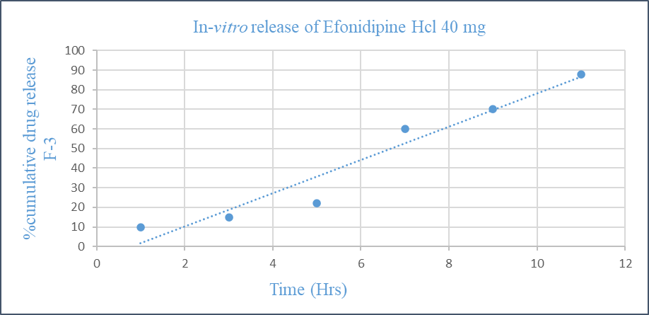
Fig. In-vitro drug release of efonidipine hydrochloride 40 mg
Encapsulation Efficiency Determination (EE) [21-23]
A pestle and mortar were used to precisely weigh and smash 75 mg of microparticles carrying 25 mg of medication. The resulting fine particles were mixed with 50 millilitres of disodium phosphate buffer solution to create a dispersion, sonicated on a sonicator for 30 minutes, and then centrifuge the mixture at high speed 4000 rpm for 10-15 minute, then separate the dissolved polymer and capsule debris. A volume of 1 ml was diluted with 50 ml of phosphate buffer solution. The standard curve was created using the UV Visible Spectrophotometer (IRMACO Germany) and the optimal absorbance measured at 250 nm. EE was calculated using the following equation [21-23].
The actual amount of drug
% of Encapsulation Efficiency = X 100
Conceptual drug content
Microencapsulation yielded an efficiency of 85%, indicating effective drug entrapment.
Particle Size Distribution
|
Formulation code |
Na-alginate concentration (%) |
Particle size (µm) |
|
F-1 |
1.0 |
98 – 105 |
|
F-2 |
2.0 |
161 – 192 |
|
F-3 |
2.5 |
110 – 117 |
|
F-4 |
3.0 |
167 – 169 |
The average particle size was ranging from 98 to 169 µm ± 15 µm, at different concentration of sodium alginate polymer and it is suitable for oral administration and controlled release.
RESULT AND DISCUSSION
The morphology of microspheres was found to be highly dependent on the concentration of sodium alginate used during ionic gelation. At concentrations (2.5% w/v), the microspheres exhibited a nearly spherical shape with smooth surfaces, indicating favorable droplet formation and cross-linking. However, as the polymer concentration increased beyond 2.5% w/v, the viscosity of the solution rose significantly, impairing the ability of droplets to form uniformly. This resulted in irregular shapes, surface roughness, and aggregation of particles. At concentrations of 2% w/v and below, microspheres became irregular, elongated, clumped, or distorted, reflecting poor sphericity and reduced reproducibility. The excessive viscosity at higher alginate levels hindered proper dispersion in the cross-linking medium, leading to non-uniform gelation and loss of discrete microsphere formation. These findings suggest that while sodium alginate is an effective encapsulating polymer, its concentration must be carefully optimized to balance encapsulation efficiency with desirable morphological characteristics. Taken together, these results establish that ionic gelation using sodium alginate at 2.5% w/v provides a robust encapsulation system, ensuring both chemical compatibility and desirable particle morphology. This optimization is essential for achieving high encapsulation efficiency, controlled release, and reproducibility, which are critical parameters for pharmaceutical applications.
ACKNOWLEDGEMENT
Authors’ special thanks to Evonik India Ltd Mumbai, for providing polymer samples, Aventus Labs LLP, Mahape, Navi Mumbai, for providing the drug sample of Efonidipine hydrochloride ethanolate for research purposes and Atul Ltd, Atul, Valsad for characterization support
REFERENCES


Dileep Rathod, Dhananjay More, Ionic Gelation Approach for Microencapsulation of Efonidipine Hydrochloride Ethanolate: A Novel Drug Delivery System, Int. J. of Pharm. Sci., 2026, Vol 4, Issue 6, 6142-6150, https://doi.org/10.5281/zenodo.20828056
 10.5281/zenodo.20828056