

We use cookies to ensure our website works properly and to personalise your experience. Cookies policy
JES SND institute of pharmacy Babhulgaon Yeola -423401.
The non-enzymatic reaction between reducing sugars (or the products of their reactive carbonyl breakdown) and proteins, lipids, or nucleic acids is known as glycation. AGEs, or advanced glycation end products, are produced by it. Long-lasting dermal proteins in the skin, such collagen and elastin, accumulate AGEs. Cross-linking, rigidity, yellowing, and elasticity loss result from this. Additionally, AGEs trigger the receptor for AGEs (RAGE), which increases inflammation and oxidative stress [1,2]. As a result, anti-glycation actives are becoming more and more popular as cosmetic additives. Since the reference inhibitor, aminoguanidine, has not been used in clinical settings [1], plant-derived polyphenols and flavonoids are being investigated as potential substitutes [10–12]. In traditional medicine, plants in the genus Jatropha (Euphorbiaceae) are frequently used to treat wounds, inflammation, and skin conditions. Vitexin and isovitexin, two C-substances prevent the production of AGEs [14–16]. The literature on (i) skin glycation and anti-glycation tactics, (ii) plant-based anti-glycation evidence and assay techniques, (iii) Jatropha leaf phytochemistry, antioxidant, anti-inflammatory, and wound-healing activity, (iv) current Jatropha topical gels, and (v) hydrogels as cosmeceutical carriers is compiled in this review. Glycosylflavones are one of the flavonoids present in their leaves, and an ion framework is suggested for further studies. In different plant systems, these.
Both extrinsic (UV radiation, pollution, smoking, diet) and internal (genetics, chronological ageing) factors contribute to skin ageing. Because they result from both internal metabolism and external exposures, AGEs are regarded as a "mixed" component [1]. Glycation is significantly increased in diabetes and increases with age, high-carb diets, and UV exposure [1]. Over the past ten years, interest in "anti-AGE" cosmeceuticals has increased. Glycation is a major but little-known cause of cutaneous ageing, according to a narrative review published in the Journal of Cosmetic Dermatology [4]. It also points out that there is still no clinical data supporting various anti-glycation treatments. Initial evaluations had Early reviews had already highlighted the pharmaceutical industry's interest in anti-AGE strategies for new anti-ageing cosmeceutical compounds [3].
Antioxidants and phenolics, which frequently exhibit anti-glycation activity, are abundant in medicinal plants [10,11]. About 170 species in the genus Jatropha are used in traditional medicine to treat ailments such skin disorders, ulcers, and inflammation [21]. Jatropha has not been investigated as an anti-glycation drug despite this ethnomedical characteristics. This review's objective is to determine whether the available data warrants the creation of a hydrogel made from Jatropha leaf extract for this use.
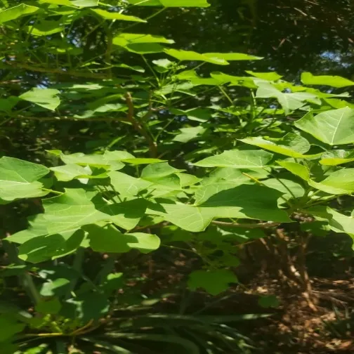
Figure 1: Image of Jatropha Curcas Plant
Table 1: Taxonomical Classification
| Kingdom: | Plantae |
|---|---|
| Sub - Kingdom | Tracheophytes |
| Class | Angiosperms |
| Clade | Eudicots |
| Sub-Clade | Rosids |
| Order | Malpighiales |
| Family | Euphorviaceae |
| Genus | Jatropha |
| Species | J. curcas |
| Biological name | Jatropha curcas |
| Marathi name | Mogali |
2. Glycation and the Skin
2.1 Chemistry of AGE formation
Schiff bases are created when free amino groups combine with reducing carbohydrates like fructose and glucose. These reorganise into Amadori and Heyns products, which are early glycation products. These compounds undergo oxidative degradation, producing reactive dicarbonyls (glyoxal, methylglyoxal, and 3-deoxyglucosone) that propel the production of AGEs [1,5]. AGEs consist of:
The most common AGE discovered in vivo is said to be CML [11]. Since oxidation and glycation are closely related, many antioxidants may theoretically also lessen glycation stress [1].
2.2 Consequences in skin
2.3 Strategies against glycation-induced skin ageing
Choi et al. outline five strategies [1]:
1. stop the creation of new AGEs (glycation inhibitors, dicarbonyl scavengers, antioxidants);
2. eliminate existing AGEs (AGE breakers, deglycating enzymes);
3. regulate RAGE-mediated signalling; 4. bolster cellular antioxidant defences;
5. restore cells and matrix.
The typical positive control in laboratory research is aminoguanidine. Because of worries about the safety/efficacy balance, it is not used in clinical settings. There are also restrictions on the AGE-breaker alagebrium (ALT-711) and similar substances [1]. Rutin, quercetin, apigenin, rosmarinic acid, ferulic acid, and chlorogenic acid are examples of natural phenolics that have been thoroughly investigated as glycation inhibitors with possible anti-aging effects on the skin [1].
Numerous topical treatments containing anti-glycation or antioxidant agents have shown beneficial in clinical settings. A gel cream with Dunaliella salina extract, a lotion with Cirsium japonicum flower extract, a serum with sunflower sprout extract, and an essence with supramolecular carnosine are a few examples. In certain instances, this decreased skin AGE levels and enhanced skin-aging metrics [1]. Nevertheless, there are still few clinical trials, and it is challenging to compare natural products directly [1].
2.4 Standard in vitro methods for screening anti-glycation activity
Table 2: Standard in vitro Methods for Screening Anti-Glycation Activity
| Parameter | Typical approach | Ref. |
|---|---|---|
| Model protein | Bovine serum albumin (BSA), or collagen/elastin for skin relevance | [1] |
| Glycating agent | Glucose, fructose, ribose, glyoxal, methylglyoxal (MGO) | [1,19] |
| Conditions | pH 7.4, 37 °C, days to weeks; sodium azide added to prevent microbial growth | [1] |
| Readouts | Fluorescent AGEs; NBT assay; ELISA/CML; SDS-PAGE; fructosamine; protein carbonyls and thiols | [1,19] |
| Positive control | Aminoguanidine | [1,19] |
| Cell/skin models | Human dermal fibroblasts, reconstructed skin, skin explants | [1] |
An important caution is that the same compound can behave differently depending on the sugar used to induce glycation. Sadowska-Bartosz et al. tested 25 natural compounds against BSA glycoxidation by glucose, fructose and ribose. They found differing effects across sugars and advised caution in extrapolating from a single-sugar model [12]. Screening should therefore use more than one glycating system.
3. Plant-Derived Anti-Glycation Agents
Searches for glycation inhibitors, particularly in vitro, have frequently identified polyphenols and other natural antioxidants as effective [11]. Plants rich in phenolics and triterpenoids commonly demonstrate combination antioxidant and anti-glycation properties [10]. A study of medicinal plant extracts indicated that suppression of AGE production linked with phenolic content and antioxidant activity [13]. Selected evidence relevant to a Jatropha leaf product is summarised below.
Table 3: Selected Plant Extracts and Flavonoids with Reported Anti-Glycation Activity
| Material | Key finding | Ref. |
|---|---|---|
| Mung bean extract; vitexin and isovitexin | Both compounds inhibited AGE formation (glucose- or MGO-induced) with efficacy above 85% at 100 µM. They did not directly trap MGO, so activity was attributed mainly to free-radical scavenging. | [14] |
| Mung bean water extract | Vitexin and isovitexin identified in the extract. The extract inhibited AGE formation (IC~50~ = 2.28 mg/mL). | [18] |
| Bamboo-leaf flavonoids (orientin, isoorientin, vitexin, isovitexin) | All four significantly reduced fluorescent AGEs and protein oxidation. Mechanisms included inhibiting protein oxidation, lowering AGE-precursor concentration and blocking dicarbonyl–protein binding. | [15] |
| Duchesnea chrysantha constituents | Isovitexin and several other flavonoids and ellagic acids showed excellent AGE-formation inhibition. | [16] |
| Flavone C-monoglucosides (orientin, homoorientin, vitexin, isovitexin) | Compared with quercetin for antioxidant, ACE-inhibitory, AChE-inhibitory and AGE-inhibitory activity in model systems. | [17] |
| Moringa oleifera aqueous leaf extract | Total phenolics 38.56 mg GAE/g. Dose-dependent (0.5–2.0 mg/mL) reduction of fluorescent AGEs and CML in glucose- and fructose-BSA systems, with reduced protein carbonyls. Aminoguanidine was the comparator. | [19] |
| Varronia curassavica ethanolic leaf extract and two isolated compounds | Significantly reduced AGE formation and protein oxidative damage in an MGO model. | [20] |
This evidence is consistent with the flavone C-glycosides isovitexin and vitexin. Additionally, they are reported from Jatropha leaves (Section 4), which serves as the primary chemical foundation for the review's theory.
4. The Genus Jatropha: Botany, Phytochemistry and Relevant Bioactivities
4.1 Overview
In the seasonally dry tropics, there are roughly 170 species of trees, shrubs, subshrubs, and plants that belong to the Euphorbiaceae family. Inflammation, ulcers, skin conditions, diabetes, and oedema are among the common uses [21]. Cyclic peptides, alkaloids, diterpenes, flavonoids, phenols, lignans, and coumarins are among the substances that have been reported. Anti-inflammatory, antioxidant, antibacterial, anticoagulant, cytotoxic, and other properties are demonstrated by extracts and pure substances [21, 22]. In Africa, Asia, South America, and the Middle East, J. curcas has been utilised to treat about 100 distinct conditions [22]. There are also systematic evaluations of its toxicity and pharmacology [23].
Bellyache bush, or J. gossypiifolia, is examined independently. Its leaves are said to have insecticidal, anti-inflammatory, and antibacterial qualities; evaluations suggest little toxicity in certain in vitro and in vivo experiments [24, 25].
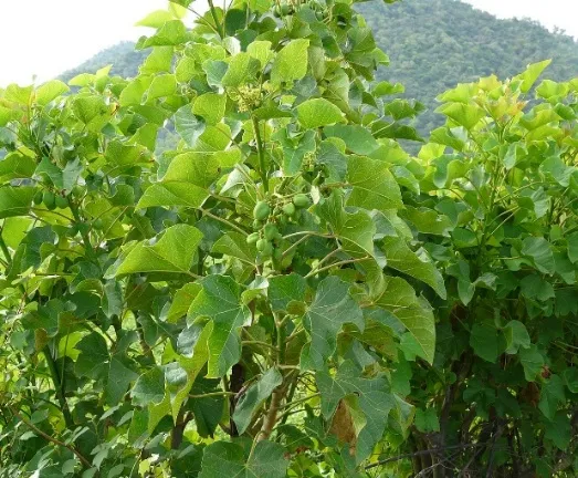
Figure 2: Image of Jatropha Curcas Leaves
4.2 Leaf flavonoids and antioxidant activity
Table 4: Leaf Phytochemistry and Antioxidant Data Relevant to Anti-Glycation Potential
| Species / extract | Finding | Ref. |
|---|---|---|
| J. curcas leaves (grown in Southern Italy) | HPLC-DAD showed a flavone C-glycoside profile: vitexin most abundant, followed by vicenin-2, stellarin-2, rhoifolin, with traces of isovitexin and isorhoifolin. Extracts showed antioxidant and cytoprotective activity in lymphocytes. | [28] |
| J. curcas leaf ethanol extract | DPPH IC~50~ 7.47 µg/mL and ABTS IC~50~ 6.6 µg/mL. HPLC-DAD-MS^n^ tentatively identified orientin, vitexin, isovitexin and rhoifolin. | [29] |
| J. gossypiifolia leaf and stem methanolic extracts | Leaves had higher total phenolics (65.66 mg GAE/g) than stems (33.33 mg GAE/g). Antioxidant and membrane-stabilising activity were reported. | [30] |
| J. gossypiifolia leaf crude extract and aqueous fraction | Antioxidant activity including iron- and copper-chelating activity was demonstrated in vitro. | [26] |
| J. gossypiifolia aqueous leaf extract | Contained sugars, alkaloids, flavonoids, tannins, terpenes/steroids and proteins; showed anti-inflammatory and antiophidic effects. | [27] |
| J. mollissima hydroethanolic leaf extract | 11 compounds characterised by LC-MS, with emphasis on flavonoids. | [32] |
Metal chelation matters because transition metals catalyse the oxidative steps that convert early glycation products into AGEs (glycoxidation) [1].
4.3 Anti-inflammatory, wound-healing and antidiabetic activity
. A J. mollissima leaf-extract gel demonstrated notable wound-healing efficacy, decreased inflammatory and oxidative indices, and prevented carrageenan-induced paw oedema as well as acute and chronic croton-oil ear oedema [32].A J. variegata extract gel demonstrated wound-healing efficacy in rats and had MIC values of 0.05% against S. aureus, P. aeruginosa, and E. coli [33].At 30, 50, and 100 mg/kg, J. gossypiifolia leaf extract reduced glycaemia in streptozotocin-induced diabetic rats [31]. Diabetes is the clinical setting in which glycation is most relevant
5. Rationale for Jatropha Leaf Extract as an Anti-Glycation Agent
Note: This section is an inference from indirect evidence. It is not a report of experimental results.
Three lines of evidence support the hypothesis:
1. overlap of chemicals. Vitexin and isovitexin are found in J. curcas leaves [28, 29]. In the glucose and MGO systems, these flavonoids prevent the production of AGEs [14–16]. Extracts containing these flavones also demonstrated anti-glycation action in other plants [18]. 2. Overlap of mechanisms. Phenolic extracts lower glycoxidation by metal chelation, radical scavenging, and antioxidant action [1,14,15]. Strong radical scavenging and metal chelation are demonstrated by Jatropha leaf extracts for J. gossypiifolia [26, 29]. 3. Activities that complement the skin. Beyond glycation inhibition, cosmeceutical benefit may be enhanced by anti-inflammatory and wound-healing properties [32,33] and antibacterial activity [33,35,36]. This aligns with the "multi-target" strategy suggested for anti-aging skin care products [1].
Limitations of this reasoning.
From single-compound data, extract activity is unpredictable. It is possible for other components, such as pigments, to oppose or interfere. There are currently no experimental Jatropha anti-glycation studies available.
6. Safety Considerations
Safety is the most important consideration for any topical Jatropha product. Toxicity in the genus is mainly associated with latex and seeds. The toxins of J. curcas include phorbol esters (irritant, co-carcinogenic diterpenoids) and curcin (a ribosome-inactivating protein), and skin contact with the plant sap can cause irritation [21,22,24,37]. J. gossypiifolia has shown low toxicity in some studies, but reviews still note the toxic potential of the genus [24,25].
7. Hydrogels as Cosmeceutical Delivery Systems
Hydrogels are networks of synthetic and/or biopolymers that are cross-linked and have a high capacity to absorb water. Both biomedicine and cosmetics make extensive use of them [38, 39]. Reviews point to a number of benefits for cosmetology and dermatology: Easy application and few side effects [38], biocompatibility, particularly for natural polymers [39], suitability as bioadhesive skin-care formulations [39], and the capacity to deliver actives in a controlled manner, though delivery is dependent on skin-barrier characteristics and the physicochemical properties of the active and its carrier [3]
8. Existing Jatropha Topical Gel Studies
Table 5: Reported Jatropha Gel Formulations
| Species / part | Formulation and evaluation | Main outcome | Ref. |
|---|---|---|---|
| J. mollissima hydroethanolic leaf extract | Topical gel; physicochemical characterisation; dermal irritation test; mouse inflammation, wound-healing and antiophidic models | Reduced oedema; accelerated wound healing; reduced venom-induced dermonecrosis by 55% | [32] |
| J. variegata methanolic leaf/stem extract | Gel tested on rats (12 female albino rats, three wounds each) | MIC 0.05% against three bacteria; wound healing superior to control (33% healed in control) | [33] |
| J. curcas leaf extract | Gels with 10, 15 and 20% extract on rat burns; positive control Bioplacenton | 20% gel most effective; not significantly different from positive control (P > 0.05) | [34] |
| J. curcas leaf 70% ethanolic extract | Hand-sanitiser gel using 100 mg/mL extract; organoleptic, pH, stability and spreadability tests | Gel physically stable; inhibition zone 15.6 mm against S. aureus | [35] |
| J. curcas sap | Hand-sanitiser gel; best formula had 5% sap, pH 4.89, viscosity 4.60 ± 0.14 Pa·s | Homogeneous, clear whitish gel with antibacterial activity | [36] |
These investigations demonstrate the ability to create stable topical gels using Jatropha extracts. It is possible to immediately adopt the evaluation criteria they employed (pH, viscosity, spreadability, homogeneity, stability, and irritation). They didn't look at glycation.
9. Proposed Development and Evaluation Framework
Note: This framework is the author's proposal built on the literature above. It is not a published protocol.
Stage 5: Physicochemical analysis. appearance, colour, smell, homogeneity, storage stability, and spreadability [32, 35, 36]. Since delivery is dependent on the skin barrier and the carrier, including extract content (marker chemicals) and release or permeation testing [38].
Stage 6: The gel's effectiveness. On the prepared gel, repeat the glycation tests in comparison to the blank base. Extend to glycated fibroblast or reconstructed-skin models when facilities permit, and eventually assess skin autofluorescence in human research [1].
10. Research Gaps and Future Directions
1. No Jatropha species have direct anti-glycation data (the main gap found here). 2. Inconsistent species identification across studies and a lack of standardised leaf . 3. It is necessary to determine whether activity results from dicarbonyl trapping. 4. There is a dearth of safety information about the topical application of leaf extracts. It is necessary to rule out latex contamination and irritating diterpenes. 5. Skin models have not been used to test current Jatropha gels for AGE-related outcomes. 6. Since there is little clinical evidence for anti-glycation cosmeceuticals in general [1,4], any new product would require carefully planned human investigations.
CONCLUSION
Plant polyphenols are attractive, seemingly harmless anti-glycation possibilities. Glycation is known to contribute to skin ageing. Jatropha leaves exhibit potent antioxidant activity, include flavone C-glycosides such vitexin and isovitexin, and have been developed into topical gels with anti-inflammatory and wound-healing properties. When taken as a whole, these results provide a solid scientific foundation for researching a hydrogel made from Jatropha leaf extract as an anti-glycation cosmeceutical. The genus's toxicity profile necessitates cautious safety work, and the anti-glycation activity itself has not yet been tested. A useful path for experimental verification is provided by the suggested framework.
REFERENCES


Aniket Chavan, Gayatri Jadhav, Shital Chavan, Ankush Khanjode, Manish Ahire, Sagar Vaidya, Jatropha Leaf Extract as an Anti-Glycation Cosmeceutical Hydrogel: A Review of the Evidence, Rationale and Formulation Prospects, Int. J. of Pharm. Sci., 2026, Vol 4, Issue 11, 65-73. https://doi.org/10.5281/zenodo.23876654
 10.5281/zenodo.23876654