
View Article

Abstract

Microneedle (MN)-based drug delivery systems have emerged as a promising approach for transdermal drug administration by overcoming the barrier function of the stratum corneum. These minimally invasive systems offer several advantages, including improved drug permeation, reduced pain, enhanced patient compliance, and the potential for controlled drug release. This review discusses the classification of microneedles, including solid, coated, dissolving, hollow, and hydrogel-forming systems, along with their mechanisms of drug delivery. Various materials, such as metals, silicon, and biodegradable polymers, and fabrication techniques, including micromoulding, photolithography, and three-dimensional (3D) printing, are also explored. Furthermore, the review highlights important characterization parameters and pharmaceutical applications in vaccination, diabetes management, dermatological treatment, wound healing, cancer therapy, and biosensing. Recent innovations, including stimuli-responsive microneedles, bioinspired designs, wearable sensing devices, and personalized drug delivery platforms, may further expand their therapeutic potential. Despite these advances, challenges related to mechanical strength, drug loading, stability, dose accuracy, manufacturing scale-up, sterility, patient acceptability, and regulatory requirements remain. Overall, microneedle technology represents a promising platform for advanced drug delivery; however, further research, standardized evaluation, and clinical studies are necessary to establish its safety, efficacy, and successful commercial translation.

Keywords

Microneedles, Transdermal drug delivery, Controlled drug release, 3D printing, Stimuli-responsive microneedles, Drug delivery challenges

Introduction

× Popup Image

Over the past few decades, transdermal drug delivery systems (TDDS) have attracted significant interest as a viable alternative to oral and injectable 'formulations due to their ability to provide sustained drug release, bypass first-pass metabolism, and enhance patient compliance. However, the efficacy of traditional transdermal delivery is greatly hindered by the stratum corneum, the skin's outermost layer, which serves as a highly effective barrier to the penetration of most therapeutic agents [1]. Transdermal drug delivery has developed from old topical treatments to advanced medical devices that allow for controlled and effective delivery of medication through the skin [2]. The past few decades, advances in microfabrication and material science have driven the emergence of microneedle (MN) technologies, representing a transformative leap that overcomes the limitations of oral and conventional parenteral drug delivery methods [3]. This technology enables minimally invasive, pain-free delivery of a wide range of drugs and biologics, improving patient compliance and therapeutic outcomes [4].

1.1 History of MN

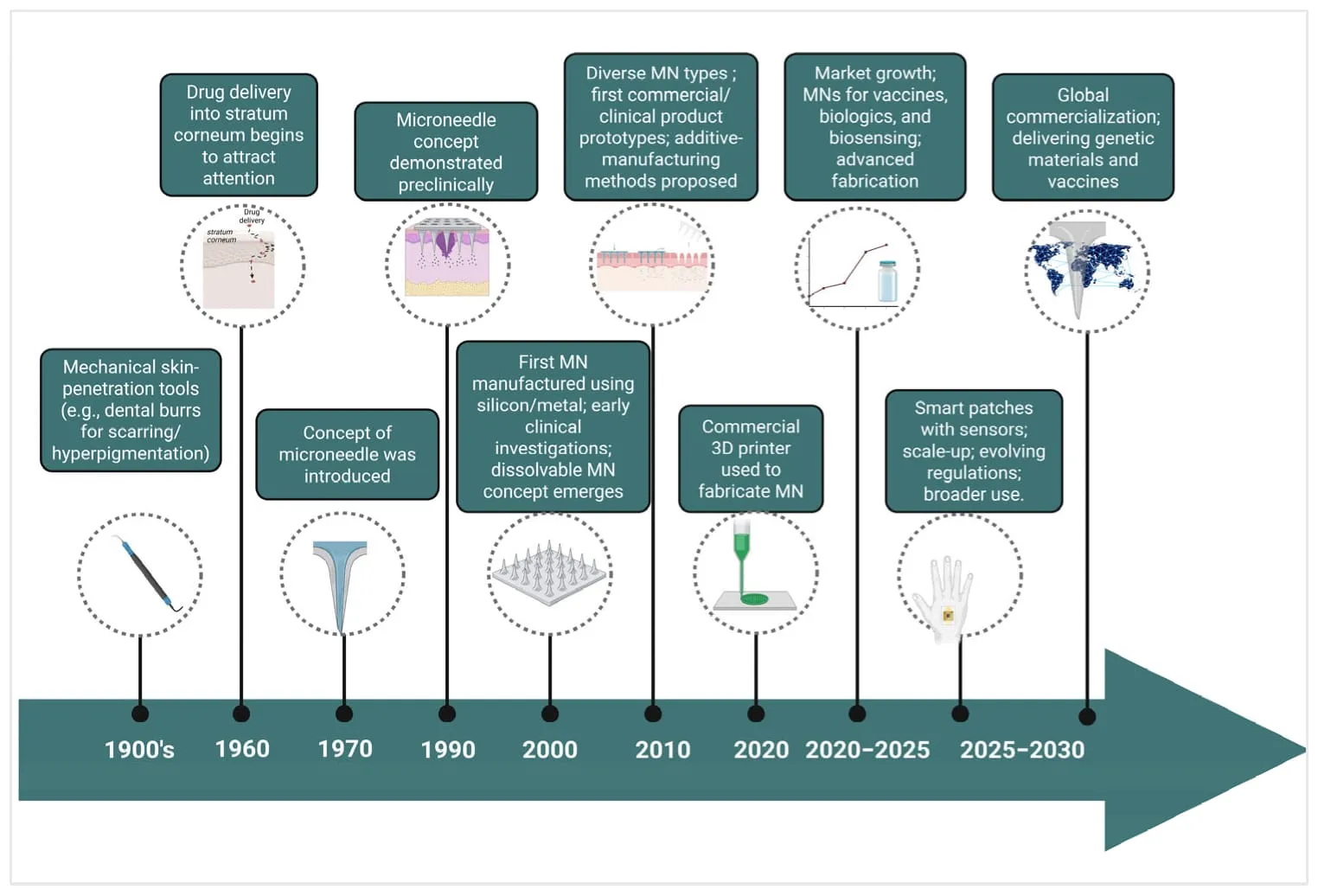
MN technology has advanced considerably since its inception in the 1970s, with key innovations occurring in the 1990s, when microfabricated silicon MNs were introduced as less-invasive instruments for transdermal drug delivery. This period initiated advanced manufacturing techniques utilizing materials including silicon, metals, and polymers [5]. The 2000s witnessed diversification into numerous MN forms, providing distinct benefits for the delivery of vaccines, small compounds, and biomacromolecules. Clinical translation advanced through trials that exhibited less pain, enhanced patient adherence, and expanded therapeutic possibilities, encompassing cosmetic uses [6.7].

Over the past decade, significant progress has been achieved through the integration of innovative materials and manufacturing methods, such as 3D printing, to facilitate scalable, precise production of MNs. Concurrently, research efforts have expanded toward multifunctional MN platforms incorporating sensing elements, stimuli-responsive materials, and closed-loop control capabilities. However, such smart MN systems largely remain at the preclinical or early translational stage [8]. The period from 2020 to 2030 characterizes the transformation of MN technology from a laboratory concept into a clinically viable platform, as the field addresses remaining hurdles in drug loading capacity, scale-up, and regulatory compliance. The continuous developments underscore the evolution of MNs from experimental apparatus to multifunctional instruments pivotal to next-generation medicines and diagnostics [9] (Figure 1).
figure

Figure 1: Timeline Showing the Evolution and Projected Future of MN Technology from Research to Global Commercialization (Created Using Biorender, Godugu, D. (2025) Https://BioRender.Com/0X4V3Gz, Accessed on 2 November 2026).

2. Skin structure and Barrier Function

The skin is the largest organ of the human body and serves as a protective barrier against environmental, chemical, and biological insults. Structurally, the skin consists of three primary layers:

2.1 Epidermis

The epidermis is the outermost layer and is composed of five sublayers, of which the stratum corneum is the most significant in terms of drug delivery. The stratum corneum is approximately 10–20 µm thick and consists of dead, flattened keratinocytes embedded in a lipid matrix, often described by the “brick and mortar” model.

2.2 Dermis

The dermis lies beneath the epidermis and contains blood vessels, lymphatic vessels, nerves, and connective tissue. Drug molecules reaching the dermis can readily enter systemic circulation.

2.3 Hypodermis

The hypodermis is composed mainly of adipose tissue and provides insulation and mechanical protection.

Microneedles are designed to penetrate the stratum corneum and upper epidermis while avoiding deeper dermal nerves and blood vessels, thus enabling painless drug administration [10].

3. Classification of Microneedle

Microneedles are broadly classified into four major types based on their design and mechanism of drug delivery [11].

3.1 Solid Microneedles

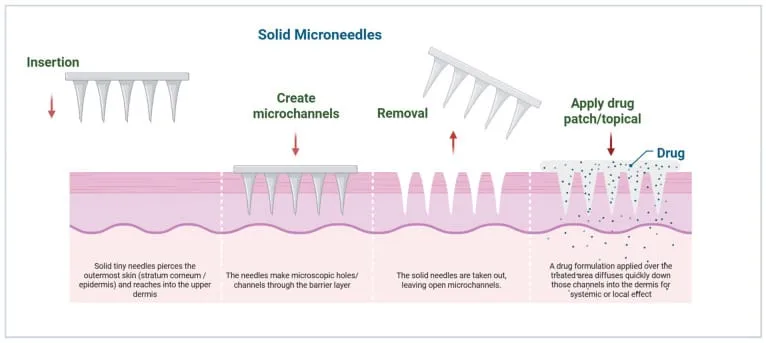
Solid microneedles are fabricated from metals or silicon and are primarily used to create microchannels in the skin. After microneedle application, a drug formulation is applied topically, allowing enhanced permeation through the created pathways. This approach is commonly referred to as the “poke-and-patch” method.

• Advantages:

  • Simple design
  • Reusable arrays
  • Effective skin permeabilization

• Limitations:

  • Two-step application
  • Risk of microbial contamination

3.2 Coated Microneedles

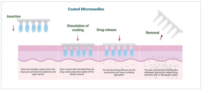
In coated microneedles, the drug is coated onto the surface of the microneedle shafts using techniques such as dip-coating or spray-coating. Upon insertion, the drug rapidly dissolves in interstitial fluid.

• Advantages:

  • Rapid drug release
  • Suitable for vaccines and potent drug
  • Limitations:
  • Limited drug loading
  • Uniform coating challenges

3.3 Dissolving Microneedles

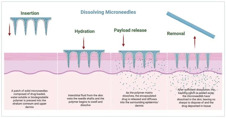
Dissolving microneedles are fabricated from water-soluble or biodegradable polymers that encapsulate the drug within the microneedle matrix. Upon insertion into the skin, the microneedles dissolve completely, releasing the drug.

• Advantages:

  • No sharp waste
  • High patient safety
  • Ideal for mass vaccination

• Limitations:

  • Mechanical strength constraints
  • Moisture sensitivity

3.4 Hollow Microneedles

Hollow microneedles contain a central lumen through which liquid drug formulations can be delivered under pressure. They function similarly to miniature hypodermic needles.

• Advantages:

  • Precise dosing
  • Suitable for continuous infusion

• Limitations:

  • Complex fabrication
  • Risk of clogging

Classification of MNs: mechanism, fabrication, advantages, and limitations.

Table 1:

Type Fabrication Materials/Methods Mechanism Advantages Limitations Reference
Solid Silicon, metals, polymers, etching, molding Creates microchannels for passive diffusion Simple design, low cost Poor control of dosing [11]
Coated Dip-coating, spray-coating, and inkjet printing Drug layered on the surface, dissolves upon insertion Rapid release, suited for vaccines Limited drug load [11]
Dissolving Polymers (polyvinylpyrrolidone, hyaluronic acid) via micro molding The biodegradable matrix dissolves in the skin, releasing the drug No waste, suitable for biologics Fragility, limited penetration [11]
Hollow Silicon, glass, stainless steel; laser micromachining Drug infused through the central lumen Larger volumes, controlled infusion Complex design, higher cost [11]
Hydrogel-forming Crosslinked polymers (PEG, PHEMA) Swellable polymers form drug-permeable conduits Sustained release, reusable reservoir Removal required, slower onset [11]
Hybrid/Next-gen Composite polymers, 3D printing, NPs Combines multiple features; smart materials High versatility, personalized therapy Still experimental, scalability issues [11]

4. Mechanisms of Drug Delivery via Microneedles

MNs enhance drug permeation across the skin by physically or chemically modulating the stratum corneum, the primary barrier to transdermal delivery. The mechanism of delivery depends on the MN type, formulation, and physicochemical properties of the drug.[12,13]

4.1 Passive Diffusion via Solid Microneedles

Solid MNs work primarily by forming microchannels through the stratum corneum, facilitating the subsequent diffusion of topically applied drugs. The “poke-and-patch” approach relies on passive diffusion gradients, hydrophilicity, and hydration [14]. The formation of microchannels is rapid and reversible; microchannels usually close within hours due to the skin’s elasticity and its mechanisms for repairing damaged skin. Studies have demonstrated enhanced delivery of small molecules, peptides, and vaccines using solid MN-mediated microchannels, with permeability enhancements ranging from 10 to 1000-fold compared to intact skin [15]. (figure 2)
figure

Figure 2: Schematic Representation of Solid MNs (Poke-And-Patch) and Their Mechanisms of Drug Delivery Through the Skin (Created Using Biorender, Godugu, D. (2025) Https://BioRender.Com/Y50Mebj, Accessed on 7 November 2025).

4.2 Coating Dissolution Kinetics in Coated Microneedles

Coated microneedles (MNs) deliver drugs through a thin layer of active pharmaceutical ingredient (API) deposited on the needle surface [16]. After the MNs are inserted into the skin, the coating dissolves quickly, allowing the drug to be released within a few minutes. The rate of drug release mainly depends on the thickness of the coating, the type and composition of polymeric excipients, and the movement of interstitial fluid within the skin [17]. This rapid and efficient drug delivery is particularly useful for vaccines and potent biologics, where accurate dosing and a quick therapeutic response are important. For consistent and reproducible drug delivery, it is essential that the coating is uniform and remains firmly attached to the microneedle during insertion [18].(Figure 3)
figure

Figure 3: Illustrative Mechanism of Drug Diffusion and Release from Coated MNs into the Dermal Layer (Created Using Biorender, Godugu, D. (2025) Https://BioRender.Com/Mbtqmrm, Accessed on 7 November 2025).

4.3 Biodegradable Matrix Dissolution in Dissolving Microneedles

Dissolving microneedles (MNs) incorporate the active pharmaceutical ingredient (API) into a biodegradable polymer matrix, such as polyvinylpyrrolidone, hyaluronic acid, or carboxymethyl cellulose [19]. After the MNs are inserted into the skin, interstitial fluid (ISF) enters the polymer matrix and gradually dissolves it, releasing the drug into the skin. The rate of drug release can be adjusted by changing the polymer molecular weight, degree of crosslinking, and microneedle design [20]. A major advantage of dissolving MNs is that they dissolve completely after application, leaving no sharp waste and reducing biohazardous waste. They can also help improve the stability of temperature-sensitive biologics by maintaining them in a solid form [21]. Clinical studies using insulin and influenza vaccines have shown promising results, highlighting the potential of dissolving MNs for convenient administration and better patient compliance [22].(Figure 4)
figure

Figure 4: Representation of the Insertion, Hydration, and Drug Release Mechanisms of Dissolving MNs for Controlled Drug Delivery Through the Skin (Created Using Biorender, Godugu, D. (2025) Https://BioRender.Com/J1Hiys8, Accessed on 7 November 2025).

4.4 Infusion Through Hollow Microneedles

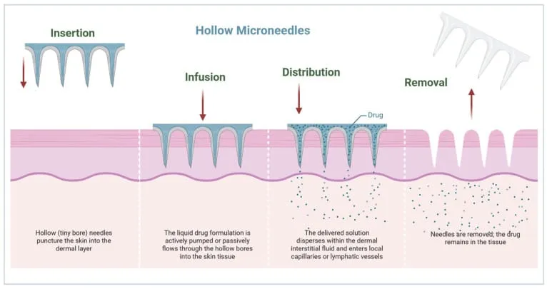
Hollow microneedles (MNs) have a small hollow channel, or lumen, through which liquid drug formulations can be delivered directly into the dermal layer of the skin. Depending on the treatment requirement, the drug can be administered as a single bolus, continuous infusion, or pulsatile dose, allowing better control over drug exposure and pharmacokinetics [23]. The rate of drug delivery is influenced by several factors, including the needle diameter, lumen length, insertion depth, and resistance or backpressure from the skin. Hollow MNs are particularly useful for delivering relatively large volumes or viscous formulations and may be suitable for biologics, monoclonal antibodies, and certain chemotherapeutic agents [24].(Figure 5)
figure

Figure 5: Diagram Depicting the Structure and Drug Distribution Process of Hollow MNs Across the Skin Barrier (Created Using Biorender, Godugu, D. (2025) Https://BioRender.Com/Ksv0Rll, Accessed on 7 November 2025).

4.5 Swelling and Sustained Release via Hydrogel-Forming Microneedles

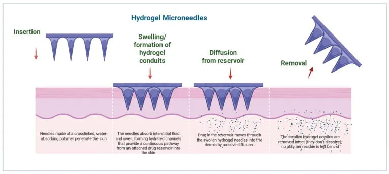
Hydrogel-forming microneedles (MNs) are made from crosslinked, water-absorbing polymers such as PEG and polyHEMA. After insertion into the skin, these polymers absorb interstitial fluid (ISF) and swell, forming pathways that allow the drug to move from an attached drug reservoir into the surrounding tissue [25]. The rate and duration of drug release can range from several hours to a few days, depending on factors such as the polymer composition and degree of crosslinking [26]. Unlike dissolving microneedles, hydrogel-forming MNs do not dissolve completely during treatment, which can help minimize the accumulation of polymeric material within the skin [27]. These systems have shown promising results for the sustained delivery of insulin, vaccines, and small-molecule drugs. Their ability to provide controlled drug release may improve pharmacokinetic profiles and reduce the need for frequent dosing [28].(Figure 6)
figure

Figure 6: Schematic Illustration of Hydrogel-Forming MNs and Their Transdermal Drug Delivery Mechanism (Created Using Biorender, Godugu, D. (2025) Https://BioRender.Com/Hqv7Bh1, Accessed on 7 November 2025).

4.6 Hybrid and Stimuli-Responsive Mechanisms

Next-generation microneedle (MN) systems are being developed by combining different delivery approaches within a single platform. For example, dissolving microneedle tips can be combined with strong, mechanically stable bases to improve both drug delivery and structural integrity. Some advanced MNs also use stimuli-responsive polymers that can control drug release in response to specific physiological or external conditions, such as changes in temperature, pH, or enzymatic activity [29]. In addition, nanocarriers such as nanoparticles (NPs), liposomes, and polymeric micelles can be incorporated into the MN matrix to improve drug targeting and regulate the rate and location of drug release. These hybrid systems can provide different release patterns, including sustained, pulsatile, or on-demand drug delivery. When integrated with wearable electronic devices, they may also monitor physiological changes in real time and adjust drug administration accordingly, offering a promising approach toward personalized and closed-loop drug delivery [30].(Figure 7)

5. Microneedle structure design

Several factors should be considered when designing microneedles for skin penetration:

(1) geometric features, such as length

(2) diameter, tip size, and shape

(3) physical form: solid, hollow, side-opened, conical, bevelled tip

(4) material selection

(5) fabrication feasibility

(6) application

(7) layout of the arrays

(8) density

(9) total number of microneedles

10) surface layer state

In addition, microneedles are defined according to their array density, length or height, and shaft and tip shape. The final design will depend on the limitations of the fabrication method and the mechanical properties, physical, and chemical stability of the material.

Most microneedles to date have been fabricated with heights below 1 mm, yet sufficient to access ISF or capillary blood or to deliver therapeutic agents. Some obstacles to insertion of microneedles into the skin are the presence of dermatoglyphics (small wrinkles) and hair, while the passage of cells through hollow microneedles depends on their internal lumen diameter, which must be large enough to ensure the flow of microscopic entities, including larger objects such as white blood cells (leukocytes), which are tens of microns in diameter. Other parameters determining flow in a microchannel include blood viscosity, contact angle, hydrodynamic diameter, and driving forces such as surface tension. In addition, due to the elastic nature of the skin and its irregular surface, varying from person to person, and with age and position on the body, the efficient penetration of microneedles to the desired depth, without fracture and with high accuracy, may require an applicator to facilitate skin penetration in a controlled and reproducible manner.

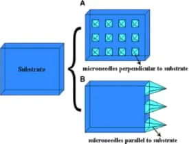
The microneedle design will vary depending on the application and fabrication method used (e.g., solid, hollow, open groove) but all microneedles can be classified as in-plane or out-of-plane In-plane microneedles have the longitudinal axis of the shaft parallel to the surface of the substrate while out-of-plane microneedles have their longitudinal axis perpendicular to the substrate surface. Fabrication of two-dimensional arrays of in-plane microneedles is very difficult, but it is easier to integrate in-plane microneedles with micropumps, sensors, microfluid chips, and electronic circuitry. On the other hand, it is significantly more convenient to fabricate arrays of out-of-plane microneedles in high-density two-dimensional arrays, although the resulting microneedles are restricted to lower aspect ratio and shorter height compared to in-plane microneedles if traditional microfabrication methods, such as wet and dry etching, are used [31].
figure

Figure 7: Schematic Showing In-Plane and Out-Of-Plane Microneedle Arrays [31]. Figure 2 Was Reproduced from [31] (© 2019 X. He Et Al., Published by SAGE, Distributed Under the Terms of Creative Commons Attribution-NonCommercial 4.0 License (Http://Www.Creativecommons.Org/Licenses/By-Nc/4.0/). This Content Is Not Subject to CC By 4.0.).

6. Structure and Design Principles

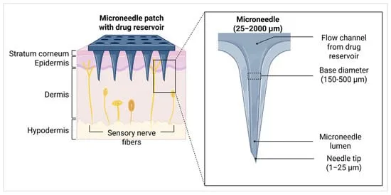
MNs are arrays of microscale needles typically ranging in length from 25 micrometers (μm) to 2000 μm (2 mm), which corresponds approximately to the thickness of the human epidermis, including the stratum corneum . The needle tip radius is designed to be sharp, ranging from 1 μm to 25 μm, to ensure efficient penetration through the skin barrier with minimal pain. The base diameter of individual MNs typically ranges from 150 μm to 500 μm [32]. These are arranged in arrays commonly sized around 8 mm by 8 mm, with spacing (pitch) between the needles approximately 500 μm [33]. The needle height is carefully engineered to breach the outer skin layers without reaching pain receptors or blood vessels in the dermis, thereby providing a minimally invasive and painless drug delivery experience. These dimensions are crucial for achieving a balance between effective skin penetration and patient comfort and safety [34].
figure

Figure 8: MN Patch Penetrating Skin Layers for Transdermal Drug Delivery. Inset Shows Microneedle Structure with Lumen and Delivery Channel (Created Using Biorender, Godugu, D. (2025) Https://BioRender.Com/R3Qefwt, Accessed on 7 November 2026).

7. Matarials Used for Microneedles

7.1 Fabrication and use of metal microneedles

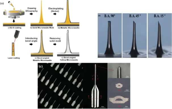
In addition to silicon and polymers, metals such as nickel, titanium, stainless steel, and palladium are also used for microneedle fabrication. These metals offer good mechanical strength and biocompatibility, making them suitable for producing microneedles, although certain metals may raise concerns regarding toxicity. Common fabrication techniques include photochemical etching, electroplating, and laser cutting, which can be used to produce both solid and hollow microneedles. For example, hollow nickel microneedles with heights of approximately 300–450 μm have been successfully fabricated using electroplating in a 6 × 6 array. In this approach, a two-layer SU-8 epoxy resin structure was used as the master mould for preparing the microneedles [35]. (Figure 9)
figure

7.2 Fabrication and use of silicon microneedles

Silicon microneedles have been widely used for many years and remain one of the most commonly studied types of microneedles. Their fabrication generally relies on microfabrication techniques originally developed for the microelectronics industry [36]. However, these methods often involve several processing steps and require sophisticated and expensive equipment. Among the different fabrication approaches, subtractive techniques, particularly wet and dry etching, are commonly employed. In wet etching, a single-crystal silicon wafer is immersed in a suitable chemical etchant to remove material from the surface. Depending on the etchant and processing conditions, the process can produce either isotropic etching, where material is removed at a similar rate in all directions, or anisotropic etching, where the etching rate varies according to the orientation of the silicon crystal planes [37].

7.3 Fabrication and use of polymer microneedles

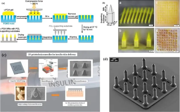
Polymeric materials have attracted increasing attention in microneedle fabrication because of their good biocompatibility, suitable mechanical properties, low cost, and biodegradability. Compared with silicon-based microneedles, polymers also offer the advantage of simpler and more economical fabrication. These features make polymers promising materials for the development of next-generation microneedle-based point-of-care diagnostic systems and drug delivery patches. Common fabrication techniques include photolithography, replica moulding, three-dimensional (3D) printing, and micromachining, which allow microneedles to be produced with different shapes, structures, and drug-loading capabilities [38]. (Figure 9)
figure

8. Fabrication Techniques

Conventional MN fabrication includes micromachining, photolithography, laser-based processing, micro-molding, drawing lithography, droplet-based approaches and centrifugation-assisted molding. Polymer casting is widely used for dissolving MNs because it is relatively accessible and can accommodate sensitive biological cargo when processing conditions are mild. [39]

3D printing is one of the most important recent developments. Stereolithography (SLA), digital light processing (DLP), two-photon polymerization and other micro/nano-printing approaches can generate complex geometries with high dimensional control. Recent reviews emphasize potential advantages in customization, geometry optimization and personalized devices, while also identifying limitations involving printable biomaterials, residual photoinitiators, resolution, production speed, cost and regulatory qualification. [40]

For pharmaceutical translation, fabrication should be considered as a complete process rather than simply a method of making the needle. Critical process parameters may include polymer concentration, viscosity, mold filling, drying temperature, drying time, humidity, compression/curing conditions, printing exposure and post-processing. These parameters can influence critical quality attributes such as needle height, base width, tip sharpness, fracture resistance, drug content and release profile.

9. Characterization and Evaluation

A strong review paper should distinguish between geometric, mechanical, insertion, pharmaceutical and biological characterization.

Geometrical characterization: needle height, base width, tip radius, interneedle spacing, array density and overall patch dimensions using microscopy or 3D imaging.

Mechanical characterization: compression testing, insertion force and fracture/buckling behavior. Mechanical strength must be sufficient for consistent skin insertion without excessive deformation.

Insertion performance: dye staining, optical coherence tomography, histological methods, confocal microscopy or validated ex vivo skin models can be used to demonstrate microchannel formation.

Insertion performance is a critical quality attribute because drug loading alone does not guarantee effective delivery [41]

Pharmaceutical evaluation: drug content, content uniformity, moisture content, in vitro dissolution/release, ex vivo permeation, skin retention and stability.

Biological evaluation: cytocompatibility, irritation, sensitization, inflammatory response, microbial considerations and, where relevant, pharmacokinetic/pharmacodynamic studies.

Recent regulatory-science work specifically highlights delivered dose, puncture performance and dissolution as areas where conventional compendial approaches may require adaptation for MN dosage forms [42].

10. Applications

MNs have been investigated for local dermatological therapy, systemic drug delivery, vaccination, insulin and other peptide delivery, cancer therapy, wound management, cosmetic delivery, diagnostics and biosensing [43,44,45].

Vaccination: Delivery into the skin can exploit the immunological environment of the epidermis and dermis. MN patches may also improve logistical aspects of vaccination by supporting simplified administration and potentially reducing sharps waste.

Diabetes: Insulin and glucose-responsive MN systems have received extensive research attention. Responsive designs aim to regulate release in relation to glucose concentration, although safety, dose accuracy, reliability and clinical translation remain major challenges [46].

Dermatology: MNs can improve delivery of agents for acne, psoriasis, pigmentation disorders, inflammatory skin diseases and other localized conditions by bypassing the stratum corneum. Recent reviews specifically emphasize their potential for skin disease treatment [47].

Wound healing: Hydrogel and responsive MNs can deliver therapeutic agents into difficult-to-treat wound environments. Designs responding to pH, glucose, reactive oxygen species, bacteria, temperature or other microenvironmental cues are being investigated [48].

Biosensing and diagnostics: MNs can access interstitial fluid while minimizing discomfort. Integrated biosensing platforms may support continuous or repeated monitoring of biomarkers and may eventually enable closed-loop therapeutic systems [49].

11. Recent and Emerging Innovations

The newest direction of the field is the integration of delivery with sensing, responsive release, advanced manufacturing and personalized design.

3D-printed MNs: Advanced printing allows precise control of needle geometry and may enable patient-specific arrays or complex multilayer structures. However, print resolution, material compatibility, residual chemicals, throughput and validation remain important concerns [50, 51].

Stimuli-responsive MNs: These systems can respond to glucose, pH, reactive oxygen species, enzymes, temperature, light or ultrasound. Their goal is to provide release that better matches the biological environment [52].

Bioinspired MNs: Inspired by structures such as insect mouthparts, plant surfaces or biological adhesion mechanisms, these designs seek improved insertion, adhesion or controlled drug deposition.

MN biosensing: MNs can sample interstitial fluid or integrate sensing elements, creating opportunities for minimally invasive monitoring. The long-term vision is a wearable platform that combines sensing with controlled therapy [53].

Combination enhancement: MNs may be combined with iontophoresis, electroporation, sonophoresis or other physical enhancement methods to further improve delivery of difficult molecules. Such combinations may increase performance but also increase device complexity and regulatory burden [54].

12. Major challenges microneedles

Despite substantial laboratory progress, MN technology faces several barriers before widespread pharmaceutical adoption.

1 Mechanical failure: Needles must penetrate skin without bending or fracturing. Increasing strength can conflict with rapid dissolution or biodegradability.

2 Dose limitations: Coated MNs can have limited payload, while dissolving matrices may be constrained by polymer volume, drug solubility and acceptable patch size.

3 Variable insertion: Human skin varies with age, anatomical site, hydration, thickness and disease. Application force and angle also influence performance.

4 Drug stability: Drying, heat, moisture, light, solvents and mechanical processing may reduce stability of proteins, vaccines and nucleic acids.

5 Scale-up: Laboratory molds and small-batch casting may not translate directly to high-throughput manufacturing. Uniform filling, drying and release across large batches must be demonstrated.

6 Sterility and microbial control: A device that breaches the skin requires stringent control of microbial contamination. Sterilization can be challenging when the matrix contains sensitive biological cargo.

7 Patient acceptability: Although MNs are designed to reduce pain, perception of needles, application time, patch adhesion and confidence in correct use can influence acceptance.

8 Regulatory uncertainty: Drug-device combination characteristics may complicate classification, testing and approval. Current regulatory science remains immature for MN dosage forms [55,56].

9 Sustainability: Materials, packaging, manufacturing energy and disposal should be considered early. Dissolving MNs can reduce sharps waste, but polymer sourcing and multilayer packaging still require life-cycle consideration.

13. Regulatory and Quality-by-Design Considerations

Regulatory translation requires the MN patch to be treated as a pharmaceutical product, device, or combination product according to its intended function and jurisdiction. The U.S. FDA has issued guidance concerning microneedling products, but this guidance primarily addresses microneedling devices and should not be interpreted as a complete approval framework for drug-loaded MN dosage forms. [57]

A 2024 international white paper on regulatory science for MN-based dosage forms identified the need for harmonized terminology and highlighted more than 20 potential critical quality attributes. It emphasizes that delivered dose, puncture performance and dissolution may require bespoke or adapted analytical methods. [58]

A Quality-by-Design framework can be applied by defining the quality target product profile, identifying critical material attributes and critical process parameters, performing risk assessment, and establishing a control strategy. Candidate CQAs include needle dimensions, mechanical strength, insertion performance, drug content, content uniformity, moisture, dissolution/release, residual solvents where relevant, microbial quality and stability.

For future products, standardized reference methods and inter-laboratory reproducibility will be essential.

Regulatory submissions should connect every manufacturing parameter to measurable product performance rather than relying only on proof-of-concept imaging.

14. Future perspectives

Microneedle (MN)-based drug delivery systems have considerable potential to improve the delivery of therapeutic agents through minimally invasive administration. Future developments are expected to focus on smart materials, controlled drug release, wearable technologies, nanotechnology, personalized medicine, and advanced manufacturing. Integrating these approaches may transform conventional microneedle patches into multifunctional platforms capable of drug delivery, disease monitoring, and individualized treatment. However, further research is required to establish their safety, reproducibility, cost-effectiveness, and clinical applicability [59,60].

CONCLUSION

Microneedle-based drug delivery is a rapidly evolving platform that combines physical skin-barrier disruption with advanced pharmaceutical formulation. Solid, coated, hollow, dissolving and hydrogel-forming MNs have established the foundation, while 3D-printed, bioinspired, porous and stimulus-responsive systems are expanding the design space. The technology offers major opportunities for transdermal delivery of molecules that are difficult to administer through conventional patches.

However, successful translation depends on more than proof of enhanced permeation. Mechanical reliability, dose accuracy, drug stability, insertion reproducibility, manufacturing scale-up, sterility, patient handling, sustainability and regulatory science must be addressed together. Recent work on critical quality attributes and quality-by-design provides a pathway toward more standardized development [61,62].

Overall, MNs have the potential to become versatile platforms for drug delivery, vaccination, diagnostics and personalized therapy. The next stage of the field should prioritize reproducibility, clinically relevant endpoints, standardized characterization and manufacturable designs so that promising laboratory systems can progress into safe, effective and commercially viable products.

REFERENCES

  1. Alkilani AZ, Nasereddin J, Hamed R, et al. Beneath the skin: a review of current trends and future prospects of transdermal drug delivery systems. Pharmaceutics. 2022;14(6):1152. https://doi.org/10.3390/pharmaceutics14061152
  2. Jung, J.H.; Jin, S.G. Microneedle for transdermal drug delivery: Current trends and fabrication. J. Pharm. Investig. 2021, 51, 503-517. [Google Scholar] [CrossRef] [Scilit] https://doi.org/10.1007/s40005-021-00512-4
  3. Chen, K.; Sun, X.; Liu, Y.; Li, S.; Meng, D. Advances in clinical applications of microneedle. Front. Pharmacol. 2025, 16, 1607210. [Google Scholar] [CrossRef] [Scilit] https://doi.org/10.3389/fphar.2025.1607210
  4. Hulimane Shivaswamy, R.; Binulal, P.; Benoy, A.; Lakshmiramanan, K.; Bhaskar, N.; Pandya, H.J. Microneedles as a Promising Technology for Disease Monitoring and Drug Delivery: A Review. ACS Mater. Au 2025, 5, 115-140. [Google Scholar] [CrossRef] [Scilit] https://doi.org/10.1021/acsmaterialsau.4c00125
  5. Aldawood, F.K.; Andar, A.; Desai, S. A Comprehensive Review of Microneedles: Types, Materials, Processes, Characterizations and Applications. Polymers 2021, 13, 2815. [Google Scholar] [CrossRef] [Scilit] [PubMed] https://doi.org/10.3390/polym13162815
  6. Kulkarni, D.; Damiri, F.; Rojekar, S.; Zehravi, M.; Ramproshad, S.; Dhoke, D.; Musale, S.; Mulani, A.A.; Modak, P.; Paradhi, R.; et al. Recent Advancements in Microneedle Technology for Multifaceted Biomedical Applications. Pharmaceutics 2022, 14, 1097. [Google Scholar] [CrossRef] [Scilit] [PubMed] https://doi.org/10.3390/pharmaceutics14051097
  7. Vitore, J.G.; Pagar, S.; Singh, N.; Karunakaran, B.; Salve, S.; Hatvate, N.; Rojekar, S.; Benival, D. A comprehensive review of nanosuspension loaded microneedles: Fabrication methods, applications, and recent developments. J. Pharm. Investig. 2023, 53, 475-504. [Google Scholar] [CrossRef] [Scilit] https://doi.org/10.1007/s40005-023-00622-1
  8. Joshi, N.; Azizi Machekposhti, S.; Narayan, R.J. Evolution of Transdermal Drug Delivery Devices and Novel Microneedle Technologies: A Historical Perspective and Review. JID Innov. 2023, 3, 100225. [Google Scholar] [CrossRef] [Scilit] https://doi.org/10.1016/j.xjidi.2023.100225
  9. Wang, C.; Yang, Y.; Zhang, J.; Zhang, H.; Wang, Q.; Ma, S.; Zhao, P.; Li, Z.; Liu, Y. Microneedles at the Forefront of Next Generation Theranostics. Adv. Sci. 2025, 12, e2412140. [Google Scholar] [CrossRef] [Scilit] https://doi.org/10.1002/advs.202412140
  10. Dharadhar S, Majumdar A, Dhoble S, Patravale V. Microneedles for transdermal drug delivery: A systematic review. Drug Dev Ind Pharm. 2019;45(2):188-201. https://doi.org/10.1080/03639045.2018.1543269 https://doi.org/10.1080/03639045.2018.1539497
  11. Kim YC, Park JH, Prausnitz MR. Microneedles for drug and vaccine delivery. Adv Drug Deliv Rev. 2012;64(14):1547-1568. https://doi.org/10.1016/j.addr.2012.04.005 https://doi.org/10.1016/j.addr.2012.04.005
  12. Liu, T.; Chen, M.; Fu, J.; Sun, Y.; Lu, C.; Quan, G.; Pan, X.; Wu, C. Recent advances in microneedles-mediated transdermal delivery of protein and peptide drugs. Acta Pharm. Sin. B 2021, 11, 2326-2343. [Google Scholar] [CrossRef] [Scilit] https://doi.org/10.1016/j.apsb.2021.03.003
  13. Ahmed Saeed Al-Japairai, K.; Mahmood, S.; Hamed Almurisi, S.; Reddy Venugopal, J.; Rebhi Hilles, A.; Azmana, M.; Raman, S. Current trends in polymer microneedle for transdermal drug delivery. Int. J. Pharm. 2020, 587, 119673. [Google Scholar] [CrossRef] [Scilit] [PubMed] https://doi.org/10.1016/j.ijpharm.2020.119673
  14. Waghule, T.; Singhvi, G.; Dubey, S.K.; Pandey, M.M.; Gupta, G.; Singh, M.; Dua, K. Microneedles: A smart approach and increasing potential for transdermal drug delivery system. Biomed. Pharmacother. 2019, 109, 1249-1258. [Google Scholar] [CrossRef] [Scilit] https://doi.org/10.1016/j.biopha.2018.10.078
  15. Shi, S.; Wang, Y.; Mei, R.; Zhao, X.; Liu, X.; Chen, L. Revealing drug release and diffusion behavior in skin interstitial fluid by surface-enhanced Raman scattering microneedles. J. Mater. Chem. B 2023, 11, 3097-3105. [Google Scholar] [CrossRef] [Scilit] https://doi.org/10.1039/D2TB02600G
  16. Damiri, F.; Kommineni, N.; Ebhodaghe, S.O.; Bulusu, R.; Jyothi, V.; Sayed, A.A.; Awaji, A.A.; Germoush, M.O.; Al-Malky, H.S.; Nasrullah, M.Z.; et al. Microneedle-Based Natural Polysaccharide for Drug Delivery Systems (DDS): Progress and Challenges. Pharmaceuticals 2022, 15, 190. [Google Scholar] [CrossRef] [Scilit] https://doi.org/10.3390/ph15020190
  17. Shi, S.; Wang, Y.; Mei, R.; Zhao, X.; Liu, X.; Chen, L. Revealing drug release and diffusion behavior in skin interstitial fluid by surface-enhanced Raman scattering microneedles. J. Mater. Chem. B 2023, 11, 3097-3105. [Google Scholar] [CrossRef] [Scilit] https://doi.org/10.1039/D2TB02600G
  18. Damiri, F.; Kommineni, N.; Ebhodaghe, S.O.; Bulusu, R.; Jyothi, V.; Sayed, A.A.; Awaji, A.A.; Germoush, M.O.; Al-Malky, H.S.; Nasrullah, M.Z.; et al. Microneedle-Based Natural Polysaccharide for Drug Delivery Systems (DDS): Progress and Challenges. Pharmaceuticals 2022, 15, 190. [Google Scholar] [CrossRef] [Scilit] https://doi.org/10.3390/ph15020190
  19. Mao, Y.A.; Xu, S.; Shi, X.; Jin, Y.; Pan, Z.; Hao, T.; Li, G.; Chen, X.; Wang, H.; Wang, Y.; et al. Bioengineered microneedles and nanomedicine as therapeutic platform for tissue regeneration. J. Nanobiotechnol. 2025, 23, 573. [Google Scholar] [CrossRef] [Scilit] https://doi.org/10.1186/s12951-025-03623-4
  20. Toews, P.; Bates, J. Influence of drug and polymer molecular weight on release kinetics from HEMA and HPMA hydrogels. Sci. Rep. 2023, 13, 16685. [Google Scholar] [CrossRef] [Scilit] https://doi.org/10.1038/s41598-023-42923-3
  21. Moawad, F.; Pouliot, R.; Brambilla, D. Dissolving microneedles in transdermal drug delivery: A critical analysis of limitations and translation challenges. J. Control. Release 2025, 383, 113794. [Google Scholar] [CrossRef] [Scilit] [PubMed] https://doi.org/10.1016/j.jconrel.2025.113794
  22. Ye, W.; Wu, W.; Peng, S.; Jiang, Z.; Wang, W.; Wang, G.; Yang, B.; Jia, F.; Lu, A.; Lu, C.; et al. Microneedle Technology for Overcoming Biological Barriers: Advancing Biomacromolecular Delivery and Therapeutic Applications in Major Diseases. Research 2025, 8, 0879. [Google Scholar] [CrossRef] [Scilit] https://doi.org/10.34133/research.0879
  23. Kim, J.; Jeong, J.; Jo, J.K.; So, H. Hollow microneedles as a flexible dosing control solution for transdermal drug delivery. Mater. Today Bio 2025, 32, 101754. [Google Scholar] [CrossRef] [Scilit] https://doi.org/10.1016/j.mtbio.2025.101754
  24. Abdullah, A.C.; Ahmadinejad, E.; Tasoglu, S. Optimizing Solid Microneedle Design: A Comprehensive ML-Augmented DOE Approach. ACS Meas. Sci. Au 2024, 4, 504-514. [Google Scholar] [CrossRef] [Scilit] [PubMed] https://doi.org/10.1021/acsmeasuresciau.4c00021
  25. Donnelly, R.F.; Singh, T.R.; Garland, M.J.; Migalska, K.; Majithiya, R.; McCrudden, C.M.; Kole, P.L.; Mahmood, T.M.; McCarthy, H.O.; Woolfson, A.D. Hydrogel-Forming Microneedle Arrays for Enhanced Transdermal Drug Delivery. Adv. Funct. Mater. 2012, 22, 4879-4890. [Google Scholar] [CrossRef] [Scilit] https://doi.org/10.1002/adfm.201200864
  26. Shriky, B.; Babenko, M.; Whiteside, B.R. Dissolving and Swelling Hydrogel-Based Microneedles: An Overview of Their Materials, Fabrication, Characterization Methods, and Challenges. Gels 2023, 9, 806. [Google Scholar] [CrossRef] [Scilit] [PubMed] https://doi.org/10.3390/gels9100806
  27. Fu, X.; Xian, H.; Xia, C.; Liu, Y.; Du, S.; Wang, B.; Xue, P.; Wang, B.; Kang, Y. Polymer homologue-mediated formation of hydrogel microneedles for controllable transdermal drug delivery. Int. J. Pharm. 2024, 666, 124768. [Google Scholar] [CrossRef] [Scilit] https://doi.org/10.1016/j.ijpharm.2024.124768
  28. Turner, J.G.; White, L.R.; Estrela, P.; Leese, H.S. Hydrogel-Forming Microneedles: Current Advancements and Future Trends. Macromol. Biosci. 2021, 21, e2000307. [Google Scholar] [CrossRef] [Scilit] https://doi.org/10.1002/mabi.202170003
  29. Majumder, J.; Minko, T. Multifunctional and stimuli-responsive nanocarriers for targeted therapeutic delivery. Expert Opin. Drug Deliv. 2021, 18, 205-227. [Google Scholar] [CrossRef] [Scilit] https://doi.org/10.1080/17425247.2021.1828339
  30. Wang, C.; Yang, Y.; Zhang, J.; Zhang, H.; Wang, Q.; Ma, S.; Zhao, P.; Li, Z.; Liu, Y. Microneedles at the Forefront of Next Generation Theranostics. Adv. Sci. 2025, 12, e2412140. [Google Scholar] [CrossRef] [Scilit] https://doi.org/10.1002/advs.202412140
  31. He, X.; Sun, J.; Zhuang, J.; Xu, H.; Liu, Y.; Wu, D. Dose-Response 2019, 17, 155932581987858. doi:10.1177/1559325819878585 Return to citation in text: [1] [2] [3] https://doi.org/10.1177/1559325819878585
  32. Wu, C.; Yu, Q.; Huang, C.; Li, F.; Zhang, L.; Zhu, D. Microneedles as transdermal drug delivery system for enhancing skin disease treatment. Acta Pharm. Sin. B 2024, 14, 5161-5180. [Google Scholar] [CrossRef] [Scilit] https://doi.org/10.1016/j.apsb.2024.08.013
  33. Zhang, X.; Gu, Q.; Sui, X.; Zhang, J.; Liu, J.; Zhou, R. Design and optimization of hollow microneedle spacing for three materials using finite element methods. Sci. Rep. 2025, 15, 652. [Google Scholar] [CrossRef] [Scilit] https://doi.org/10.1038/s41598-024-82564-8
  34. Lyu, S.; Dong, Z.; Xu, X.; Bei, H.P.; Yuen, H.Y.; James Cheung, C.W.; Wong, M.S.; He, Y.; Zhao, X. Going below and beyond the surface: Microneedle structure, materials, drugs, fabrication, and applications for wound healing and tissue regeneration. Bioact. Mater. 2023, 27, 303-326. [Google Scholar] [CrossRef] [Scilit] https://doi.org/10.1016/j.bioactmat.2023.04.003
  35. Yu, H.; Du, R.; Guo, Y.; Wang, Y.; Di, S. Fabrication of taper hollow metallic microneedle array for portable drug delivery system. In 5th International Conference on Nano/Micro Engineered and Molecular Systems, Xiamen, China, Jan 20-23, 2010; IEEE, 2010; pp 676-679. doi:10.1109/nems.2010.5592495 Return to citation in text: [1] https://doi.org/10.1109/NEMS.2010.5592495
  36. Elliott, D. J. Integrated Circuit Fabrication Technology; McGraw-Hill, 1982. Return to citation in text: [1] [2] [3] [4]
  37. Madou, M. J. From MEMS to Bio-MEMS and Bio-NEMS: Manufacturing Techniques and Applications; CRC Press: Boca Raton, FL, USA, 2011. doi:10.1201/9781439895245 Return to citation in text: [1] [2] [3] https://doi.org/10.1201/9781439895245
  38. Elliott, D. J. Integrated Circuit Fabrication Technology; McGraw-Hill, 1982. Return to citation in text: [1] [2] [3] [4]
  39. Dissolving and swelling hydrogel-based microneedles: an overview of their materials, fabrication, characterization methods, and challenges. Gels. 2023;9:806. doi:10.3390/gels9100806. https://doi.org/10.3390/gels9100806
  40. Han X, Yang Y, Xu Y, et al. 3D micro-nano printing technology as a transformative tool applied to microneedle drug delivery. Journal of Drug Delivery Science and Technology. 2024;96:105709. doi:10.1016/j.jddst.2024.105709. https://doi.org/10.1016/j.jddst.2024.105709
  41. White paper: Understanding, informing and defining the regulatory science of microneedle-based dosage forms that are applied to the skin. Journal of Controlled Release. 2024;378:402-415. doi:10.1016/j.jconrel.2024.11.056. https://doi.org/10.1016/j.jconrel.2024.11.056
  42. White paper: Understanding, informing and defining the regulatory science of microneedle-based dosage forms that are applied to the skin. Journal of Controlled Release. 2024;378:402-415. doi:10.1016/j.jconrel.2024.11.056. https://doi.org/10.1016/j.jconrel.2024.11.056
  43. Current advancements in microneedle technology for therapeutic and biomedical applications. Science International. 2024. doi:10.1016/j.sintl.2024.100325. https://doi.org/10.1016/j.sintl.2024.100325
  44. Advancements in microneedle technology: current status and next-generation innovations. 2024. PubMed PMID: 39475226.
  45. Smart responsive microneedles for drug delivery and therapeutic applications. Colloids and Surfaces B: Biointerfaces. 2026. [Recent review of responsive, bioinspired and wearable MN systems].
  46. Zhao J, Xu G, Yao X, Zhou H, et al. Microneedle-based insulin transdermal delivery system: current status and translation challenges. Drug Delivery and Translational Research. 2021;12:2403-2427. doi:10.1007/s13346-021-01077-3. https://doi.org/10.1007/s13346-021-01077-3
  47. . Microneedles as transdermal drug delivery system for enhancing skin disease treatment. Acta Pharmaceutica Sinica B. 2024;14(12):5161-5180. doi:10.1016/j.apsb.2024.08.013. https://doi.org/10.1016/j.apsb.2024.08.013
  48. Liu M, Jiang J, Wang Y, et al. Smart drug delivery and responsive microneedles for wound healing. Materials Today Bio. 2024;29:101321. doi:10.1016/j.mtbio.2024.101321. https://doi.org/10.1016/j.mtbio.2024.101321
  49. Research progress of hydrogel microneedles in wound management. ACS Biomaterials Science & Engineering. 2024;10(8):4771-4790. doi:10.1021/acsbiomaterials.4c00972. https://doi.org/10.1021/acsbiomaterials.4c00972
  50. Han X, Yang Y, Xu Y, et al. 3D micro-nano printing technology as a transformative tool applied to microneedle drug delivery. Journal of Drug Delivery Science and Technology. 2024;96:105709. doi:10.1016/j.jddst.2024.105709. https://doi.org/10.1016/j.jddst.2024.105709
  51. Advancements in materials for 3D-printed microneedle arrays: enhancing performance and biocompatibility. Micromachines. 2024;15:1433. doi:10.3390/mi15121433. https://doi.org/10.3390/mi15121433
  52. Smart responsive microneedles for drug delivery and therapeutic applications. Colloids and Surfaces B: Biointerfaces. 2026. [Recent review of responsive, bioinspired and wearable MN systems].
  53. Research progress of hydrogel microneedles in wound management. ACS Biomaterials Science & Engineering. 2024;10(8):4771-4790. doi:10.1021/acsbiomaterials.4c00972. https://doi.org/10.1021/acsbiomaterials.4c00972
  54. Prabhu A, Baliga V, Shenoy R, et al. 3D printed microneedles: revamping transdermal drug delivery systems. Drug Delivery and Translational Research. 2025;15:436-454. doi:10.1007/s13346-024-01679-7. https://doi.org/10.1007/s13346-024-01679-7
  55. White paper: Understanding, informing and defining the regulatory science of microneedle-based dosage forms that are applied to the skin. Journal of Controlled Release. 2024;378:402-415. doi:10.1016/j.jconrel.2024.11.056. https://doi.org/10.1016/j.jconrel.2024.11.056
  56. Advanced transdermal drug delivery system: a comprehensive review of microneedle technologies, novel designs, diverse applications, and critical challenges. 2024/2025. PubMed PMID: 39710310.
  57. U.S. Food and Drug Administration. Regulatory Considerations for Microneedling Products: Guidance for Industry and FDA Staff. 2020.
  58. White paper: Understanding, informing and defining the regulatory science of microneedle-based dosage forms that are applied to the skin. Journal of Controlled Release. 2024;378:402-415. doi:10.1016/j.jconrel.2024.11.056. https://doi.org/10.1016/j.jconrel.2024.11.056
  59. Zheng M, Sheng T, Yu J, Gu Z, Xu C, et al. Microneedle biomedical devices. Nature Reviews Bioengineering. 2024;2:324-342. https://doi.org/10.1038/s44222-023-00141-6 https://doi.org/10.1038/s44222-023-00141-6
  60. Microneedle Technologies for Drug Delivery: Innovations, Applications, and Commercial Challenges. 2026. PubMed record.
  61. Sustainable design: Biodegradable materials, reduced packaging and low-energy manufacturing should be considered during product development.
  62. Clinical translation: More comparative clinical studies are required to establish meaningful benefits over existing transdermal, oral or injectable therapies.

Reference

  1. Alkilani AZ, Nasereddin J, Hamed R, et al. Beneath the skin: a review of current trends and future prospects of transdermal drug delivery systems. Pharmaceutics. 2022;14(6):1152. https://doi.org/10.3390/pharmaceutics14061152
  2. Jung, J.H.; Jin, S.G. Microneedle for transdermal drug delivery: Current trends and fabrication. J. Pharm. Investig. 2021, 51, 503-517. [Google Scholar] [CrossRef] [Scilit] https://doi.org/10.1007/s40005-021-00512-4
  3. Chen, K.; Sun, X.; Liu, Y.; Li, S.; Meng, D. Advances in clinical applications of microneedle. Front. Pharmacol. 2025, 16, 1607210. [Google Scholar] [CrossRef] [Scilit] https://doi.org/10.3389/fphar.2025.1607210
  4. Hulimane Shivaswamy, R.; Binulal, P.; Benoy, A.; Lakshmiramanan, K.; Bhaskar, N.; Pandya, H.J. Microneedles as a Promising Technology for Disease Monitoring and Drug Delivery: A Review. ACS Mater. Au 2025, 5, 115-140. [Google Scholar] [CrossRef] [Scilit] https://doi.org/10.1021/acsmaterialsau.4c00125
  5. Aldawood, F.K.; Andar, A.; Desai, S. A Comprehensive Review of Microneedles: Types, Materials, Processes, Characterizations and Applications. Polymers 2021, 13, 2815. [Google Scholar] [CrossRef] [Scilit] [PubMed] https://doi.org/10.3390/polym13162815
  6. Kulkarni, D.; Damiri, F.; Rojekar, S.; Zehravi, M.; Ramproshad, S.; Dhoke, D.; Musale, S.; Mulani, A.A.; Modak, P.; Paradhi, R.; et al. Recent Advancements in Microneedle Technology for Multifaceted Biomedical Applications. Pharmaceutics 2022, 14, 1097. [Google Scholar] [CrossRef] [Scilit] [PubMed] https://doi.org/10.3390/pharmaceutics14051097
  7. Vitore, J.G.; Pagar, S.; Singh, N.; Karunakaran, B.; Salve, S.; Hatvate, N.; Rojekar, S.; Benival, D. A comprehensive review of nanosuspension loaded microneedles: Fabrication methods, applications, and recent developments. J. Pharm. Investig. 2023, 53, 475-504. [Google Scholar] [CrossRef] [Scilit] https://doi.org/10.1007/s40005-023-00622-1
  8. Joshi, N.; Azizi Machekposhti, S.; Narayan, R.J. Evolution of Transdermal Drug Delivery Devices and Novel Microneedle Technologies: A Historical Perspective and Review. JID Innov. 2023, 3, 100225. [Google Scholar] [CrossRef] [Scilit] https://doi.org/10.1016/j.xjidi.2023.100225
  9. Wang, C.; Yang, Y.; Zhang, J.; Zhang, H.; Wang, Q.; Ma, S.; Zhao, P.; Li, Z.; Liu, Y. Microneedles at the Forefront of Next Generation Theranostics. Adv. Sci. 2025, 12, e2412140. [Google Scholar] [CrossRef] [Scilit] https://doi.org/10.1002/advs.202412140
  10. Dharadhar S, Majumdar A, Dhoble S, Patravale V. Microneedles for transdermal drug delivery: A systematic review. Drug Dev Ind Pharm. 2019;45(2):188-201. https://doi.org/10.1080/03639045.2018.1543269 https://doi.org/10.1080/03639045.2018.1539497
  11. Kim YC, Park JH, Prausnitz MR. Microneedles for drug and vaccine delivery. Adv Drug Deliv Rev. 2012;64(14):1547-1568. https://doi.org/10.1016/j.addr.2012.04.005 https://doi.org/10.1016/j.addr.2012.04.005
  12. Liu, T.; Chen, M.; Fu, J.; Sun, Y.; Lu, C.; Quan, G.; Pan, X.; Wu, C. Recent advances in microneedles-mediated transdermal delivery of protein and peptide drugs. Acta Pharm. Sin. B 2021, 11, 2326-2343. [Google Scholar] [CrossRef] [Scilit] https://doi.org/10.1016/j.apsb.2021.03.003
  13. Ahmed Saeed Al-Japairai, K.; Mahmood, S.; Hamed Almurisi, S.; Reddy Venugopal, J.; Rebhi Hilles, A.; Azmana, M.; Raman, S. Current trends in polymer microneedle for transdermal drug delivery. Int. J. Pharm. 2020, 587, 119673. [Google Scholar] [CrossRef] [Scilit] [PubMed] https://doi.org/10.1016/j.ijpharm.2020.119673
  14. Waghule, T.; Singhvi, G.; Dubey, S.K.; Pandey, M.M.; Gupta, G.; Singh, M.; Dua, K. Microneedles: A smart approach and increasing potential for transdermal drug delivery system. Biomed. Pharmacother. 2019, 109, 1249-1258. [Google Scholar] [CrossRef] [Scilit] https://doi.org/10.1016/j.biopha.2018.10.078
  15. Shi, S.; Wang, Y.; Mei, R.; Zhao, X.; Liu, X.; Chen, L. Revealing drug release and diffusion behavior in skin interstitial fluid by surface-enhanced Raman scattering microneedles. J. Mater. Chem. B 2023, 11, 3097-3105. [Google Scholar] [CrossRef] [Scilit] https://doi.org/10.1039/D2TB02600G
  16. Damiri, F.; Kommineni, N.; Ebhodaghe, S.O.; Bulusu, R.; Jyothi, V.; Sayed, A.A.; Awaji, A.A.; Germoush, M.O.; Al-Malky, H.S.; Nasrullah, M.Z.; et al. Microneedle-Based Natural Polysaccharide for Drug Delivery Systems (DDS): Progress and Challenges. Pharmaceuticals 2022, 15, 190. [Google Scholar] [CrossRef] [Scilit] https://doi.org/10.3390/ph15020190
  17. Shi, S.; Wang, Y.; Mei, R.; Zhao, X.; Liu, X.; Chen, L. Revealing drug release and diffusion behavior in skin interstitial fluid by surface-enhanced Raman scattering microneedles. J. Mater. Chem. B 2023, 11, 3097-3105. [Google Scholar] [CrossRef] [Scilit] https://doi.org/10.1039/D2TB02600G
  18. Damiri, F.; Kommineni, N.; Ebhodaghe, S.O.; Bulusu, R.; Jyothi, V.; Sayed, A.A.; Awaji, A.A.; Germoush, M.O.; Al-Malky, H.S.; Nasrullah, M.Z.; et al. Microneedle-Based Natural Polysaccharide for Drug Delivery Systems (DDS): Progress and Challenges. Pharmaceuticals 2022, 15, 190. [Google Scholar] [CrossRef] [Scilit] https://doi.org/10.3390/ph15020190
  19. Mao, Y.A.; Xu, S.; Shi, X.; Jin, Y.; Pan, Z.; Hao, T.; Li, G.; Chen, X.; Wang, H.; Wang, Y.; et al. Bioengineered microneedles and nanomedicine as therapeutic platform for tissue regeneration. J. Nanobiotechnol. 2025, 23, 573. [Google Scholar] [CrossRef] [Scilit] https://doi.org/10.1186/s12951-025-03623-4
  20. Toews, P.; Bates, J. Influence of drug and polymer molecular weight on release kinetics from HEMA and HPMA hydrogels. Sci. Rep. 2023, 13, 16685. [Google Scholar] [CrossRef] [Scilit] https://doi.org/10.1038/s41598-023-42923-3
  21. Moawad, F.; Pouliot, R.; Brambilla, D. Dissolving microneedles in transdermal drug delivery: A critical analysis of limitations and translation challenges. J. Control. Release 2025, 383, 113794. [Google Scholar] [CrossRef] [Scilit] [PubMed] https://doi.org/10.1016/j.jconrel.2025.113794
  22. Ye, W.; Wu, W.; Peng, S.; Jiang, Z.; Wang, W.; Wang, G.; Yang, B.; Jia, F.; Lu, A.; Lu, C.; et al. Microneedle Technology for Overcoming Biological Barriers: Advancing Biomacromolecular Delivery and Therapeutic Applications in Major Diseases. Research 2025, 8, 0879. [Google Scholar] [CrossRef] [Scilit] https://doi.org/10.34133/research.0879
  23. Kim, J.; Jeong, J.; Jo, J.K.; So, H. Hollow microneedles as a flexible dosing control solution for transdermal drug delivery. Mater. Today Bio 2025, 32, 101754. [Google Scholar] [CrossRef] [Scilit] https://doi.org/10.1016/j.mtbio.2025.101754
  24. Abdullah, A.C.; Ahmadinejad, E.; Tasoglu, S. Optimizing Solid Microneedle Design: A Comprehensive ML-Augmented DOE Approach. ACS Meas. Sci. Au 2024, 4, 504-514. [Google Scholar] [CrossRef] [Scilit] [PubMed] https://doi.org/10.1021/acsmeasuresciau.4c00021
  25. Donnelly, R.F.; Singh, T.R.; Garland, M.J.; Migalska, K.; Majithiya, R.; McCrudden, C.M.; Kole, P.L.; Mahmood, T.M.; McCarthy, H.O.; Woolfson, A.D. Hydrogel-Forming Microneedle Arrays for Enhanced Transdermal Drug Delivery. Adv. Funct. Mater. 2012, 22, 4879-4890. [Google Scholar] [CrossRef] [Scilit] https://doi.org/10.1002/adfm.201200864
  26. Shriky, B.; Babenko, M.; Whiteside, B.R. Dissolving and Swelling Hydrogel-Based Microneedles: An Overview of Their Materials, Fabrication, Characterization Methods, and Challenges. Gels 2023, 9, 806. [Google Scholar] [CrossRef] [Scilit] [PubMed] https://doi.org/10.3390/gels9100806
  27. Fu, X.; Xian, H.; Xia, C.; Liu, Y.; Du, S.; Wang, B.; Xue, P.; Wang, B.; Kang, Y. Polymer homologue-mediated formation of hydrogel microneedles for controllable transdermal drug delivery. Int. J. Pharm. 2024, 666, 124768. [Google Scholar] [CrossRef] [Scilit] https://doi.org/10.1016/j.ijpharm.2024.124768
  28. Turner, J.G.; White, L.R.; Estrela, P.; Leese, H.S. Hydrogel-Forming Microneedles: Current Advancements and Future Trends. Macromol. Biosci. 2021, 21, e2000307. [Google Scholar] [CrossRef] [Scilit] https://doi.org/10.1002/mabi.202170003
  29. Majumder, J.; Minko, T. Multifunctional and stimuli-responsive nanocarriers for targeted therapeutic delivery. Expert Opin. Drug Deliv. 2021, 18, 205-227. [Google Scholar] [CrossRef] [Scilit] https://doi.org/10.1080/17425247.2021.1828339
  30. Wang, C.; Yang, Y.; Zhang, J.; Zhang, H.; Wang, Q.; Ma, S.; Zhao, P.; Li, Z.; Liu, Y. Microneedles at the Forefront of Next Generation Theranostics. Adv. Sci. 2025, 12, e2412140. [Google Scholar] [CrossRef] [Scilit] https://doi.org/10.1002/advs.202412140
  31. He, X.; Sun, J.; Zhuang, J.; Xu, H.; Liu, Y.; Wu, D. Dose-Response 2019, 17, 155932581987858. doi:10.1177/1559325819878585 Return to citation in text: [1] [2] [3] https://doi.org/10.1177/1559325819878585
  32. Wu, C.; Yu, Q.; Huang, C.; Li, F.; Zhang, L.; Zhu, D. Microneedles as transdermal drug delivery system for enhancing skin disease treatment. Acta Pharm. Sin. B 2024, 14, 5161-5180. [Google Scholar] [CrossRef] [Scilit] https://doi.org/10.1016/j.apsb.2024.08.013
  33. Zhang, X.; Gu, Q.; Sui, X.; Zhang, J.; Liu, J.; Zhou, R. Design and optimization of hollow microneedle spacing for three materials using finite element methods. Sci. Rep. 2025, 15, 652. [Google Scholar] [CrossRef] [Scilit] https://doi.org/10.1038/s41598-024-82564-8
  34. Lyu, S.; Dong, Z.; Xu, X.; Bei, H.P.; Yuen, H.Y.; James Cheung, C.W.; Wong, M.S.; He, Y.; Zhao, X. Going below and beyond the surface: Microneedle structure, materials, drugs, fabrication, and applications for wound healing and tissue regeneration. Bioact. Mater. 2023, 27, 303-326. [Google Scholar] [CrossRef] [Scilit] https://doi.org/10.1016/j.bioactmat.2023.04.003
  35. Yu, H.; Du, R.; Guo, Y.; Wang, Y.; Di, S. Fabrication of taper hollow metallic microneedle array for portable drug delivery system. In 5th International Conference on Nano/Micro Engineered and Molecular Systems, Xiamen, China, Jan 20-23, 2010; IEEE, 2010; pp 676-679. doi:10.1109/nems.2010.5592495 Return to citation in text: [1] https://doi.org/10.1109/NEMS.2010.5592495
  36. Elliott, D. J. Integrated Circuit Fabrication Technology; McGraw-Hill, 1982. Return to citation in text: [1] [2] [3] [4]
  37. Madou, M. J. From MEMS to Bio-MEMS and Bio-NEMS: Manufacturing Techniques and Applications; CRC Press: Boca Raton, FL, USA, 2011. doi:10.1201/9781439895245 Return to citation in text: [1] [2] [3] https://doi.org/10.1201/9781439895245
  38. Elliott, D. J. Integrated Circuit Fabrication Technology; McGraw-Hill, 1982. Return to citation in text: [1] [2] [3] [4]
  39. Dissolving and swelling hydrogel-based microneedles: an overview of their materials, fabrication, characterization methods, and challenges. Gels. 2023;9:806. doi:10.3390/gels9100806. https://doi.org/10.3390/gels9100806
  40. Han X, Yang Y, Xu Y, et al. 3D micro-nano printing technology as a transformative tool applied to microneedle drug delivery. Journal of Drug Delivery Science and Technology. 2024;96:105709. doi:10.1016/j.jddst.2024.105709. https://doi.org/10.1016/j.jddst.2024.105709
  41. White paper: Understanding, informing and defining the regulatory science of microneedle-based dosage forms that are applied to the skin. Journal of Controlled Release. 2024;378:402-415. doi:10.1016/j.jconrel.2024.11.056. https://doi.org/10.1016/j.jconrel.2024.11.056
  42. White paper: Understanding, informing and defining the regulatory science of microneedle-based dosage forms that are applied to the skin. Journal of Controlled Release. 2024;378:402-415. doi:10.1016/j.jconrel.2024.11.056. https://doi.org/10.1016/j.jconrel.2024.11.056
  43. Current advancements in microneedle technology for therapeutic and biomedical applications. Science International. 2024. doi:10.1016/j.sintl.2024.100325. https://doi.org/10.1016/j.sintl.2024.100325
  44. Advancements in microneedle technology: current status and next-generation innovations. 2024. PubMed PMID: 39475226.
  45. Smart responsive microneedles for drug delivery and therapeutic applications. Colloids and Surfaces B: Biointerfaces. 2026. [Recent review of responsive, bioinspired and wearable MN systems].
  46. Zhao J, Xu G, Yao X, Zhou H, et al. Microneedle-based insulin transdermal delivery system: current status and translation challenges. Drug Delivery and Translational Research. 2021;12:2403-2427. doi:10.1007/s13346-021-01077-3. https://doi.org/10.1007/s13346-021-01077-3
  47. . Microneedles as transdermal drug delivery system for enhancing skin disease treatment. Acta Pharmaceutica Sinica B. 2024;14(12):5161-5180. doi:10.1016/j.apsb.2024.08.013. https://doi.org/10.1016/j.apsb.2024.08.013
  48. Liu M, Jiang J, Wang Y, et al. Smart drug delivery and responsive microneedles for wound healing. Materials Today Bio. 2024;29:101321. doi:10.1016/j.mtbio.2024.101321. https://doi.org/10.1016/j.mtbio.2024.101321
  49. Research progress of hydrogel microneedles in wound management. ACS Biomaterials Science & Engineering. 2024;10(8):4771-4790. doi:10.1021/acsbiomaterials.4c00972. https://doi.org/10.1021/acsbiomaterials.4c00972
  50. Han X, Yang Y, Xu Y, et al. 3D micro-nano printing technology as a transformative tool applied to microneedle drug delivery. Journal of Drug Delivery Science and Technology. 2024;96:105709. doi:10.1016/j.jddst.2024.105709. https://doi.org/10.1016/j.jddst.2024.105709
  51. Advancements in materials for 3D-printed microneedle arrays: enhancing performance and biocompatibility. Micromachines. 2024;15:1433. doi:10.3390/mi15121433. https://doi.org/10.3390/mi15121433
  52. Smart responsive microneedles for drug delivery and therapeutic applications. Colloids and Surfaces B: Biointerfaces. 2026. [Recent review of responsive, bioinspired and wearable MN systems].
  53. Research progress of hydrogel microneedles in wound management. ACS Biomaterials Science & Engineering. 2024;10(8):4771-4790. doi:10.1021/acsbiomaterials.4c00972. https://doi.org/10.1021/acsbiomaterials.4c00972
  54. Prabhu A, Baliga V, Shenoy R, et al. 3D printed microneedles: revamping transdermal drug delivery systems. Drug Delivery and Translational Research. 2025;15:436-454. doi:10.1007/s13346-024-01679-7. https://doi.org/10.1007/s13346-024-01679-7
  55. White paper: Understanding, informing and defining the regulatory science of microneedle-based dosage forms that are applied to the skin. Journal of Controlled Release. 2024;378:402-415. doi:10.1016/j.jconrel.2024.11.056. https://doi.org/10.1016/j.jconrel.2024.11.056
  56. Advanced transdermal drug delivery system: a comprehensive review of microneedle technologies, novel designs, diverse applications, and critical challenges. 2024/2025. PubMed PMID: 39710310.
  57. U.S. Food and Drug Administration. Regulatory Considerations for Microneedling Products: Guidance for Industry and FDA Staff. 2020.
  58. White paper: Understanding, informing and defining the regulatory science of microneedle-based dosage forms that are applied to the skin. Journal of Controlled Release. 2024;378:402-415. doi:10.1016/j.jconrel.2024.11.056. https://doi.org/10.1016/j.jconrel.2024.11.056
  59. Zheng M, Sheng T, Yu J, Gu Z, Xu C, et al. Microneedle biomedical devices. Nature Reviews Bioengineering. 2024;2:324-342. https://doi.org/10.1038/s44222-023-00141-6 https://doi.org/10.1038/s44222-023-00141-6
  60. Microneedle Technologies for Drug Delivery: Innovations, Applications, and Commercial Challenges. 2026. PubMed record.
  61. Sustainable design: Biodegradable materials, reduced packaging and low-energy manufacturing should be considered during product development.
  62. Clinical translation: More comparative clinical studies are required to establish meaningful benefits over existing transdermal, oral or injectable therapies.

Photo
Shruti Bhagwat
Corresponding author

Department of Pharmaceutical, JES’s SND Collage Of Pharmacy, Babhulgaon (Yeola), India.

Photo
Amol Gayke
Co-author

Department of Pharmaceutical, JES’s SND Collage Of Pharmacy, Babhulgaon (Yeola), India.

Photo
Vikas Shinde
Co-author

Department of Pharmaceutical, JES’s SND Collage Of Pharmacy, Babhulgaon (Yeola), India.

Shruti Bhagwat, Dr. Amol Gayke, Vikas Shinde, Microneedle- Based Drug Delivery Systems: Challenges and Future Perspectives, Int. J. of Pharm. Sci., 2026, Vol 4, Issue 10, 4053-4069. https://doi.org/10.5281/zenodo.23684265

More related articles
An Overview of Conventional, Controlled, Targeted ...
Priyanka Bandu Hire, Gayatri Yadav, Jyoti More, Nikita Avhad, Vik...
Development and Validation of a UV-Visible Spectro...
Patil Pritesh, Shailendra Suryawanshi, Pallavi Rananavare, Yogesh...
Association of Quality of Life and Dietary Pattern...
Raja Waleed Sajjad, Sardar Tabish Mehmood, Muhammad Subhan Ahmad,...
Precision Medicine in Pharmacology: Current Trends, Innovations,and Future Persp...
Pragati Rothe, Anita Karnar, Ashwini Sangle, Arati Salunke, Monika Valvi, Sakshi Rajwade, Amol Darwa...
Related Articles
Gastric Ulcer 2030: Insights from Yesterday, Today and Future Trends...
Spoorti Malki, Snehal Ogale, Sonali Shende, Vishnu Kangralkar...
Herbs Used in the Treatment of Periodontal Disease: A Review...
Sonali shende, Spoorti Malki, Snehal Ogale, Dr. Vishnu Kangralkar, Shridhar Deshpande...
An Overview of Conventional, Controlled, Targeted and Nanotechnology-Based Appr...
Priyanka Bandu Hire, Gayatri Yadav, Jyoti More, Nikita Avhad, Vikas Kunde...
More related articles
An Overview of Conventional, Controlled, Targeted and Nanotechnology-Based Appr...
Priyanka Bandu Hire, Gayatri Yadav, Jyoti More, Nikita Avhad, Vikas Kunde...
Development and Validation of a UV-Visible Spectrophotometric Method for the Qua...
Patil Pritesh, Shailendra Suryawanshi, Pallavi Rananavare, Yogesh Kolekar, Saniya Jambotkar, Harshad...
Association of Quality of Life and Dietary Pattern with Iron Deficiency Anemia i...
Raja Waleed Sajjad, Sardar Tabish Mehmood, Muhammad Subhan Ahmad, Raja Ahmed, Saba Manzoor, Khalil A...