

We use cookies to ensure our website works properly and to personalise your experience. Cookies policy
Dept. of Zoology, Govt. P. G. College Maldevta (Raipur) Dehradun.
Hexavalent chromium [Cr (VI)] is an important environmental contaminant associated with industrial activities such as leather tanning, electroplating, mining, pigments and metal processing. Unlike the relatively less bioavailable Cr (III) and Cr (VI) can readily enter biological cells and undergo intracellular reduction, generating reactive oxygen species (ROS) and reactive chromium intermediates. These processes may result in oxidative stress, lipid peroxidation, DNA damage and disruption of cellular antioxidant defence mechanisms. Hence, manmade environmental pollution by trace metals suggests that the world may be facing a silent epidemic of environmental metal poisoning. In the present study four months old inbred Wistar strain male rats (Rattus rattus albino) were selected from laboratory conditions for experiments and different methods for analysis of biochemical assay were used. In present study, observations reveal heavy metals induced ROS damage on tissues i.e. oxidative stress including lipid peroxidation and the ROS mediated alternation of scavenging through non – enzymatic antioxidants as selenium (Se), glutathione (GSH) and ? – tocopherol were studied and find out positive results. These findings suggest that antioxidant interventions may protect against Cr (VI) toxicity not only by directly scavenging ROS but also by regulating cellular antioxidant and inflammatory signalling pathways. Hence, one of the significant benefits of the present study on ROS will perhaps be in designing a suitable antioxidant therapy to control ROS mediated disorders and diseases processes.
Hexavalent chromium [Cr (VI)] is a toxic heavy metal which primarily exists in two inorganic forms Cr (VI) and Cr (III). Highly soluble Cr (VI) is carcinogenic due to its oxidizing nature. Current concept about Cr (VI) toxicity implicates the formation of reduced oxygen metabolites such as the superoxide radicals (O•‾), hydrogen peroxide (H2O2) and the hydroxyl radicals (OH•) as mechanisms producing cell damage the so called "oxidative stress". Antioxidants are molecules which can safely interact with free radicals (FR) and the terminate the chain reaction before vital molecules are damaged. Although, there are several enzyme system within the body that scavenge FR. The principal micronutrient antioxidants which have been used in this study are α-tocopherol, selenium and GSH. The body cannot manufacture these micronutrients so they must be supplied in the diet. Like other xenobiotics, heavy metals are potent inducers of oxidative stress (Rana, 1997).
Transitional metal especially Cr (VI) act as catalysts in the deterioration of biological macro-molecules and therefore, toxicity associated with this metal may be due at least part to oxidative stress. Oxidative stress includes DNA damage, depletion of ATP, GSH oxidation, rise in intracellular free Ca++, enzyme inactivation and lipid peroxidation (Orrenious et.al. 1989, Halliwell 1998). Generated ROS react with compounds containing >C=C< bonds and lead to lipid peroxidation (oxidation of polyunsaturated lipids founds in membrane) chain reaction (Rana and Kumar 1997). Oxidative stress leads to potential cellular damage by several metabolic dysfunction and form a number of disorders in the form of dreaded diseases (Halliwel 1997).
Hexavalent chromium [Cr (VI)] is an important environmental toxicant and established human carcinogen. Its high toxicity is largely associated with its ability to enter cells and undergo intracellular reduction, generating reactive chromium intermediates and reactive oxygen species (ROS). These ROS disturb cellular redox homeostasis and induce lipid, protein and DNA oxidation. Depletion or alteration of cellular antioxidants such as glutathione, catalase, superoxide dismutase and glutathione peroxidase further enhances Cr (VI) - induced oxidative injury. The U.S. EPA IRIS toxicological assessment indicates a consistent and biologically plausible relationship between Cr (VI) exposure, oxidative stress, DNA damage and inflammatory signalling.
Hexavalent chromium [Cr (VI)] is an important environmental contaminant associated with industrial activities such as leather tanning, electroplating, mining, pigments and metal processing. Unlike the relatively less bioavailable Cr (III), Cr (VI) can readily enter biological cells and undergo intracellular reduction, generating reactive oxygen species (ROS) and reactive chromium intermediates. These processes may result in oxidative stress, lipid peroxidation, DNA damage and disruption of cellular antioxidant defence mechanisms.
The present study proposes to investigate the toxicological effects of environmentally relevant concentrations of Cr (VI) in albino laboratory rats. Experimental groups will be exposed to different concentrations of chromium for defined periods, followed by assessment of biochemical and physiological responses. Oxidative stress will be evaluated using biomarkers such as malondialdehyde (MDA) and catalase (CAT).
The study is expected to demonstrate a concentration- and exposure-dependent alteration in antioxidant defence and oxidative damage following Cr (VI) exposure. The findings may provide useful insights into the biological consequences of chromium-contaminated aquatic environments and contribute to understanding the mechanisms of heavy metal toxicity and environmental risk assessment.
Chromium is one of the most toxic chemical compound because of its increased level in the environment as a result of metallurgies refractory, chemical and tannery industries as well as by agricultural practices. It has become one of the most abundant pollutant in aquatic and terrestrial ecosystems (Costa et al., 2003). Hexavalent Cr (VI) and trivalent Cr (III) are two stable chromium oxidation states found in nature. As an analogue of sulphate, chromate can enter mammalian cells readily via sulphate transport system (Ackerley et al., 2004). Hexavalent chromium toxicity produced in pancreas studied by Savici et al., (2026).
Cr (III), which is less toxic and less soluble than Cr (VI), is readily being converted into Cr (VI) under natural conditions through various oxidation processes and this oxidized Cr (VI) reacts with nucleic acid and other cell components to produce mutagenic and carcinogenic effects on biological systems (McLean and Beveridge, 2001). Long term exposure of Cr (VI) affect reproductive and neurotoxicity in zebrafish (Zhao et al., 2024). Effects on lethal concentration 50% hematological parameters and plasma components of Starry flounder, Platichthys stellatus exposed to hexavalent chromium (Ryu et al., 2025).
However, Cr (III) is considered to be a trace element essential for the proper functioning of living organisms (Zayed and Terry 2003, Wang, et al., 2009). Our results show that Cr (VI) readily and selectively accumulate in soft tissues like liver and kidney. The retention was higher in liver than kidney. Cr (VI) which is taken up by mammalian cell is ultimately converted to Cr (III) species by sulphydryl groups. Effect of subchronic exposure to chromium affect hematological and biochemical parameters of male albino rat (Saha et al., 2017). Reduction of Cr (VI) involves membrane bound chromate reduce during anaerobic respiration or a soluble cytosolic chromate reductase under aerobic conditions and activity of which is enhanced by NADPH or glutathione as enzyme co-factors (Elangovan et al., 2006). This intracellular reduction effectively traps Cr inside cell. Cr (III) species thus formed can not easily cross cell membrane. The end result is the massive build up of Cr (III) inside cell with intracellular and intranuclear levels reaching several nmoles/l.
Antioxidants protection was found to mobilize Cr these organs. Antioxidants stimulate metallothionein against metal toxicity (Kumar, 2012). GSH has a high affinity for several metals and the complexes formed are excreted in bile. Formation of a transportable Cr-GSH complex seems to be an attractive hypothesis. GSH can chelate Cr as suggested by Chaberel and Martell (1959). Selenium has been found to protect rats against cadmium (Rana and Verma, 1997) and copper (Rana and Verma,1996). Hexavalent chromium-induced toxic effects on the antioxidant levels, histopathological alterations and expression of Nrf2 and MT2 genes in the branchial tissue of Ctenopharyngodon idellus (Jindal et al., 2019).
Furthermore, it is suggested that Se-Cr interaction is brought by endogenous glutathine which reduces selenite to selenide compounds (Iwata et al., 1981). The high lipo-affinity of this compound might alter or reduce its distribution and toxicity in critical tissues. Nevertheless, α-tocopherol seems to have no direct effect. It might indirectly promote metal ions sequestration by proteins. The relative importance of an antioxidant is determined by physical factors such as effective concentration and mobility at the specific environment. α-tocopherol binding proteins found in hepatic cytosol may mediate intermembrane transport in cells (Murphy and Moris, 1981).
Reddy and colleague (1988) suggested that α-tocopherol is required for expression of antioxidant activity by GSH-dependent factor. The involvement of oxidative mechanism in chromate induce mutagenic and carcinogenic processes has been proposed by a few workers (Standeven and Watterhahn, 1991 and Sugiyama, 1992).
Present results also suggest that Cr (VI) induce lipid peroxidation in liver and kidney of rats which is manifested by ROS particularly H2O2 producing OH• ions in a Fenton-type mechanism (Shi and Dalal,1990 a, b). α-tocopherol could act as OH• scavengers and O2 quenchers at sites where lipid peroxidation is likely to occur. Mattagaja-Singh and Mishra (1997) suggested that Cr (VI) induce the generation of oxidants in the cell, probably through the direct interaction of a reduced form of Cr with molecular oxygen.
The administration of these antioxidants together with Cr inhibited lipid peroxidation. α-tocopherol was more protective in liver whereas selenium was more protective in kidney. GSH acts as a reductant in the metabolism of hydroperoxides and FR. Selenium protection can be offered by the induction of glutathione dependent enzymes as reported earlier (Rana and Verma, 1996). In biological system, α-tocopherol scavenges peroxy radicals. Peroxy radicals endogenously oxidize α-tocopherol to the tocopheryl radical that does not allow further propagation of the radical chain (Burton and Ingold, 1981).
The accumulation of Cr in soft tissues and resultant oxidative stress disturbed liver function. Hexavalent chromium-induced toxic effects on the hematology, redox state, and apoptosis in Cyprinus carpio (Shi et al., 2022). The range is wide in terms of threshold concentration that result in inhibition by the various heavy metals, but they are in the order of100 mg/litre (Chen et al., 2008). Heavy metals are recognized as a strong biotoxicants, because of their persistent nature and cumulative action to the living life (Sharma and Agrawal 2005). However, an improvement was indicated by diminished activities of AST and ALT recorded after antioxidant protection. Inhaled Cr (VI) dust might not affect the enzyme markers of liver functions (Lee et al., 1988) but ingested Cr did injure the liver parenchyma.
2. OBJECTIVES:
2.1 To evaluate the effect of hexavalent chromium exposure on oxidative stress
2.2 To investigate the changes in antioxidant enzyme activities such as catalase following hexavalent chromium exposure.
2.3 To assess the extent of lipid peroxidation by measuring malondialdehyde (MDA) levels in chromium exposed Wistar rats.
2.4 To evaluate the protective effects of antioxidant therapy against chromium induced oxidative damage in Wistar rats.
2.5 To compare oxidative stress biomarkers between control rats, chromium exposed rats and receiving antioxidant treatments
METHODOLOGY
3.1 Animals: Four months old Wistar strain rats (150 ± 10 gm) were obtained from Central Animal Facility of All India Institute of Medical Sciences (AIIMS), New Delhi. Each rat was housed in a polypropylene cage and maintained under standard laboratory conditions (Room temperature 25 ± 5 o C and relative humidity 50 ± 10%). They were fed commercial pellets (Lipton, India) and tap water ad libitum. After acclimatization to standard laboratory conditions for two weeks, the rats were divided into five groups for further treatments.
3.2 Treatments: The selected rats were separated at random into 5 groups. Rats of group A were administrated predetermined (Kumar and Rana 1984) sublethal dose of K2CrO4 (5 mg /100 gm body wt) by gavage on each alternate day for 30 days. Rats of group B were fed on K2CrO4 as rats of group A but also injected GSH (0.25 ml / 100 gm body wt) intraperitoneally on each alternate day o K2CrO4 f Rats of group C were treated with K2CrO4 and also injected α - tocopherol (5.2 I.U. / 100 gm body wt) intramuscular on each alternate day of K2CrO4 for 30 days. Similarly, the rats of group D were treated with Cr and supplemented with GSH (0.25 mg /100 gm body weight) as described earlier (Rana and Verma, 1996). Rats of group E injected saline only to serve as control.
3.2 Chemicals: A-tocopherol GSH, TBARS, 1.1.3 tetramethoxy propane, 1-chloro -2-4-dinitrobenzene were procured from Sigma chemical company (St. Lous, Mo, USA). K2CrO4 and sodium selenite were procured from Glaxo (Mumbai, India). All other reagents and chemicals used in this study were either of analytical grade or highest purity.
3.3 Biochemical assay: On 31st day all the rats were starved overnight and sacrificed next morning by light anesthesia for further studies.
4. ESTIMATION
Estimation of hexavalent chromium (Cr (VI)) Microsomal Lipid Peroxidation and Catalase has been done with following method:
4.1 Chromium estimation: Cr (VI) was estimated through Induction Plasma Emission Spectroscopy at USIC, IIT, Roorkee, Uttarakhand, India.
4.2 Microsomal Lipid Peroxidation: The assay of microsomal lipid peroxidation was performed following the method of Jordan and Schenkman (1982). The microsomes were separated following the method of Schenkman and Cinti (1978). The formation of TBARS was recorded at 535 nm using a spectrophotometer. 1,1,3 tetramethoxypropane was used as the standard.
4.3 Catalase: Catalase was assayed through a spectrophotometric method based on disappearance of H2O2. Absorbance was recorded at 240 nm. The enzyme activity was measured in unit / mg proteins.
5. STASTICAL ANALYSIS
All data are represented as mean ± standard error (SEM). Analysis of variance (ANOVA) with posthock Scheffe test used for multiple comparisons.
RESULTS AND DISCUSSION
Cr (VI) like other heavy metals also accumulate in soft tissues as in liver and kidney. This accumulative behavior of Cr (VI) has been investigated in present study. Present results reveals significant accumulation of Cr (VI) in liver and kidney both. Supplementary co-treatments with antioxidants mobilized Cr (VI) in both organs. Antioxidants in general known to inhibit DNA protein cross-link induced by Cr (VI) (Mattagajasingh and Mishra 1994). In liver and kidney maximum mobilization was observed after α-tocopherol treatment. However, minimum protection was shown by selenium in both organs.
Studies made by Rana and Kumar (1984), Stohs et. al. (1984) and Sunderman (1996) have shown that heavy metals exhibit the ability to produce ROS resulting in oxidative stress as well as lipid peroxidation. A number of in –vitro studies have proposed the involvement of ROS, in particular H2O2 in Cr (VI) induced mutagenic and carcinogenic processes. Mattagajasingh and Mishra (1997) showed for the first time that Cr (VI) induce the generation of H2O2 or other peroxides in the cells. It can be speculated that reduced species of Cr (VI) such as and Cr (III) may react with O2 given rise to superoxide anions.
Influence of antioxidants on accumulation of Cr (VI) (μ / gm dry tissue) in liver and
kidney of rats.
Table 01:
| Treatments | Liver | Kidney |
|---|---|---|
| Cr(VI) | 1.230 ± 0.023* | 0.550 ± 0.20* |
| Cr(VI) + GSH | 0.718 ± 0.002** | 0.336 ± 0.008** |
| Cr(VI) + α - Tocopherol | 0.530 ± 0.003** | 0.318 ± 0.006** |
| Cr(VI) + Se | 0.849 ± 0.006** | 0.374 ± 0.006** |
| Control | 0.230 0.009 | 0.174 0.004 |
Results are expressed as mean ± SE (n = 5).
* Significantly different (p < 0.05) when compared with saline fed rats.
* * Significantly different (p < 0.05) when compared with Cr (VI) fed rats.
Analysis of variance F (ANOVA) was significant among all these groups, for liver (561.51, p< 0.05) and kidney (362.00, p < 0.05).
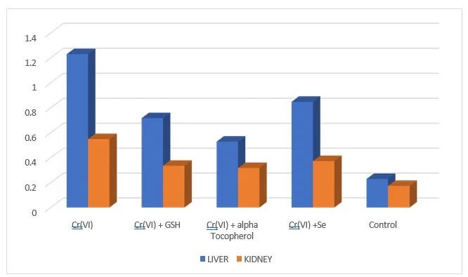
Influence of antioxidants on accumulation of Cr (VI) (μ / gm dry tissue) in liver and
kidney of rats.
The involvement of oxidative mechanism induced mutagenic and carcinogenic processes has been proposed by a few workers (Standeven and Watterhahn 1991, Sugiyma 1992). Present investigations also suggest that Cr (VI) induced oxidative stress as well as lipid peroxidation in liver and kidney of rats, which is manifested by ROS particularly H2O2 producing OH ions in a Fenton –type mechanism (Shi and Dalal 1990, A,B). However, it was inhibited in both the organs by the co-treatments with antioxidants. In liver, α -tocopherol offered maximum protection but in kidney selenium was highly protective. Minimum protection was offered by GSH in both organs. Mattagajasingh and Mishra (1997) suggested that Cr (VI) induces the generation of oxidants in the cells, probably, through the direct interaction of a reduced form of Cr (VI) with molecular O2. The administration of followed antioxidants with Cr (VI) inhibited oxidative stress as well lipid peroxidation.
GSH acts a reductant in the metabolism of H2O2 and other FR (Bellomo et. al. 1987). Selenium protection can be offered by the induction of GSH – dependent enzymes as reported earlier (Rana and Verma 1996). While, in biological system α – tocopherol scavenges peroxyl radicals. Peroxy radicals endogenously oxidize α – tocopherol to tocopheryl radical that does not allow further propagation of FR chain (Burton and Ingold 1981).
Influence of antioxidants on microsomal lipid peroxidation – oxidative stress (n moles
MDA / mg protein) in Cr (VI) fed rats.
Table 02:
| Treatments | Liver | Kidney |
|---|---|---|
| Cr(VI) | 0.250 ± 0.011* | 0.253 ± 0.007* |
| Cr(VI) + GSH | 0.222 ± 0.004** | 0.241 ± 0.011** |
| Cr(VI) + α- Tocopherol | 0.197 ± 0.005** | 0.204 ± 0.004** |
| Cr(VI) + Se | 0.228 ± 0.007** | 0.200 ± 0.003** |
| CONTROL | 0.211 ± 0.007 | 0.208 ± 0.005 |
Results are expressed as mean ± SE (n = 5).
* Significantly different (p < 0.05) when compared with saline fed rats.
* * Significantly different (p < 0.05) when compared with Cr (VI) fed rats.
Analysis of variance F (ANOVA) was significant among all these groups, for liver (561.51, p< 0.05) and kidney (362.00, p < 0.05).
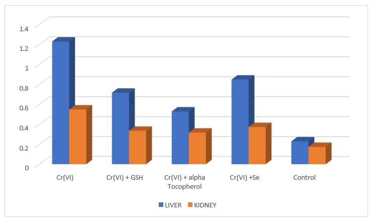
Influence of antioxidants on microsomal lipid peroxidation – oxidative stress (n moles
MDA / mg protein) in Cr (VI) fed rats.
SOD, Catalase and glutathione peroxidase constitute a mutually team of defence against generate ROS. Catalase is antioxidant enzymes that catalyzes and remove the H2O2 from tissues (Deisseroth and Dounce 1970). Catalase usually present in cytosol (Moslen 1992). Oxidative stress caused by Cr (VI) in liver and kidney might be attributed to significantly inhibited catalase activity. Usually, the peroxide (H2O2 and other peroxides) produced during the normal metabolic processes are broken down by enzyme catalase in the blood and tissues. It is present within the peroxisomal matrix that degrades H2O2 either by catalytic or peroxidative mechanism.
Recent experimental evidence has strengthened this mechanistic understanding. Zhou et al. (2023) demonstrated that Cr (VI) exposure in largemouth bass produced oxidative stress in liver tissue, characterized by decreased SOD, CAT, GSH-Px and total antioxidant capacity and increased MDA, together with inflammation, apoptosis and DNA damage. These effects were associated with disruption of the Nrf2–Keap1 antioxidant signalling pathway
Present investigation indicate that catalase activity was inhibited by Cr (VI) in liver and kidney both and enhance oxidative stress in cellular system. However, antioxidants restored catalase activity. Maximum restoration of catalase was brought about by α -tocopherol in liver, while in kidney GSH expressed best effects. Minimum protection was offered by selenium in both organs. Catalase present in almost all the mammalian cells is localized in the peroxisomes or microperoxisomes (Chance et. al. 1979). It has a heam – protein and catalyses the decomposition of H2O2 to H2O and O2 and thus protects the cell from oxidative damage by H2O2 and OH (Deisseroth and Dounce 1970).
Influence of antioxidants on Catalase (K / mg protein) in Cr (VI) fed rats.
Table 03:
| Treatments | Liver | Kidney |
|---|---|---|
| Cr(VI) | 0.125 ± 0.025* | 0.228 ± 0.012* |
| Cr(VI) + GSH | 0.180 ± 0.031** | 0.397 ± 0.015** |
| Cr(VI) + α -Tocopherol | 0.341± 0.100** | 0.351 ± 0.016** |
| Cr(VI) +Se | 0.166 ± 0.017** | 0.299 ± 0.018** |
| Control | 0.401 ± 0.033 | 0.559 ± 0.019 |
Results are expressed as mean ± SE (n = 5).
* Significantly different (p < 0.05) when compared with saline fed rats.
* * Significantly different (p < 0.05) when compared with Cr (VI) fed rats.
Analysis of variance F (ANOVA) was significant among all these groups, for liver (561.51, p< 0.05) and kidney (362.00, p < 0.05).
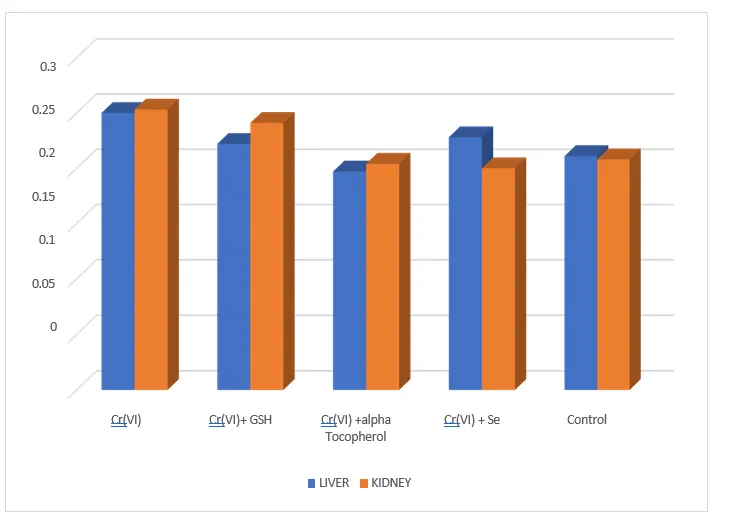
Influence of antioxidants on Catalase (K / mg protein) in Cr (VI) fed rats.
CONCLUSION
Antioxidants protection offered by three nutrients viz: α-tocopherol (vitamin E), reduced glutathione (GSH) and selenium (Se) against hexavalent chromium [Cr (VI)] toxicity has been studied in laboratory rats (Rattus rattus albino). Chromium like other transitional metal ions induced oxidative stress in liver and kidney. However, treatments with antioxidants [α-tocopherol (vitamin E), reduced glutathione (GSH) and selenium (Se)] inhibited lipid peroxidation and activated serum transminases and catalase activity in both the organs. Selective preferences were shown by these antioxidants for these parameters in each organ. Our results confirm that antioxidants offer protection by different mechanisms. Disturbances in cellular equilibrium of anti and pro-oxidants caused by hexavalent chromium can be overcome by antioxidants with certain preferences.
8. ACKNOWLEDGEMENT
The author is grateful to all the faculty members of Department Zoology and Principal of Govt. P. G. College Maldevta (Raipur) Dehradun for providing all basic and applied facilities to conduct research in the College.
REFERENCES

Dharmendra Kumar, Mitigating Hexavalent Chromium Induced Oxidative Stress Through, Int. J. of Pharm. Sci., 2026, Vol 4, Issue 10, 4920-4929. https://doi.org/10.5281/zenodo.23830349
 10.5281/zenodo.23830349