

We use cookies to ensure our website works properly and to personalise your experience. Cookies policy
PG Department of Pharmacology and Research Division, College of Pharmaceutical Sciences, Government Medical College, Kannur, Kerala-670503.
Tuberculosis (TB), caused by Mycobacterium tuberculosis (Mtb), remains a global health challenge due to drug resistance and the long duration of current treatments. A promising target for novel anti-TB therapies is shikimate kinase (MtSK), an essential enzyme in the shikimate pathway, which is crucial for Mtb survival and virulence but absent in mammalian homologue. Recent advances in structural biology and computational chemistry have identified several novel MtSK inhibitors with diverse structural features. Shikimic acid analogs, despite their structural similarity to endogenous shikimate, inhibit MtSK by preventing the transfer of phosphate from ATP to shikimate. Other inhibitors include compounds with Chalcones, pyrazolone, oxadiazole-amide, and 2-aminobenzothiazole frameworks. Notably, the dipeptide +H3N-Arg-Asp-COO– (RD) has shown superior binding affinity compared to shikimate, forming unique interactions with MtSK in both open- and closed-lid conformations. This review delves into the structural and functional insights of these novel inhibitors, shikimate kinase as a potential target emphasizing their potential in developing more effective and targeted anti-TB therapies.
In 2022, tuberculosis (TB) claimed the lives of 1.3 million individuals, including 167,000 people living with HIV. Globally, TB ranks as the second deadliest infectious disease, following COVID-19 and surpassing HIV/AIDS. An estimated 10.6 million people contracted TB in 2022, comprising 5.8 million men, 3.5 million women, and 1.3 million children. TB affects all countries and age groups, but it is both curable and preventable 1. Approximately 1.7 billion people, or 23% of the global population, are believed to have a latent TB infection, putting them at risk of developing active disease at some point in their lives. Drug-susceptible (DS) tuberculosis is managed with a six-month regimen of anti-TB medications, including isoniazid, rifampicin, pyrazinamide, and ethambutol. These drugs have been in use for over six decades. To date, 576 proteins from M. tuberculosis H37Rv have been identified as potential druggable targets and have entered the structure determination pipeline. These proteins are involved in various metabolic pathways, including dehydrogenases, PE-PGRS family proteins, oxidoreductases, transporters, lipases, kinases, PPE family proteins, proteases, hydrolases, hydratases, RNA polymerases, ligases, and others. Unfortunately, anti-TB drug discovery has predominantly focused on a limited number of clinically validated targets2. The drugs currently in clinical use primarily include inhibitors of fatty acid biosynthesis such as isoniazid (targets KatG, InhA, and AhpC), pyrazinamide (target PncA), ethionamide (target InhA), prothionamide (target InhA), and thioacetazone (target CmaA2). Additionally, there are inhibitors of nucleic acid synthesis like rifampicin (target RpoB), ofloxacin, levofloxacin, moxifloxacin, and ciprofloxacin (all targeting GyrA and GyrB). Furthermore, inhibitors of protein synthesis include streptomycin (targets RpsL, Rrs, and GidB), kanamycin (targets Rrs and Eis), amikacin (target Rrs), capreomycin (targets Rrs and TylA), and linezolid (target RplC). The mechanisms of action of these drugs have been extensively reviewed by Tiwari and Ryoo3-4 . One of the challenges in combating M. tuberculosis with antibiotics is its highly hydrophobic and less permeable cell envelope. Additionally, its ability to evade the host’s immune system and persist within alveolar macrophages in a hypometabolic, non-replicating form makes it a successful intracellular pathogen. Consequently, the primary difficulty in TB therapy is targeting and eliminating the drug-tolerant and persistent M. tuberculosis residing in TB lesions5- 6.The global health challenge is compounded by multidrug-resistant (MDR) and extensively drug-resistant (XDR) TB, where treatment can last up to 2 years with limited success7. It is necessary to look for alternative therapeutic approaches to enhance the current course of treatment and get around Mtb's resistance mechanisms. One such target that can be investigated to create novel antitubercular drugs is the shikimate pathway. Parish et al. have shown that the survival of microbes depends on shikimate kinase8. This gene is equally relevant in MDR-TB since its expression is not altered in the drug-susceptible or MDR strains of M. tuberculosis9. The shikimate (SKM) pathway involves seven enzymatic steps that transform erythrose 4-phosphate and phosphoenolpyruvate into chorismate. Chorismate serves as a precursor for the biosynthesis of aromatic amino acids, vitamins B9 and K1, ubiquinone, and salicylate. This pathway exists in plants, fungi, bacteria, and apicomplexan parasites, but is absent in metazoans, including humans. Consequently, the enzymes within this pathway are attractive targets for developing non-toxic antimicrobials and herbicides10. The shikimate kinase (SK) enzyme facilitates the fifth step of the shikimate pathway by phosphorylating shikimate (SKM) with ATP as the phosphate donor, producing shikimate-3-phosphate (S3P) and adenosine diphosphate (ADP). SK is part of the Nucleoside Monophosphate Kinase (NMP) family, a crucial group of enzymes responsible for the reversible transfer of a phosphate group from a nucleoside triphosphate to a specific nucleoside diphosphate. During catalysis, NMP kinases undergo significant conformational changes11.
TB chemotherapy – a brief history
The first successful TB medication, streptomycin (SM), was identified by Schatz and Waksman in 1943, marking the beginning of the history of TB chemotherapy. The British Medical Research Council conducted the first-ever randomized clinical trial shortly after, demonstrating the medication's effectiveness, but resistance to SM was soon found. Subsequent research revealed that para-aminosalicylic acid (PAS) added to SM prevented drug resistance, thereby developing the crucial idea of the drug combination. In the 1950s, it was discovered that adding INH to the SM/PAS regimen can successfully cure TB patients in 18 to 24 months. Treatment was further decreased to nine months with the discovery of RIF in the 1960s and its subsequent usage in conjunction with INH. The current 6-month TB chemotherapy was developed in the 1970s and 1980s as a result of a re-evaluation of pyrazinamide (PZA) in conjunction with INH and RIF. The World Health Organization has approved directly observed therapy-short course (DOTS) as the standard TB chemotherapy since 1991. The DOTS protocol takes six months to finish. It consists of two months of daily INH, RIF, PZA, and ethambutol (EMB) administration during the initial phase, and four months of INH and RIF administration during the continuation phase12. Drug-susceptible tuberculosis (TB) is currently being treated with 23 different medications; multidrug-resistant TB is being studied in phase I, II, and III studies. These medications include seven repurposed medications, three more medications (Bedaquiline, Delamanid, and Pretomanid) that have already received regulatory approval and thirteen novel compounds. For the treatment of multidrug-resistant tuberculosis (MDR-TB), the FDA approved the TB medication Bedaquiline (Sirturo)13.Mycobacterial adenosin 5-triphosphate synthase is an enzyme required for the last stage of ATP synthesis through oxidative phosphorylation, and situro specifically inhibits it14.
Shikimate pathway
The shikimate pathway is a promising target for developing herbicides and antimicrobial agents since it is crucial for algae, higher plants, fungi, bacteria, and apicomplexan parasites, yet it is not found in mammals15. In the mycobacterial shikimate pathway, chorismic acid is synthesized and subsequently transformed by various enzymes into several key compounds. These include prephenate (a precursor for phenylalanine and tyrosine), anthranilate (a precursor for tryptophan), aminodeoxychorismate (a precursor for para-aminobenzoic acid, which leads to tetrahydrofolate synthesis), para-hydroxybenzoic acid (a precursor for ubiquinone or Coenzyme Q), and isochorismate (a common precursor for naphthoquinones, menaquinones, and mycobactins).The host defense, which aims to starve mycobacteria of tryptophan by a CD4 T cell-mediated killing mechanism, is overcome by these cells due to the mycobacterial genetic determinants (the "counteractome"). However, M. tuberculosis can produce tryptophan under stressful conditions (from chorismate precursor), therefore starvation is ineffective against it. Hence, tryptophan production should be prevented by inhibiting any enzyme in the mycobacterial shikimate pathway, increasing the probability of starvation as a highly effective means of death provided by the human host16
The initial enzyme of the shikimate pathway is 3-deoxy-7-phosphoheptulonate synthase (DAHPS) (EC 2.5.1.54). In Mycobacterium tuberculosis, the gene encodes this enzyme with the ID 13318866. The aroF gene produces the enzyme synthesizing the metabolite 3-deoxy-D-arabinoheptulosonic acid-7-phosphate (DAHP)17. Second, 3-dehydroquinate synthase (DHQS), is responsible for the generation of the first carbocyclic compound of the pathway, converting DAHP into 3-dehydroquinate (DHQ). 3-dehydroquinate dehydratase catalyses the third step in the shikimate pathway, the dehydration of DHQ to give 3-dehydroshikimate (DHS), introducing the first double bond of the aromatic ring18. The fourth step in the shikimate pathway involves the reversible conversion of DHS and NADPH to produce shikimate, a 3,4,5-trihydroxycyclohexene-1-carboxylic acid, and NADP+. This reaction is catalyzed by shikimate 5-dehydrogenase (SD), which was initially known as DHS reductase 19 . The second half of the shikimate pathway initiates (fifth step) with the reaction catalyzed by shikimate kinase (SK). The enzyme catalyzes a phosphate transfer from ATP co-substrate to the carbon3 hydroxyl group of shikimate resulting in the formation of shikimate-3-phosphate (S3P) and ADP20 . The sixth (penultimate) step in the shikimate pathway involves the incorporation of a second PEP into the pathway. This PEP, along with S3P, is condensed to form 5-enolpyruvylshikimate-3-phosphate (EPSP) and Pi. This reaction is catalyzed by EPSP synthase. Levin and Sprinson were the first to demonstrate the formation of EPSP as a product of E. coli EPSP synthase activity21 . The seventh and final reaction in the main trunk of the shikimate pathway introduces a second double bond into the aromatic ring system, producing chorismic acid and Pi through a trans-1,4 elimination of phosphate from EPSP. This reaction is catalyzed by chorismate synthase (CS) and requires reduced flavin mononucleotide (FMN) as a coenzyme for activity. Despite this, the overall reaction is redox neutral, similar to DHQ synthase, making it unusual because the redox state of the functional cofactor flavin remains unchanged22.
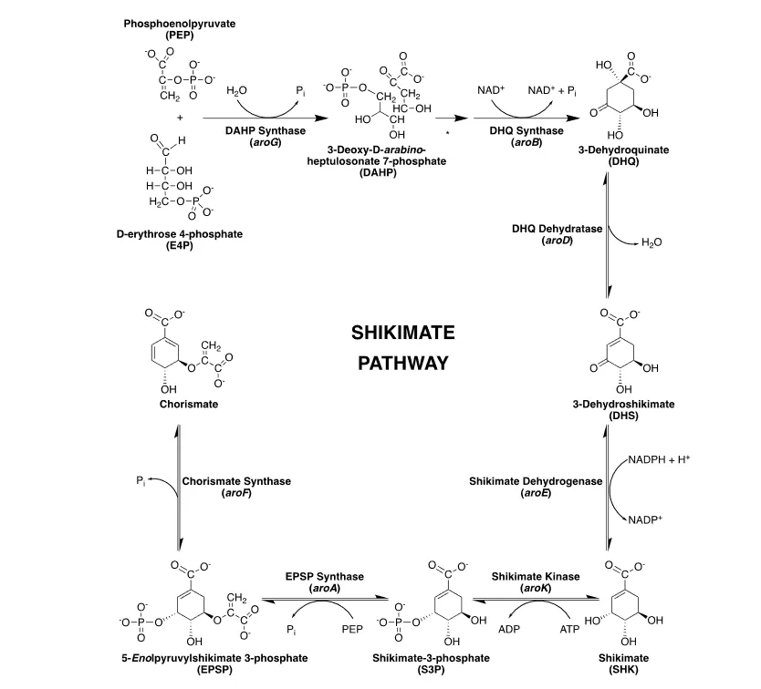
Figure 1: Shikimate Pathway in Mycobacterium tuberculosis 23
Shikimate kinase
Shikimate kinase is a key enzyme in the biosynthesis of aromatic amino acids, specifically involved in the fifth step of the shikimate pathway. It catalyzes the transfer of a phosphoryl group from ATP to shikimate, producing ADP and shikimate-3-phosphate in Mycobacterium tuberculosis (Mtb). This enzyme is a promising target for drug discovery, particularly against pathogens like Mtb that depend on this pathway. The genomic structure of the aro genes in the Mycobacterium TB shikimate pathway includes one operon, aroCKBQ, and three separate aro genes: aroE, aroG, and aroA.In M. tuberculosis, the aroK gene is crucial. It carries the shikimate kinase enzyme's gene8. The crystal structures of SK I type shikimate kinases from several different organisms, such as M. tuberculosis, Campylobacter jejuni24, Helicobacter pylori25, and E. coli26 , have been determined. Currently, 33 crystal structures are available in rcsb protein data bank27(Table 1). The MtSK structures that are currently available depict the enzyme in a variety of liganded states, such as binary complexes of MtSK with SO4 or MgADP (PDB code- 1L4Y)28 and ternary complexes of MtSK with SO4 (2G1K), ADP (1U8A), MgADP, or the ATP analogue AMPPCP (1ZYU) as one ligand and shikimate (SKM) as the second ligand. The putative structures of an "apo-MtSK" and a "MtSK.SKM" complex had a sulfate ion bound at the nucleotide-binding site, significantly affecting the enzyme conformation.
Table 1: Crystal Structures of Shikimate Kinase
| Structure id. | Ligand | Organism | Resolution(Å) |
|---|---|---|---|
| 1L4U | Shikimate kinase and MGADP | Mycobacterium tuberculosis | 1.8 |
| 1L4Y | Shikimate kinase and MGADP | Mycobacterium tuberculosis | 2 |
| 1VIA | Shikimate kinase | Campylobacter jejuni | 1.57 |
| 1WE2 | Shikimate kinase with MGADP and shikimic acid | Mycobacterium tuberculosis | 2.3 |
| 1U8A | Shikimate kinase with Shikimate and ADP | Mycobacterium tuberculosis | 2.15 |
| 1ZUH | Shikimate kinase | Helicobactor pylori | 1.8 |
| 1ZUI | Shikimate kinase | Helicobactor pylori | 2.3 |
| 1ZYU | Shikimate kinase with amppcp | Mycobacterium tuberculosis | 2.9 |
| 2DFN | Shikimate kinase with ADP and shikimate | Mycobacterium tuberculosis | 1.93 |
| 2DFT | Shikimate kinase with ADP and Mg | Mycobacterium tuberculosis | 2.8 |
| 2G1J | Shikimate kinase | Mycobacterium tuberculosis | 2 |
| 2G1K | Shikimate kinase | Mycobacterium tuberculosis | 1.75 |
| 2IYQ | Shikimate kinase with ADP | Mycobacterium tuberculosis H37Rv | 1.7 |
| 2IYR | Shikimate kinase with shikimate | Mycobacterium tuberculosis H37Rv | 1.98 |
| 2IYS | Shikimate kinase with shikimate(open LID) | Mycobacterium tuberculosis H37Rv | 1.4 |
| 2IYT | Shikimate kinase in unliganded state (open LID) | Mycobacterium tuberculosis H37Rv | 1.47 |
| 2IYU | Shikimate kinase with ADP (open LID) | Mycobacterium tuberculosis H37Rv | 1.85 |
| 2IYV | Shikimate kinase with ADP (open LID,conf.B) | Mycobacterium tuberculosis H37Rv | 1.35 |
| 2IYW | Shikimate kinase with MgATP, open LID, conf.B) | Mycobacterium tuberculosis H37Rv | 1.85 |
| 2IYX | Shikimate and SO4 | Mycobacterium tuberculosis H37Rv | 1.49 |
| 2IYY | Shikimate kinase with shikimate-3-phosphate | Mycobacterium tuberculosis H37Rv | 1.62 |
| 2IYZ | Shikimate kinase with shikimate-3-phosphate and ADP | Mycobacterium tuberculosis H37Rv | 2.3 |
| 2PT5 | Shikimate kinase | Aquifex aeolicus VF5 | 2.1 |
| 3BAF | Shikimate kinase with AMP-PNP | Mycobacterium tuberculosis | 2.25 |
| 3HR7 | Shikimate kinase-sulfate complex | Helicobacter pylori 26695 | 1.8 |
| 3MRS | Shikimate kinase mutant(R57A) | Helicobacter pylori 26695 | 2.4 |
| 3MUF | Shikimate kinase with shikimate-3-phosphate and ADP | Helicobacter pylori | 2.3 |
| 3N2E | Shikimate kinase with NSC162535 | Helicobacter pylori 26695 | 2.53 |
| 3TRF | Shikimate kinase(aroK) | Coxiella burnetii | 2.6 |
| 3VAA | Shikimate kinase | Bacteroides thetaiotaomicron | 1.7 |
| 4BQS | Shikimate kinase with ADP and shikimic acid derivative | Mycobacterium tuberculosis | 2.15 |
The three major domains of SK are depicted in Figure 2: a binding domain SB that binds to the SK substrate, a flexible LID with an opening and closing feature also seen in other protein kinases, and a stiffer CORE domain made up of α-β-α motives. Some conserved residues, including D33, R57, R116, and R132, are determined to be essential for catalysis in the binding pocket of SK because mutations affecting these amino acids result in inactive enzymes29.
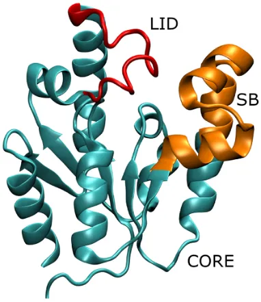
Figure 2: The Three Main Domains in the Shikimate Kinase (SK) Enzyme Structure: The Binding SB (in Orange), the LID (in Red), and the CORE Domains (in Turquoise).
MtSK has an α/β architecture with five parallel β-strands in the center that are flanked by α-helices. Four domains make up the overall structure30 (i) the ESB domain (residues 32–93), which permits stiff body movements; this includes the SHK binding (SB) subdomain (residues 32–61), which corresponds to the binding of NMP in NMP kinases; (ii) the nucleotide-binding (NB) domain, which is in charge of the flexible regions that make up the nucleotide binding; these are formed by the phosphate binding P-loop (Walker-A motif; residues 9–17), (31) the adenine-binding (AB)–loop (residues 148–155) and the segment corresponding to residues 11–110, including α6 (residues 104–110); (iii) the LID domain (residues Gly112 to Asp124), which is in charge of opening and closing the catalytic site and permitting the enzyme to interact with substrates and products; and (iv) the reduced core (RC) domain, covering NB domain and the residues 62–93 of the ESB section. The LID and NB domains are the most flexible parts of the enzyme, whereas the RC domain is the most rigid of all16.
Ojeda-May P. examined the SK enzyme's dynamics through μs time scale. The enzyme was examined in three different forms: the WT case, the R116A, and the R116K mutants. Through RMSF calculations, the dynamics of the WT enzyme were analyzed. The results indicated that the CORE domain's 310 helix, the LID and SB domains, and a tiny region had the biggest variations, while the remaining sections stayed nearly stiff. discovered that, in comparison to the WT population, the mutant population ensembles had changed, particularly in the direction of more "closed" states. These changed populations may make it more difficult for substrates to bind to the enzyme and may also account for the enzyme's lower binding activity. In this sense, the mutants ruin the SK's choreography achieved during protein evolution32 .
Shikimate kinase target validation
The recombinant protein used in these tests is made by researchers using the same amino acid sequence as the target protein. After that, a substance suggested as a possible inhibitor of the target protein is incubated with this protein. The compound's ability to prevent the target protein from functioning is then examined. If it is effective, the substance is regarded as a strong inhibitor of the intended protein. Then, to further confirm the compound's validity, scientists add it to a cell culture with the target protein. After that, the compound's impact on the target protein's activity is observed in the cell culture. If the target protein exhibits resistance to the chemical, the experiment is deemed unsuccessful in certain instances. In other situations, the substance might be effective in inhibiting the target protein's function, but it might not be specific enough to produce a medicinal substance. In this instance, the experiment is deemed to have been only partially successful, and more research may be required to maximize the compound's potential for therapeutic utilization33
To understand the binding mode and structural features, Shikimate kinase was modelled through homology modelling using Modeller 9.16. The modeled structures were validated with a Ramachandran plot34 , showing that 97.3% (142/146 residues) were in the most favorable regions [A, B, L], 2.7% (4/146 residues) were in additional allowed regions [a, b, l, p], and none were in generously allowed or disallowed regions. Validation with the ProsA server showed a Z-Score of -8.4435 , indicating a high-quality model with strong structural integrity. Molecular docking analysis36 using both synthetic ligands (from the Zinc Database) and natural ligands (from PubChem Database) revealed that the ligands exhibited effective activity against the enzyme, suggesting Shikimate kinase as a promising target for developing anti-tubercular drugs37 .
Comprehensive AS-RNA silencing investigations on the translation of target cidality from in-vitro to in-vivo were carried out by Kaur et al., and the results showed that AroK is the target that was verified in-vivo as a result of the cidality score. It was identified as a "in-vitro total" and "in-vivo" cidal target, and clinical Mtb mortality is predicted from its inhibition. According to this study, long-term TB patient treatment may be possible with the development of AS-RNA-based therapies. The utilization of AS-RNA for in-vitro cidality confirmation in experimental tuberculosis in BALB/c mice led them to determine cidal targets arranged in the rank order of rpoB>aroK>ppk>rpoC>ilvB. The control for cidality was RpoB. Research both in vitro and in vivo has shown that aroK, which codes for shikimate kinase, is a mycobactericidal target in vivo that can be used to find anti-TB drugs38 .
Chemical characteristics needed for selective inhibitors.
Inhibition of MtSK can be achieved by compounds that bind at the shikimate binding site38-39 or ATP-binding site 40. The in silico and in vitro data suggest that compounds interacting with residues D34, R58 (NMP-binding domain), and R117 exhibit inhibition of MtSK due to their similar interactions with shikimate41-42 . Structurally, MtSK inhibitors have shown considerable diversity. Due to their similarity to endogenous shikimate, shikimic acid analogs bind in a manner akin to shikimate. However, they do not facilitate the transfer of phosphate from ATP to shikimate to produce shikimate 3-phosphate. Other MtSK inhibitors that have been identified include compounds with an indole ring connected to a chromene ring by seven covalent bonds, a mercapto group, a triazole or tetrazole ring, an oxadiazole-amide, or a 2-aminobenzothiazole scaffold. A dipeptide composed of arginine and aspartic acid (RD) demonstrated a higher binding affinity for MtSK than shikimate. Furthermore, pyrazolone analogs have also shown inhibitory activity against MtSK43 .
Shikimate kinase inhibitors
1. Dipeptide inhibitor
Kumar et al. identified the dipeptide +H3N-Arg-Asp-COO– (RD) as a potential SK inhibitor due to its high predicted binding affinity.The best-known inhibitor, asxe1 has a predicted binding affinity of about 56nm according to eHiTS (electronic high-throughput screening) score. The dipeptide inhibitor RD occupies a position similar to that of shikimate. By employing the LigandFit protocol for molecular docking, they analyzed both the open- and closed-lid conformations of the enzyme44 . RD maintains hydrogen bonds with D34, R58 (NMP-binding domain), and R136, similar to SK binding. In the closed-lid conformation, RD interacts with K15, S16 (P-loop), D32 (NMP-binding domain), L78 (Walker-B motif), V116, and R117 (lid domain), which are not involved in shikimate binding, resulting in a Kd of 5.49 nM. In the open-lid conformation, RD shows additional interactions with K15 (P-loop), D32, F49 (NMP-binding domain), S77, and L78 (Walker-B motif), resulting in a Kd of 26 nM 42 . This peptide is anticipated to bind to M.tb SK at the shikimate-binding pocket in both the open and closed LID conformations, occupying nearly all the available space for binding. The predicted dissociation constant (Kd) for the binding of RD to M.tb SK is 5.49 nm, which is approximately 8000 times more effective than that of the substrate shikimate and 10 times more effective than the lead compound asxe1 identified through VTS45. Therefore, the dipeptide RD shows potential as a promising lead compound for the development of antitubercular drugs.
Figure 3: Chemical Structure of Shikimate
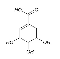
Figure 4: Chemical Structure of Asxe1
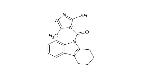
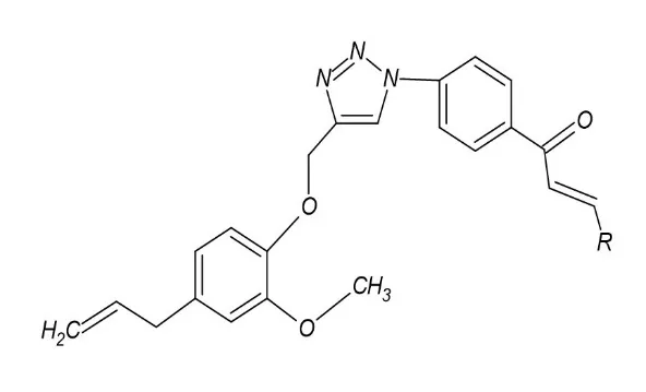
Figure 5: Eugenol-1,2,3-Triazole-Chalcone
2. Chalcones
Ardiansah B et al. successfully carried out the derivatization of eugenol natural product into the hybrid compounds containing eugenol-1,2,3-triazole-chalcone triple functionalities. Adding a chalcone moiety to eugenol-1,2,3-triazole enhanced the antitubercular potency of most compounds compared to using eugenol as the starting material. Nitrogen-rich compounds, such as dimethylaminostyryl and pyridyl, demonstrated good activity. However, the presence of a hydroxyl group did not improve the compounds' activity. Based on bioactivity evaluation, the potency order is as follows: (R) Pyridyl > 4-NMe2-styryl > 4-F-Ph > 4-O-Propargyl-3-OMe-Ph > 4-Cl-Ph > 4-OMe-Ph > 4-OH-3-OMe-Ph > 4-OH-phenyl > styryl > 4-NO2-Ph > Ph. Aldol condensation reaction was carried out with variations of aromatic aldehyde compounds to form eugenol-1,2,3-triazole-chalcone with good to excellent yields (79–99%). 2 Compounds was found as most active agents in inhibiting MtSK with IC50 1.78 and 1.66 mg/mL, respectively which exhibit an excellent binding interaction with the enzymes Arg58, Pro118, and Arg136, according to a molecular docking study 46 .
3. 2-amino benzothiazole derivatives
Mehta R et al. conducted a study on a combined approach of in silico similarity search and pharmacophore building using already reported inhibitors to screen a procured library of 20,000 compounds of the commercially available ChemBridge database. ChemAxon software was used to perform a similarity search47. From the in-silico screening, 15 hits were identified and evaluated in vitro for Mtb-SK enzyme inhibition. Two compounds presented significant enzyme inhibition with IC50 values of 10.69 ± 0.9 and 46.22 ± 1.2 μM48 . Smithy J et al. studied a potential enzymatic target identified by screening a small library of 404 synthetic antimycobacterial compounds against whole cell M. tuberculosis H37Rv using a mass spectrometry-based functional assay. These compounds were identified and supplied through the NIH Tuberculosis Antimicrobial Acquisition and Coordinating Facility (TAACF) high throughput screening program. At 50 μM, MtSK inhibitory action was demonstrated by 14 compounds with an oxadiazole-amide or 2-amino benzothiazole core scaffold; the lowest molecule had an IC50 of 1.94 μM38 Compounds containing the 2-amino benzothiazole structure exhibit several key interactions: the benzothiazole ring engages in favorable aromatic–aromatic interactions with F57 (NMP-binding domain), positioning R3 near the side chain of I45 and allowing for non-polar interactions with I37. Additionally, I45 and V35 create steric interactions with the thiophene ring, while the R2 group partakes in favourable polar interactions with R13643 .
4. Pyrazolone derivatives
Twenty-seven hybridized pyrazolone analogs were designed and synthesized in two distinct series. In the first series, 4 compounds demonstrated good antitubercular activity. Their minimum inhibition concentration (MIC) values ranged between 32.56 and 42.55 mM showing a moderate level of activity against Mycobacterium tuberculosis. The second series yielded two standout compounds, which exhibited significant antitubercular activity with MIC values of <0.37 mM and <0.44 mM, respectively. These compounds were more potent than standard antitubercular drugs such as pyrazinamide (MIC 12.99 mM), ciprofloxacin (MIC 4.82 mM), and streptomycin (MIC 5.36 mM). The selected compounds also demonstrated high selectivity indices (>630), highlighting their potential for therapeutic use with minimal toxicity49.
5. Oxadiazole-amide
A library of 404 phenotypic antimycobacterial compounds from the NIH TAACF program was screened using a functional LC-MS assay to identify inhibitors of MtSK. Potent MtSK inhibitors with low IC90 values against M. tuberculosis were identified, particularly among analogue series of oxadiazole-amide and aminobenzothiazole core scaffolds. Docking studies validated the specific interactions of these core scaffolds with MtSK, suggesting that MtSK is a potential target of these antimycobacterial compounds38 . Compounds with an oxadiazole-amide structure typically interact with the R58 residue (NMP-binding domain), which is crucial for shikimate binding, forming a positive charge–π interaction with the aromatic ring of the compounds tetrahydronaphthaline moiety. The remainder of the ring structure engages in non-polar interactions with P118 and L119 (lid). The oxadiazole ring is positioned to form favorable polar interactions with the guanidinium group in R117. Additionally, the amide linker, according to the crystal structure, participates in hydrogen bonding with water43.
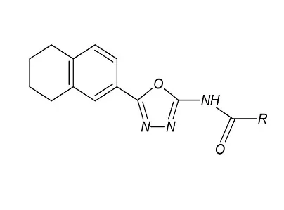
Figure 6: Oxadiazole-amide Scaffold
6. Thiobarbiturates
A library of 1000 antitubercular compounds was screened against M. tuberculosis shikimate kinase (MtSK), identifying 20 inhibitors. Five promising leads exhibited IC50 values below 10 μM, with the most potent inhibitor thiobarbiturate derivative showing an IC50 value of 5.10 μM ± 0.6. These leads were further tested for activity against multidrug-resistant TB (MDR-TB), Gram-positive and Gram-negative bacterial strains, mode of action, docking simulations, and combined with three frontline anti-TB drugs. The compound was synergistic with rifampicin50
7. Triazole and tetrazole
Triazole and tetrazole compounds likely interact with the R58, L119, and R136 residues, mimicking the key interactions of the enzyme-substrate complex, particularly the interaction of these residues with the carboxyl group of shikimate. Two nitrogen atoms in the azole system form hydrogen bonds with R58 and R136, and the proximity of L119 to the heteroaromatic ring suggests potential stabilizing CH–π interactions. The mercapto group interacts with G80, R117, and L119 through hydrophobic forces and hydrogen bonds, with the interaction with R117 being crucial for the phosphoryl transfer reaction catalyzed by MtSK51.
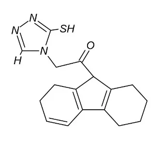
Figure 7: Triazole Scaffold
8. Indole-chromene scaffolds
An analysis of the hydrogen bond patterns in the docking results for shikimate kinase (SK) at the ATP-binding pocket of inhibitors containing an indole ring linked to a chromone ring by seven covalent bonds was conducted. This analysis revealed that residues G14, K15, and R117 consistently interact with this series of compounds. These residues are also involved in contact with the phosphate group in ADP, as observed in the crystallographic structure 1WE2 of MtSK. Additionally, van der Waals interactions were identified involving I18 (P-loop), R110 (N-terminal to the lid domain), P155, and V158 (adenine-binding loop). The binding of all five top-scoring compounds to R110 and P155 suggests that these interactions are crucial for ligand-binding affinity 52 .
CONCLUSION
In conclusion, the review focuses on the shikimate kinase enzyme which belongs to the Nucleoside Monophosphate Kinase (NMP) family, which comprises essential enzymes that facilitate the reversible transfer of a phosphate group from a nucleoside triphosphate to a specific nucleoside diphosphate. These enzymes experience substantial conformational changes during the catalytic process. Shikimate kinase is absent in mammalian homologue hence it can be a promising target against Mycobacterium tuberculosis. Resistance and serious side effects are the main drawbacks of the existing treatment regimens. Understanding the crystal structure of the shikimate kinase is also important, till date 33 crystalline structures have been identified. Several chemical molecules have undergone computational screening, in-vitro studies, and promising scaffolds, including chalcones, pyrazolone, oxadiazole-amide, 2-amino benzothiazole, and dipeptide inhibitors.Due to its high predicted binding affinity, the dipeptide +H3N-Arg-Asp-COO– (RD) is a potential SK inhibitor. The dipeptide inhibitor RD occupies a position similar to that of shikimate. The 2-amino benzothiazole compounds revealed specific interactions critical for inhibition: aromatic–aromatic interactions with the F57 residue, non-polar interactions with I37, and steric interactions with the thiophene ring involving I45 and V35. Additionally, favorable polar interactions with R136 were noted. The pyrazolone derivatives exhibited lesser MIC values surpassing the potency of standard antitubercular drugs indicating their potential for therapeutic use with minimal toxicity. Thiobarbiturates, triazoles and tetrazoles, indole-chromene scaffolds,chalcones also proved their potential to inhibit the shikimate kinase enzyme. Currently, there are no data on in vivo activity of Mycobacterium tuberculosis shikimate kinase (MtSK) inhibitors available in the literature. In this review, we present a summary of the progress of SK inhibitor discovery and evaluation, with particular attention on developing new antitubercular agents.
REFERENCES

Vandhana Vailongara Murugesan, Novel Anti-Tuberculosis Strategies: Targeting Shikimate Kinase for Effective Therapy, Int. J. of Pharm. Sci., 2026, Vol 4, Issue 10, 4186-4198. https://doi.org/10.5281/zenodo.23685558
 10.5281/zenodo.23685558