
We use cookies to ensure our website works properly and to personalise your experience. Cookies policy
Department of Pharmaceutics, Shivlingeshwar College of Pharmacy Almala, Ausa 413520
Topical drug delivery systems offer a strategic approach for administering therapeutic agents directly to the skin, bypassing hepatic first-pass metabolism and reducing systemic adverse effects. However, conventional formulations such as gels and creams often face challenges, including poor skin penetration, short residence time, and the necessity for frequent dosing. Polymeric microspheres spherical particles typically ranging from 1–1000 µm have emerged as a versatile solution to these limitations. By encapsulating active pharmaceutical ingredients within natural or synthetic polymeric matrices, these systems enable controlled, sustained drug release and improved site-specific delivery. This review examines the classification, preparation techniques (such as solvent evaporation and spray drying), and characterization methods of microspheres. Furthermore, it highlights their significant role in dermatological applications, including acne, anti-inflammatory, and antifungal therapies. Despite challenges related to manufacturing complexity and production costs, ongoing advancements in polymer science and nanotechnology continue to enhance the therapeutic efficacy and patient compliance of these advanced delivery systems, positioning them as a promising frontier in modern pharmaceutical sciences.
Topical drug delivery systems are designed to administer therapeutic agents directly onto the skin for local or systemic action. These systems provide several advantages over conventional dosage forms, including avoidance of first-pass metabolism, improved patient compliance, reduced systemic adverse effects, and targeted drug delivery at the site of action (1). However, conventional topical formulations such as creams, gels, ointments, and lotions often exhibit limitations including poor skin penetration, short residence time, rapid drug loss from the application site, and frequent dosing requirements (2).
To overcome these challenges, controlled drug delivery systems have gained considerable attention in pharmaceutical research. Controlled delivery aims to maintain drug concentration within the therapeutic window for prolonged periods while minimizing fluctuations in drug levels and reducing adverse effects (3). Among various carrier systems developed for controlled release applications, polymeric microspheres have emerged as promising drug delivery vehicles because of their ability to encapsulate active pharmaceutical ingredients and release them in a sustained and controlled manner (4).
Polymeric microspheres are small spherical particles ranging from approximately 1 µm to 1000 µm in diameter, composed of natural or synthetic polymers that encapsulate or disperse drug molecules within a polymeric matrix (5). Depending on their structural characteristics, microspheres may exist as microcapsules, where the drug is enclosed within a polymer shell, or as micromatrices, where the drug is uniformly distributed throughout the polymer network (6). Their unique properties, including controlled release capability, enhanced drug stability, improved bioavailability, and site-specific delivery, make them attractive carriers for topical formulations (7).
In topical drug delivery applications, polymeric microspheres can improve skin retention of drugs, reduce dosing frequency, enhance therapeutic efficacy, and minimize systemic absorption and irritation. Various natural polymers such as chitosan, gelatin, and alginate, as well as synthetic polymers including poly(lactic-co-glycolic acid) (PLGA), ethyl cellulose, and Eudragit, have been widely investigated for microsphere preparation (8). Advances in microsphere technology continue to provide innovative opportunities for designing novel controlled topical drug delivery systems with improved clinical outcomes (9).
Thus, polymeric microspheres represent an effective and versatile approach in modern topical drug delivery, offering significant potential to overcome limitations associated with conventional formulations and improve patient treatment outcomes (10).
2.Topical Drug Delivery Systems
Topical drug delivery systems (TDDS) are formulations intended for application onto the skin or mucosal surfaces to deliver therapeutic agents either locally or systemically. The skin serves as a protective barrier and also acts as an attractive route for drug administration due to its large surface area and easy accessibility. Topical administration enables the drug to reach the site of action directly while minimizing systemic exposure and reducing adverse effects (11).
The primary objective of topical drug delivery systems is to achieve an adequate concentration of the drug at the target site for a prolonged period while maintaining therapeutic efficacy. Topical formulations provide several advantages over oral and parenteral routes, including avoidance of hepatic first-pass metabolism, painless administration, improved patient compliance, and ease of termination of therapy whenever required (12).
Drug penetration through the skin mainly occurs via three pathways: the transcellular pathway (through cells), intercellular pathway (between cells), and appendageal pathway (through hair follicles and sweat glands). Among these, the stratum corneum acts as the major barrier to drug penetration because of its highly organized lipid structure (13).
Various conventional topical dosage forms include creams, ointments, gels, lotions, pastes, powders, and transdermal patches. However, these conventional systems often suffer from limitations such as poor skin permeation, low drug retention at the application site, short duration of action, and the need for repeated application (14).
Recent advancements in topical drug delivery have led to the development of novel carrier systems such as liposomes, niosomes, nanoparticles, microsponges, microspheres, nanoemulsions, and nanogels. These advanced systems improve drug penetration, enhance stability, provide controlled release characteristics, and increase therapeutic effectiveness (15).
Thus, topical drug delivery systems represent an important and continuously evolving field in pharmaceutical sciences, offering a promising approach for improving therapeutic outcomes and patient acceptability (16).
3.Polymeric Microspheres
Definition
Polymeric microspheres are free-flowing spherical particles composed of biodegradable or non-biodegradable polymers with sizes generally ranging from 1–1000 µm. These systems are designed to encapsulate active pharmaceutical ingredients within a polymer matrix or membrane structure for controlled and targeted drug delivery. Microspheres can protect drugs from environmental degradation, enhance stability, improve therapeutic efficacy, and provide sustained drug release characteristics (17).
Microspheres function as effective carrier systems because they can deliver drugs at a predetermined rate over an extended period while reducing fluctuations in plasma drug concentration. Due to these properties, polymeric microspheres have gained considerable attention in pharmaceutical formulations, particularly in topical drug delivery applications where prolonged retention and controlled release are required (18).
Types of Polymeric Microspheres
Polymeric microspheres can be classified based on structure, polymer type, and mechanism of drug entrapment. The major types include:
Bioadhesive microspheres possess adhesive properties that enable attachment to biological surfaces such as skin and mucosal membranes. These systems prolong residence time at the site of application and improve drug absorption and therapeutic efficacy (19).
Magnetic microspheres contain magnetic materials and can be directed toward a specific target site using an external magnetic field. These systems are mainly used for site-specific delivery and diagnostic applications (20).
Floating microspheres have lower density than gastrointestinal fluids and are designed to float over biological fluids for prolonged retention. Though primarily intended for oral delivery, the concept has been adapted for controlled delivery applications (21).
These microspheres incorporate radioactive substances and are commonly utilized in cancer therapy for targeted radiation delivery (22).
In microcapsules, the drug is enclosed within a distinct polymer membrane surrounding the core material. Drug release occurs through diffusion or degradation of the surrounding polymer shell (23).
Advantages and Limitations
Advantages
Limitations
Overall, polymeric microspheres have emerged as highly effective drug carrier systems due to their controlled release properties and ability to improve therapeutic outcomes. Despite certain limitations, continuous advancements in polymer science and formulation technologies are expanding their applications in topical and controlled drug delivery systems (27).
4. Polymers Used in Microspheres
Table 1:Natural and Synthetic Polymers Used in Microsphere Formulation
|
Category |
Polymer |
Source |
Properties |
Applications in Microspheres |
|
Natural Polymers |
Chitosan |
Chitin obtained from crustacean shells |
Biodegradable, biocompatible, bioadhesive |
Controlled release, mucoadhesive and topical formulations |
|
|
Gelatin |
Animal collagen |
Biodegradable, non-toxic, film-forming property |
Drug encapsulation and sustained release |
|
|
Alginate |
Brown seaweed |
Hydrophilic, biocompatible, gel-forming ability |
Controlled drug release and wound healing systems |
|
|
Starch |
Plant source |
Biodegradable, inexpensive, non-toxic |
Microsphere matrix and drug carrier |
|
|
Albumin |
Protein from animal source |
Biocompatible, biodegradable |
Targeted and controlled drug delivery |
|
|
Dextran |
Microbial polysaccharide |
Water soluble, stable |
Sustained release systems |
|
Synthetic Polymers |
Poly(lactic-co-glycolic acid) (PLGA) |
Synthetic copolymer |
Biodegradable, controlled degradation rate |
Sustained and targeted drug delivery |
|
|
Poly(lactic acid) (PLA) |
Synthetic polymer |
Biocompatible, biodegradable |
Controlled release formulations |
|
|
Polycaprolactone (PCL) |
Synthetic polyester |
Slow degradation, high stability |
Long-term controlled release |
|
|
Ethyl cellulose |
Cellulose derivative |
Hydrophobic, film-forming |
Sustained release microspheres |
|
|
Eudragit |
Methacrylate polymer |
pH-dependent release characteristics |
Modified and controlled release systems |
|
|
Polyvinyl alcohol (PVA) |
Synthetic polymer |
Water soluble, stabilizing property |
Emulsification and microsphere preparation |
Natural polymers are widely used because of their biodegradability, low toxicity, and biocompatibility. However, variability in composition and lower mechanical strength may limit their use in certain formulations. Synthetic polymers provide better reproducibility, controlled degradation rates, and enhanced mechanical properties, making them suitable for advanced microsphere systems (28,29). Selection of polymers mainly depends on drug properties, route of administration, release characteristics, and therapeutic requirements (30).
5. Methods of Preparation of Polymeric Microspheres
Various techniques are employed for the preparation of polymeric microspheres depending on the physicochemical properties of the drug, polymer characteristics, desired particle size, encapsulation efficiency, and release profile. Selection of an appropriate preparation method significantly influences the quality and performance of microspheres (31).
a. Solvent Evaporation Method
The solvent evaporation method is one of the most commonly used techniques for preparing polymeric microspheres. In this method, the drug and polymer are dissolved in a volatile organic solvent such as dichloromethane, chloroform, or ethanol. The resulting solution is dispersed into an external aqueous phase containing a stabilizing agent under continuous stirring to form an emulsion. Subsequently, the organic solvent evaporates, leading to polymer precipitation and formation of microspheres (32).
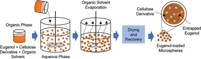
Figure 1: Solvent Evaporation Method
Advantages:
Limitations:
b. Emulsion Techniques
Emulsion methods involve the formation of single or multiple emulsions depending on drug characteristics. Single emulsion (oil-in-water, O/W) techniques are generally used for hydrophobic drugs, whereas multiple emulsion systems (water-in-oil-in-water, W/O/W) are suitable for hydrophilic drugs and proteins (33).The prepared emulsion is stabilized using surfactants and subjected to solvent removal processes, leading to microsphere formation.
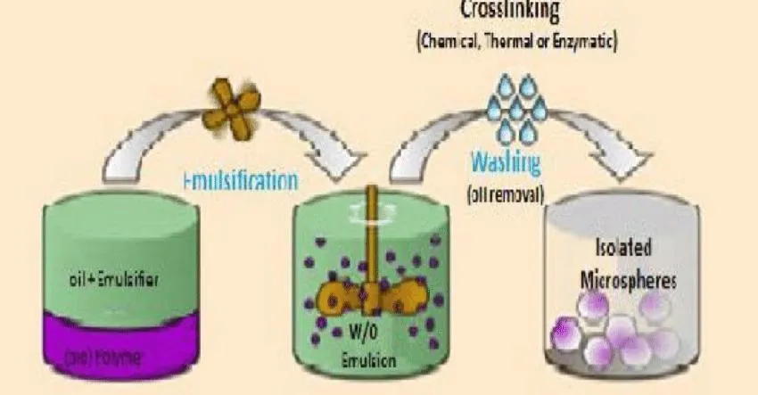
Figure 2: Emulsion technique
Advantages:
Limitations:
c. Spray Drying Method
Spray drying is a rapid and widely used one-step process for preparing microspheres. In this method, the drug and polymer solution is atomized into fine droplets and introduced into a stream of heated air. Rapid solvent evaporation results in the formation of dry microspheres that are collected through cyclone separators (34).This method is particularly suitable for large-scale industrial production due to its simplicity and rapid processing.
Advantages:
Limitations:

Figure 3: Spray drying technique
d. Coacervation Method
Coacervation or phase separation is a process involving separation of a polymer-rich phase from a polymer-poor phase under controlled conditions. Drug particles are dispersed within the polymer solution, and phase separation is induced by changing pH, temperature, or adding nonsolvents. The polymer-rich phase deposits around drug particles and forms microspheres (35).
Coacervation can be categorized as simple coacervation and complex coacervation depending on the number of polymers involved.
Advantages:
Limitations:
Different preparation methods influence particle size, surface morphology, drug loading, encapsulation efficiency, and release characteristics of polymeric microspheres. Therefore, selecting a suitable method is essential for obtaining desired formulation characteristics and therapeutic performance (36).
6. Characterization of Microspheres
Characterization of microspheres is essential for determining their quality, stability, and performance in drug delivery systems. Various parameters such as particle size, surface morphology, entrapment efficiency, drug loading, and in-vitro drug release are evaluated to ensure desired formulation properties. These parameters directly affect therapeutic efficacy, release behavior, and stability of microsphere formulations (37).
Particle Size and Morphology
Particle size and morphology significantly influence drug release characteristics, encapsulation efficiency, bioavailability, and stability of microspheres. Particle size analysis is commonly performed using optical microscopy, laser diffraction, dynamic light scattering (DLS), and scanning electron microscopy (SEM). Surface morphology and shape are usually analyzed by SEM, transmission electron microscopy (TEM), or optical microscopy (38).
Table 2: Methods for Particle Size and Morphology Analysis
|
Parameter |
Method Used |
Principle |
Significance |
|
Particle size |
Optical microscopy |
Direct measurement of particles |
Determines size distribution |
|
Particle size |
Laser diffraction |
Measures light scattering pattern |
Rapid and accurate particle analysis |
|
Particle size |
Dynamic light scattering (DLS) |
Measures Brownian movement |
Suitable for small particles |
|
Morphology |
SEM |
Surface imaging using electron beam |
Determines shape and surface texture |
|
Morphology |
TEM |
Internal structure visualization |
Provides detailed particle structure |
Smaller microspheres generally provide a larger surface area, leading to faster drug release, while larger particles may prolong drug release (39).
Entrapment Efficiency
Entrapment efficiency refers to the percentage of drug successfully encapsulated within microspheres relative to the total amount of drug used during formulation. It indicates the effectiveness of the preparation method and drug incorporation process (40).
The entrapment efficiency can be calculated using the following equation:
Entrapment Efficiency (%)=Actual drug content/Theoretical drug content ×100
Higher entrapment efficiency indicates effective incorporation of drug into the polymer matrix and better formulation performance.
Table 3: Factors Affecting Entrapment Efficiency
|
Factors |
Effect |
|
Polymer concentration |
Increased concentration may increase entrapment |
|
Drug-polymer interaction |
Strong interaction improves encapsulation |
|
Solvent type |
Influences drug diffusion and polymer precipitation |
|
Stirring speed |
Affects particle formation and drug loss |
|
Drug solubility |
Highly water-soluble drugs may exhibit leakage |
Drug Loading
Drug loading represents the amount of drug incorporated into microspheres relative to total microsphere weight. Drug loading determines the quantity of drug available for therapeutic action and influences release characteristics (41).
The percentage drug loading is calculated as:
Drug Loading (%) =Amount of drug in microsphere/Total weight of microspheres×100
Higher drug loading reduces the amount of carrier material required and improves formulation fficiency.
In-vitro Drug Release
In-vitro drug release studies are performed to determine the release profile of the drug from microspheres under simulated physiological conditions. Common methods include dissolution studies using paddle apparatus, basket apparatus, dialysis membrane methods, and diffusion cell techniques (42).
Drug release from microspheres generally occurs through diffusion, polymer swelling, erosion, or degradation mechanisms.
Table 4: Methods Used for In-vitro Drug Release Studies
|
Method |
Principle |
Applications |
|
USP Paddle apparatus |
Rotation of paddle in dissolution medium |
Controlled release studies |
|
USP Basket apparatus |
Drug release from basket-containing samples |
Solid dosage formulations |
|
Dialysis membrane method |
Drug diffusion through membrane |
Sustained release studies |
|
Franz diffusion cell |
Drug diffusion across membrane/skin |
Topical drug delivery studies |
The in-vitro release profile provides information regarding release kinetics and helps predict in-vivo drug behavior. Sustained and controlled release patterns indicate successful microsphere formulation development (43).
7. Mechanism of Controlled Drug Release
Controlled drug release from polymeric microspheres occurs through several mechanisms that regulate the rate and duration of drug release from the polymeric matrix. The release behavior depends on factors such as polymer characteristics, drug properties, particle size, degradation rate, and environmental conditions. Controlled release systems maintain drug concentrations within the therapeutic range for extended periods and reduce fluctuations in drug levels (44).
The major mechanisms involved in controlled drug release are diffusion, erosion, swelling, and degradation-controlled release.
Table 5: Mechanisms of Controlled Drug Release from Polymeric Microspheres
|
Mechanism |
Description |
Characteristics |
|
Diffusion-controlled release |
Drug molecules diffuse through pores or polymer matrix into the surrounding medium |
Provides gradual and sustained drug release |
|
Erosion-controlled release |
Polymer matrix undergoes erosion leading to drug liberation |
Release rate depends on polymer erosion |
|
Swelling-controlled release |
Polymer absorbs surrounding fluid and swells, allowing drug diffusion |
Common in hydrophilic polymers |
|
Degradation-controlled release |
Polymer degradation gradually releases entrapped drug |
Suitable for biodegradable polymers |
|
Osmotically controlled release |
Drug release occurs due to osmotic pressure differences |
Produces predictable release patterns |
Among these mechanisms, diffusion-controlled release is the most common process in polymeric microsphere systems. Biodegradable polymers such as PLGA and chitosan generally follow degradation-mediated release mechanisms, while hydrophilic polymers frequently exhibit swelling-controlled release behavior (45).
The mechanism of drug release significantly influences therapeutic efficacy and is important for designing effective topical formulations with prolonged action and reduced dosing frequency (46).
8. Applications in Topical Drug Delivery
Polymeric microspheres have gained considerable importance in topical drug delivery because of their ability to provide controlled release, improve skin retention, increase drug stability, and minimize systemic side effects. Microsphere-based formulations can enhance drug penetration through the skin and improve therapeutic effectiveness (47).
These systems are applied in various dermatological and pharmaceutical conditions.
Table 6: Applications of Polymeric Microspheres in Topical Drug Delivery
|
Application |
Role of Microspheres |
Examples |
|
Acne treatment |
Sustained release and reduced irritation |
Benzoyl peroxide, Clindamycin |
|
Antifungal therapy |
Improved skin penetration and prolonged action |
Ketoconazole, Fluconazole |
|
Anti-inflammatory therapy |
Localized delivery with reduced systemic effects |
Diclofenac, Ibuprofen |
|
Wound healing |
Enhanced tissue repair and prolonged drug action |
Silver sulfadiazine |
|
Antibacterial therapy |
Increased drug retention at infection site |
Mupirocin |
|
Cosmetic formulations |
Controlled release of active ingredients |
Vitamins, antioxidants |
|
Psoriasis treatment |
Reduced dosing frequency and improved efficacy |
Methotrexate |
Microsphere-based topical systems also improve patient compliance by decreasing application frequency and minimizing local irritation. In recent years, researchers have focused on integrating polymeric microspheres into creams, gels, ointments, and transdermal systems to improve therapeutic outcomes (48).
The application of polymeric microspheres in topical drug delivery continues to expand because of advancements in polymer technology and novel drug delivery approaches, offering promising potential for future pharmaceutical developments (49).
9.Recent Advances in Polymeric Microspheres
Recent advancements in polymeric microsphere technology have significantly improved the efficiency of controlled drug delivery systems. Continuous developments in polymer science, nanotechnology, and formulation strategies have enabled the design of microspheres with enhanced drug loading capacity, targeted delivery, and controlled release characteristics. These advancements have increased the application of polymeric microspheres in topical and other novel drug delivery systems (50).
Several innovative approaches have been introduced to improve microsphere performance, including the use of biodegradable polymers, stimuli-responsive materials, nanotechnology integration, and surface modification techniques.
Table 7: Recent Advances in Polymeric Microspheres
|
Recent advancement |
Description |
Benefits |
|
Biodegradable polymer-based microspheres |
Use of biodegradable polymers such as PLGA, PLA, and chitosan |
Reduced toxicity and improved safety |
|
Stimuli-responsive microspheres |
Drug release triggered by pH, temperature, enzymes, or light |
Site-specific and controlled release |
|
Nano-integrated microspheres |
Combination of nanoparticles and microspheres |
Enhanced penetration and bioavailability |
|
Surface-modified microspheres |
Surface functionalization with ligands or polymers |
Improved targeting efficiency |
|
Microsphere-loaded gels and creams |
Incorporation into topical formulations |
Improved patient compliance and prolonged action |
|
Smart drug delivery systems |
Systems responding to physiological changes |
Better therapeutic outcomes |
Nanotechnology-based microspheres have emerged as an important area of research because they enhance skin permeation and improve drug localization at the target site. Surface engineering techniques have also improved bioadhesion and controlled drug release properties of microspheres (51).
10. Challenges and Future Perspectives
Despite considerable advancements, polymeric microspheres still face several limitations that affect their large-scale application and commercial development. Factors such as manufacturing complexity, high production cost, stability issues, and reproducibility remain important concerns in formulation development (52).
Table 8: Challenges Associated with Polymeric Microspheres
|
Challenges |
Impact on formulation |
|
Complex manufacturing process |
Difficulty in process optimization |
|
High production cost |
Limits commercial applicability |
|
Low drug loading efficiency |
Reduced therapeutic effectiveness |
|
Initial burst drug release |
May cause dose-related adverse effects |
|
Stability issues |
Reduced shelf life |
|
Scale-up difficulties |
Challenges in industrial production |
|
Residual solvent toxicity |
Safety concerns |
Future research is expected to focus on developing safer and more efficient microsphere systems with improved targeting capability and personalized therapeutic approaches. Advanced techniques such as artificial intelligence-assisted formulation design, 3D printing technologies, and multifunctional smart polymers may provide opportunities for overcoming current limitations (53).
The future of polymeric microspheres is directed toward personalized medicine, targeted delivery systems, and environmentally sustainable manufacturing processes. Continued research and technological advancements are expected to expand their role in controlled topical drug delivery and improve patient outcomes (54).
CONCLUSION
Polymeric microspheres represent a highly effective and versatile platform for modern topical drug delivery. By providing controlled, sustained release, they successfully overcome the inherent limitations of conventional topical formulations—such as rapid drug loss and the need for frequent application—thereby enhancing therapeutic outcomes and patient compliance. While the field faces ongoing challenges concerning manufacturing complexity, scale-up difficulties, and production costs, continuous innovation in polymer science, nanotechnology, and smart delivery systems is actively addressing these barriers. As research progresses toward more efficient, targeted, and personalized therapeutic approaches, polymeric microspheres are poised to play an increasingly critical role in the future of dermatological treatments and controlled pharmaceutical delivery.
REFERENCES


Vitthal Pawar, Yashwant Lohar, Ajay Fugate, Pramod N., Vinod Hajare, Pooja Mali, Polymeric Microsphere-Based Systems for Controlled Topical Drug Delivery: A Comprehensive Review, Int. J. of Pharm. Sci., 2026, Vol 4, Issue 6, 1810-1826. https://doi.org/ 10.5281/zenodo.20590520
 10.5281/zenodo.20590520