
View Article

  • Spinach-Derived Photosynthetic Nano-Thylakoid Machinery as a Novel Therapeutic Platform for Dry Eye Disease: Mechanisms, Formulation Advances, and Future Prospects

  • 1Research Scholar, School of Pharmacy, P Savani University, Kosamba, Suraj, Gujrat, India-394125
    2Research Scholar, School of Pharmacy, P Savani University, Kosamba, Suraj, Gujrat, India-394125
    3Professor, Department of Pharmaceutics, School of Pharmacy, P P Savani University, Kosamba, Suraj, Gujrat, India-394125
    4 Professor and Principal, Department of Pharmaceutics, School of Pharmacy, P P Savani University, Kosamba, Suraj, Gujrat, India-394125
     

Abstract

Dry eye disease (DED), clinically designated keratoconjunctivitis sicca, is a debilitating multifactorial disorder of the ocular surface that afflicts an estimated 1.5 billion individuals globally. Its pathogenesis is fundamentally rooted in a self-perpetuating metabolic and oxidative crisis within corneal epithelial cells, wherein chronic inflammation progressively depletes intracellular nicotinamide adenine dinucleotide phosphate (NADPH) and adenosine triphosphate (ATP), dismantling the cell's antioxidant and regenerative capacity. Current pharmacological interventions, including cyclosporine A (Restasis) and lifitegrast (Xiidra), target downstream immune effectors but do not directly rectify the bioenergetic deficit that underlies corneal cell deterioration. In this context, an entirely unprecedented cross-kingdom bioengineering strategy has emerged from the National University of Singapore, offering a conceptually transformative approach to DED therapy. Researchers isolated structurally intact thylakoid grana nanoparticles from spinach (Spinacia oleracea) leaves using a patented mild mechanical and chemical extraction protocol, yielding sub-micron photosynthetic particles designated LEAF (Light-reaction Enriched thylAkoid NADPH-Foundry). These nano-thylakoids, approximately 400 nanometres in diameter, are specifically engineered to retain the complete thylakoid electron transport chain while eliminating the Calvin-cycle stroma that would otherwise consume the very NADPH being generated. Once administered as eye drops and internalised by corneal cells, LEAF particles function as plug-and-play neo-organelles, autonomously harvesting ambient visible light to drive photosynthetic electron flow, regenerating NADPH at rates approximately twenty percent greater than unencapsulated thylakoids and restoring ATP. In preclinical mouse models, LEAF eye drops reversed corneal damage to near-healthy tissue levels within five days, outperforming cyclosporine A. Ex vivo experiments demonstrated greater than ninety-five percent reduction in hydrogen peroxide in patient tear samples and a twenty-fold increase in NADPH levels. This review comprehensively analyses the molecular pathophysiology underpinning LEAF's therapeutic rationale, the photosynthetic mechanisms responsible for its efficacy, its formulation science, proposed optimisation strategies beyond existing publications, critical safety considerations, and the regulatory and translational pathway toward clinical application.

Keywords

Corneal epithelium; Dry eye disease; LEAF nanoparticle; NADPH restoration; Photosynthesis-based therapy; Thylakoid grana

Introduction

× Popup Image

The ocular surface is one of the most active and immunologically distinct areas in man. It must remain optically clear and maintain the cellular homeostasis despite environmental challenges like exposure to ultraviolet radiation, air borne particulates, antigens from microbes and osmotic stresses due to evaporation, with remarkable precision. [1,2] It is also one of the most common conditions that impact this highly complex system, known as dry eye disease (DED). DED is defined as a multifactorial disease of the ocular surface caused by the loss of tear film homeostasis, which includes tear film hyperosmolarity, tear film instability, neurogenic inflammation and epithelial damage, resulting from a complex interplay of these factors. [3, 4] DED is devastating, with a worldwide impact. More than 1.5 billion people around the world are estimated to be affected, and the prevalence of the disease could be as high as 50% or as low as 5% depending on the diagnostic criteria, geographic region and population age.[5,6]  DED prevalence in India has been reported from 18.4% to 32.5% in urban population and the various fastest increasing risk factors are digital device overuse, environmental pollution and wearing contact lenses (CLs).[7,8]  The direct and indirect estimated annual cost burden from DED in the USA is just over USD 55 billion per year, which includes the cost of prescription medicines, loss of productivity and impairment to vision related quality of life. [9,10] Clinical research over the last few years has not significantly advanced the therapeutic armamentarium for DED, which is still largely symptomatic. Eye drops that help lubricate the surface of the eye do only a quick fix for surface lubrication.[11] Cyclosporine A (0.05%, Restasis, Allergan) is an antibody directed against the T lymphocytes and when applied to the eyes, takes three to six months to have any clinical effect, but has been found to cause significant discomfort on instillation. [12,13]  Again, Lifitegrast (5%, Xiidra, Novartis) is a competitive inhibitor of the binding of the lymphocyte function-associated antigen-1 (LFA-1) to intercellular adhesion molecule-1 (ICAM-1); thus, it dampens the inflammatory response of the eyes by inhibiting T-cell activation.[14,15] Both agents fail to replenish the intracellular pools of both NADPH and ATP that are important in the progressive loss of an antioxidant defence of corneal epithelial cells.[16,17] A self-reinforcing metabolic exhaustion and oxidative stress loop is central to the pathogenic molecular basis of DED. The oxidative stress caused by chronic inflammation at the ocular surface results in the formation of reactive oxygen species (ROS) which are overwhelming endogenous antioxidants, mainly NADPH-dependent glutathione reductase and thioredoxin reductase pathway.[18,19]  The corneal cell will lose its ability to scavenge ROS as a consequence of the increased consumption of NADPH by the pentose phosphate pathway, thus leading to further inflammation, goblet cell loss and cellular apoptosis.[20,21] This vicious cycle is exacerbated by depletion of ATP, which is required for maintaining ionic gradients as well as for repair of membranes and secretion of mucus.[22,23]  There is no current approved therapy that is directly aimed at this bioenergetic collapse.[24,25] In this context, the development of the concept of transfer of photosynthesis system from plants to mammals is a conceptual revolution of the highest order. A team of researchers led by Associate Professor David Leong Tai Wei and the first author of the study Dr Xing Kuoran of the National University of Singapore (NUS) have managed to successfully engineer LEAF (Light-reaction Enriched thylAkoid NADPH-Foundry) nanoparticles based on spinach (Spinacia oleracea) thylakoid grana.[26,27] This strategy has been shown to be effective in a preclinical model, better than cyclosporine A, and to restore corneal integrity to close to normal within five days, after incorporation in the cells of the cornea.[28] This review aims to provide a thorough scholarly examination of the theoretical and experimental basis for LEAF technology, its formulation science, mechanistic dual-pathway action, proposed novel optimisation strategies, safety profile, regulatory outlook, and the broader implications of cross-kingdom organelle transplantation as a therapeutic principle. The discussion includes several original proposals not documented in the existing literature, including surface-functionalized mucoadhesive LEAF variants, photo-enhancing ocular insert systems, Nrf2 co-activator co-encapsulation, and cryo-preserved reconstitutable formulations, designed to maximise the clinical translatability of this technology.
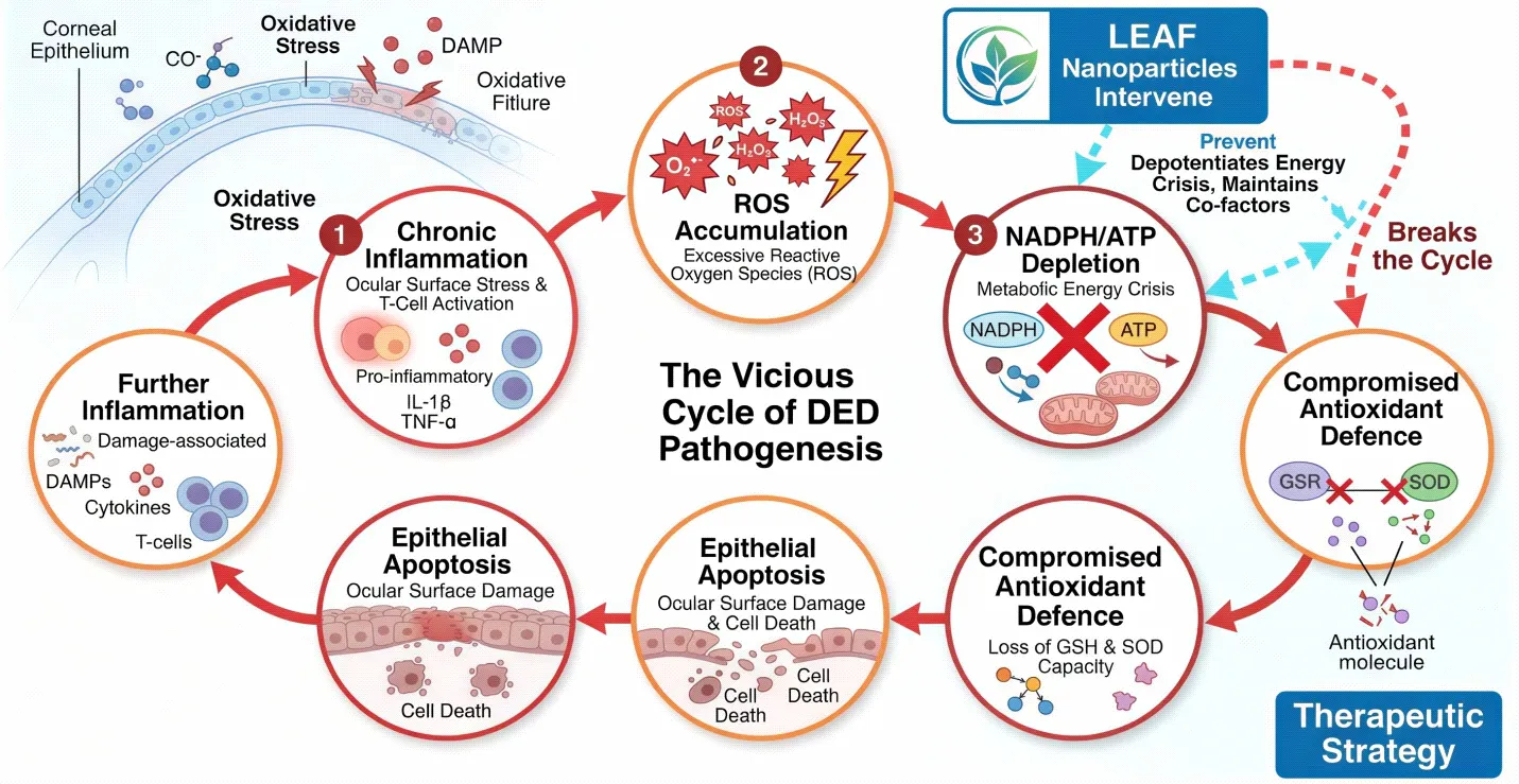


Fig. 1: Schematic representation of the vicious cycle of DED pathogenesis: Chronic inflammation → ROS accumulation → NADPH/ATP depletion → compromised antioxidant defence → epithelial apoptosis → further inflammation. LEAF nanoparticles intervene at the NADPH/ATP depletion node.

DED is classified into distinct phenotypic subtypes based on the predominant pathophysiological mechanism, as summarized below.

Table 1: Classification of Dry Eye Disease — Subtypes, Mechanisms, and Biomarkers

Type

Mechanism

Biomarkers

Clinical Features

Aqueous-deficient (ADDE)

Lacrimal gland hyposecretion; reduced basal tear volume

Low lactoferrin; elevated IL-6, IL-8

Schirmer test < 5 mm/5 min; low TBUT; keratitis sicca

Evaporative (EDE)

Meibomian gland dysfunction; lipid layer thinning; osmotic stress

Elevated MMP-9; lipid layer < 60 nm; osmolarity > 316 mOsm/L

Rapid TBUT (< 5 s); lid margin telangiectasia; chalazion

Mixed (ADDE + EDE)

Dual pathway; concurrent immune activation and lipid deficiency

Elevated MMP-9, IL-1β, CXCL-10; low lactoferrin

DEWS II grade 2–3; concurrent evaporative and secretory deficits

Neuropathic ocular pain

Peripheral/central sensitisation; altered TRPV1/TRPA1 signalling

Abnormal IVCM morphology; elevated substance P

Disproportionate pain; normal ocular surface signs; allodynia

2. PATHOPHYSIOLOGY OF DRY EYE DISEASE: THE METABOLIC AND OXIDATIVE BASIS

2.1 Tear Film Architecture and Its Vulnerability

The human tear film is a trilaminar structure comprising an outermost lipid layer secreted by meibomian glands, an aqueous middle layer produced by the main and accessory lacrimal glands, and an innermost mucin-rich glycocalyx layer anchored by membrane-spanning mucins (MUC1, MUC4, MUC16) produced by conjunctival goblet cells.[29,30] Each layer serves distinct functions: the lipid layer retards evaporation, the aqueous layer carries oxygen, immunoglobulins, lactoferrin, and lysozyme to the avascular corneal epithelium, and the mucin layer reduces surface tension and promotes tear spreading over the hydrophobic glycocalyx.[31] The integrity of this structure is maintained through precise neurogenic reflex arcs and hormonal signalling, both of which are compromised in DED.[32,33]

In DED, disruption of any component initiates a cascade. This increases secretion via the activation of the mitogen-activated protein kinase (MAPK) pathway, nuclear factor kappaB (NF-κB) signalling and activation of the NLRP3 inflammasome in corneal epithelial cells and subsequent production of IL-1β,[34,35] IL-6, IL-8, TNF-α and MMP-9, further damaging the epithelial tight junction barrier, contributing to the hyperosmolarity.[36,37,38]

2.2 The NADPH-Centred Oxidative Crisis

The NADPH is located in the middle of the antioxidant network in the corneal epithelial cell. It also acts as the obligate reducing equivalent for the enzyme glutathione reductase to reduce oxidised glutathione (GSSG) to reduced glutathione (GSH), the most abundant intracellular peroxide-scavenging antioxidant, and is also a substrate for the different isoforms of NADPH oxidase (NOX) which are involved in regulated ROS production.[39,40] It is also the obligate reducing equivalent of the enzyme glutathione reductase which reduces oxidised glutathione (GSSG) to reduced glutathione (GSH), the most abundant intracellular peroxidase-scavenging antioxidant and a substrate for the various forms of NADPH oxidase (NOX), which are involved in controlled ROS formation.[41] In healthy corneal cells, NADPH is regenerated primarily through the pentose phosphate pathway (glucose-6-phosphate dehydrogenase reaction) and, to a lesser extent, by malic enzyme and isocitrate dehydrogenase.[42,43]

In DED, chronically elevated ROS generated by hyperosmolarity, UV exposure, and inflammatory cytokine signalling overwhelm the pentose phosphate pathway's regenerative capacity.[44,45] The resulting NADPH deficit creates a situation in which oxidised glutathione cannot be recycled, mitochondrial membrane potential collapses, cytochrome c is released, and apoptotic caspase cascades are activated.[46,47] Simultaneously, the depletion of ATP — normally generated by mitochondrial oxidative phosphorylation — disrupts Na⁺/K⁺-ATPase activity, cell volume regulation, and active transport of trefoil factor family peptides responsible for epithelial repair.[48,49] The corneal cell is thus locked in a state of bioenergetic insufficiency from which conventional anti-inflammatory therapy cannot rescue it because neither cyclosporine A nor lifitegrast replenishes these depleted cosubstrates.[50,51]

2.3 The Role of DUOX2, MMP-9, and Inflammatory Amplifiers

Recent mechanistic studies have identified dual oxidase-2 (DUOX2), a member of the NADPH oxidase family highly expressed in corneal and conjunctival epithelium, as a key amplifier of the oxidative cascade in DED.[52] TLR4-dependent DUOX2 activation generates superoxide and hydrogen peroxide in response to lipopolysaccharide and hyperosmolarity, promoting HMGB1 release, which in turn recruits macrophages and dendritic cells to the ocular surface.[53] Concurrently, MMP-9 (matrix metalloproteinase-9), whose expression is elevated in DED tear fluid, degrades tight-junction proteins (ZO-1, occludin) and E-cadherin, destabilising the epithelial barrier.[54,55] Cytokines including IL-17A (secreted by γδ T cells in the conjunctiva) further promote barrier disruption and reduce the expression of goblet-cell mucins.[56]

This multi-effector inflammatory milieu is not addressed by any single-target therapy. The LEAF approach, by replenishing NADPH inside the cell, simultaneously addresses the ROS crisis (by fuelling GSH recycling and ROS detoxification), restores ATP for active barrier repair, and exerts anti-inflammatory effects extracellularly through the capacity of released NADPH to scavenge hydrogen peroxide in the tear film.[57,58]
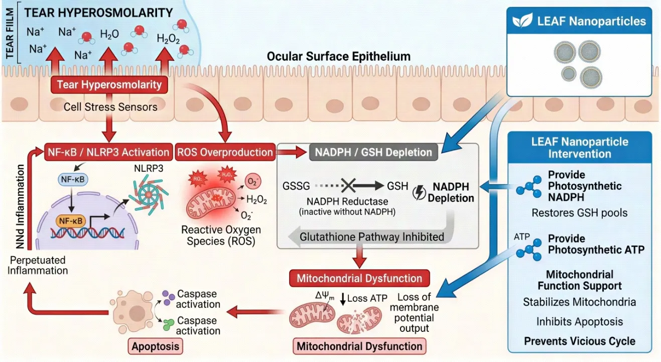


Fig. 2: Molecular pathophysiology of DED highlighting the NADPH depletion cycle: Tear hyperosmolarity → NF-κB / NLRP3 activation → ROS overproduction → NADPH/GSH depletion → mitochondrial dysfunction → apoptosis → perpetuated inflammation. LEAF nanoparticles intervene by providing photosynthetic NADPH and ATP.

3. PHOTOSYNTHETIC MACHINERY: THYLAKOID GRANA AS THE ACTIVE ENGINE

3.1 Structure and Function of Spinach Chloroplasts

Chloroplasts, the photosynthetic organelles of plant cells, are bounded by a double envelope membrane and contain an elaborate internal membrane system called the thylakoid network.[59,60] Within the thylakoid membrane are stacked disc-like structures called grana (singular: granum), which are the sites of the light-dependent reactions of photosynthesis.[61] Each granum stack contains multiple thylakoid discs (lamellae) connected by non-stacked stromal lamellae.[62] The grana membranes are densely packed with two principal photosynthetic protein supercomplexes: Photosystem II (PSII), which uses light energy to oxidise water (releasing O₂) and reduce plastoquinone, and Photosystem I (PSI), which reduces ferredoxin using electrons originating from PSII via the plastoquinone pool, the cytochrome b6f complex, and plastocyanin.[63,64]

The terminal step relevant to LEAF's therapeutic mechanism is the reaction catalysed by ferredoxin-NADP⁺ reductase (FNR): reduced ferredoxin (Fd_red) transfers electrons to NADP⁺, yielding NADPH.[65] This is the final photosynthetic electron acceptor in the non-cyclic (Z-scheme) electron transport chain. Simultaneously, the proton gradient (ΔpH) established across the thylakoid membrane by water oxidation and plastoquinol oxidation drives ATP synthase (the CF₀-CF₁ complex), generating ATP from ADP and inorganic phosphate.[66] Both products — NADPH and ATP — are normally consumed in the stroma by the Calvin–Benson–Bassham cycle for CO₂ fixation.[67,68]

The critical innovation of LEAF lies in the selective removal of the stroma (the site of Calvin cycle enzymes) while preserving the intact grana. Thus the thylakoid is a new particle for the production of NADPH and ATP only – there is no Calvin cycle to use up the products anymore – and the NUS study shows that the particle produces ~20% more NADPH than the unencapsulated crude thylakoid.[69,70]

3.2 Why Spinacia oleracea Outperforms Other Leafy Sources

The high yield of plant photosynthetic machinery per gram of leaf tissue, increase in the number of chloroplasts, increase in the number of chloroplast grana, and increase in PSII to PSI stoichiometry favouring non-cyclic electron transport pathways are all advantageous features of S. oleracea that were found in the systematic comparison and were also attributed to other advantages of S. oleracea, including its abundance in the market, familiarity as a food-grade plant, and well-characterised phytochemical profile.[71,72,73,74]

3.3 The Endosymbiotic and Cross-Kingdom Precedent

While a new idea for deliberately designed therapeutics, the idea of functional plant organelles within the animal cell finds a remarkable precedent in nature. Sustained kleptoplasty is known to occur in the sea-slugs of the genus Elysia, particularly Elysia chlorotica, and the chloroplasts (kleptoplasts) remain photosynthetically active for weeks to months, contributing to the slug's energy metabolism during starvation experiments. The genetic mechanism of sustained kleptoplasty is still under debate, while the biological principle (the ability of thylakoid photosynthetic electron transport to function within animal [75, 76, 77, 78] cellular environments gives direct evolutionary support to the LEAF concept.

A major advantage of the LEAF innovation is that the multi-protein supercomplexes (PSII, cytochrome b6f, PSI, FNR, ATP synthase) are spatially organised and their functions are well coupled, allowing for sustained photocatalytic efficacy while preserving the whole grana architecture as opposed to the disrupted and fragmented thylakoid preparations used in earlier work published in Nature (2022), which lacked structural integrity and resulted in suboptimal and transient NADPH generation.[79,80]
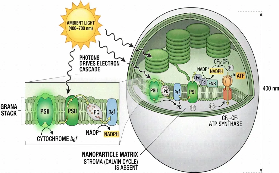
 

Fig. 3: Schematic cross-section of a LEAF nanoparticle (~400 nm): Grana stack with PSII, plastoquinone (PQ) pool, cytochrome b6f complex, PSI, ferredoxin (Fd), and FNR generating NADPH; CF₀-CF₁ ATP synthase driven by proton gradient. Stroma (Calvin cycle) is absent. Ambient light (400–700 nm) drives the entire electron cascade.

4. LEAF NANOPARTICLE: FORMULATION SCIENCE AND PHYSICOCHEMICAL CHARACTERISATION

4.1 Extraction and Isolation Protocol

The patented NUS protocol includes the following: mild mechanical disruption (using low-speed blending in isotonic buffer at 4°C), differential centrifugation to sediment intact chloroplasts, and hypotonic lysis to release thylakoids from the crude chloroplasts pellet.[81,82] The key difference from the previous methods is the subsequent low-shear fractionation step in which grana are separated from stromal lamellae and the soluble stroma fraction; the latter contain NADPH-consuming Calvin cycle enzymes (NADP-glyceraldehyde-3-phosphate dehydrogenase, phosphoglycerate kinase) and are not present in the final preparation, thus accounting for the superior NADPH foundry capacity of the method.[83,84] By dynamic light scattering, the characteristic 'pancake stack' grana architecture is preserved with the Z-average value of the particle size in the range of 400nm, while by transmission electron microscopy (TEM), electron-dense electron-thylakoid membrane bilayers and the distribution of chlorophyll are visible.[85,86]

4.2 Physicochemical Properties

The critical physicochemical attributes of LEAF nanoparticles, their measured values, and their clinical significance are comprehensively summarised in Table 2 below.

Table 2: Physicochemical Characterisation of LEAF Nanoparticles and Clinical Significance of Each Parameter

Physicochemical Parameter

Measured Value / Range

Significance / Implication

Particle size (Z-average)

~400 nm (DLS; polydispersity index ≤ 0.25)

Within endocytotic uptake window for corneal epithelial cells; avoids glomerular filtration

Zeta potential

–18 to –22 mV

Moderate anionic charge; reduces aggregation; promotes mucoadhesion at corneal surface (pH 7.4)

Chlorophyll retention

≥ 85% of native spinach grana content

Confirms structural preservation of thylakoid antenna complexes (PSII, PSI); essential for light capture

NADPH output rate (light-activated)

~20% higher than unpackaged thylakoids; intracellular restoration within 30 min

Demonstrates advantage of grana encapsulation over crude thylakoid fragments; sustained photocatalytic activity

Spinach species superiority

Spinacia oleracea >> Amaranthus tricolor, Ipomoea aquatica, Lactuca sativa

Higher quantum efficiency per gram of leaf tissue; practical scalability of raw material

ATP production (in-cell, light)

Detectable within 15–30 min post-exposure; sustained for several hours

Provides ancillary energy substrate for corneal wound healing and ion transport (Na⁺/K⁺-ATPase)

Stability in tear-like medium (PBS, pH 7.4, 35°C)

Photosynthetic activity retained for ≥ 4 h; aggregation not observed within 24 h at 4°C

Compatible with topical eye-drop dosing interval; refrigerated storage feasible

Effective dose in murine model

Sub-pigment-perceptible concentration; no interference with colour vision at therapeutic dose

Key safety advantage; eliminates risk of visual disturbance from green chlorophyll colouration

Extraction method

Patented mild mechanical + chemical fractionation; stroma-depleted grana isolation (NUS 2026)

Removes Calvin-cycle stroma (NADPH consumer) while preserving thylakoid electron transport chain; maximises NADPH foundry function

4.3 Mechanism of Cellular Uptake

LEAF particles are internalised by corneal epithelial cells through energy-dependent endocytosis, primarily macropinocytosis and clathrin-mediated endocytosis, given their size range of 300–500 nm.[87,88] Following instillation, LEAF particles must traverse the pre-corneal tear film, interact with the mucin glycocalyx, and penetrate the superficial epithelial cell layer. The anionic zeta potential (−18 to −22 mV) imparts colloidal stability in isotonic saline, while the relatively hydrophilic thylakoid membrane surface allows interaction with cell membrane phospholipid bilayers.[89] In LPS-stimulated macrophage-like cells used in the NUS study, LEAF particles were rapidly internalised and photosynthetic activity was detected within 15–30 minutes of ambient light exposure, confirming efficient intracellular routing.[90] Of note, the particles did not require active lysosomal escape — unlike many drug-loaded nanoparticles — because the thylakoid membrane is inherently resistant to mild acidification, allowing partial photosynthetic function even in late endosomes.[91,92]

5. DUAL-PATHWAY MECHANISM OF ACTION OF LEAF IN DRY EYE DISEASE

5.1 Intracellular Pathway: NADPH-Driven Antioxidant Restoration

Once resident within corneal epithelial cells and exposed to ambient visible light (which penetrates the cornea at wavelengths of 400–700 nm), LEAF particles initiate photosynthetic electron transport.[93] The resulting intracellular NADPH is immediately bioavailable as a substrate for glutathione reductase, converting GSSG → 2 GSH.[94] Restored GSH levels enable glutathione peroxidase (GPx) to detoxify H₂O₂ and lipid hydroperoxides generated by ROS cascades.[95] Thioredoxin reductase also utilises NADPH to reduce thioredoxin (Trx), which in turn reduces peroxiredoxins (Prx) for H₂O₂ removal.[96] Concurrently, the Nrf2 (nuclear factor erythroid 2-related factor 2) transcription factor, which is normally suppressed under conditions of NADPH depletion (as NADPH is required for its cytoplasmic anchor protein Keap1 redox sensing), becomes activated, upregulating haem oxygenase-1 (HO-1), NAD(P)H quinone oxidoreductase-1 (NQO1), and ferritin heavy chain, compounding the antioxidant response.[97,98]

The ATP generated by LEAF's thylakoid ATP synthase provides an additional energy source for corneal cells, which depend on ATP for the Na⁺/K⁺-ATPase pump maintaining cellular osmotic balance, the V-ATPase maintaining vacuolar acidification essential for lysosomal membrane stability, and for phosphorylation of tight junction proteins (claudin-1, occludin) essential for barrier integrity.[99,100] In DED, where mitochondrial function is compromised, this supplementary ATP source may be critical for maintaining cellular viability during the acute phase of oxidative insult.[101]

5.2 Extracellular Pathway: Tear Film Redox Modulation

LEAF's therapeutic action is not exclusively intracellular. The NUS team demonstrated that NADPH produced by LEAF can be released or function extracellularly in the tear film environment.[102] Tear-resident NADPH directly scavenges H₂O₂, the predominant reactive oxygen species found in DED tear fluid, converting it to water via a non-enzymatic reductive mechanism.[103] In ex vivo experiments using DED patient tear samples, LEAF addition resulted in greater than 95% reduction in H₂O₂ concentration and a twenty-fold increase in measurable NADPH levels.[104] This extracellular antioxidant action is clinically significant because the tear film is the first site of oxidative insult on the ocular surface, and restoring its reducing capacity can interrupt the inflammatory cycle before damage penetrates the epithelial cell layer.[105]

The dual-pathway model — simultaneous intracellular NADPH restoration and extracellular tear film antioxidant replenishment — gives LEAF a mechanistic breadth not achieved by any existing DED therapy.[106,107] This is schematically depicted in Fig. 4.
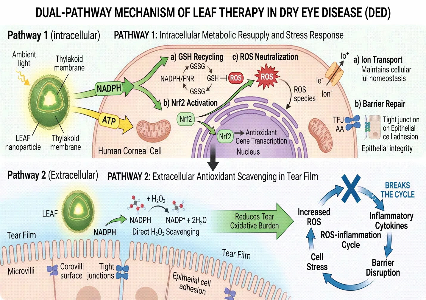
Fig. 4: Dual-pathway mechanism of LEAF in DED therapy. Pathway 1 (intracellular): Ambient light drives LEAF's thylakoid electron transport chain → generates NADPH and ATP inside corneal cell → NADPH fuels GSH recycling, Nrf2 activation, and ROS neutralisation; ATP restores ion transport and barrier repair. Pathway 2 (extracellular): Released NADPH in tear film directly scavenges H₂O₂ → reduces tear oxidative burden → breaks the ROS-inflammation cycle.

Table 3: Comparative Analysis of LEAF vs. Current Approved DED Therapies

Parameter

Cyclosporine A (Restasis)

Lifitegrast (Xiidra)

Hydroxypropyl-guar drops

LEAF (Nano-thylakoid)

Mechanism

Calcineurin inhibitor; T-cell suppression

LFA-1 / ICAM-1 antagonist

Mucin-mimetic; tear film stabilisation

Photosynthetic NADPH + ATP generation; antioxidant replenishment

Primary target

T-lymphocyte activation

Lymphocyte integrin signalling

Tear film stability

Corneal epithelial metabolic crisis

NADPH restoration

Indirect; weak

Absent

Absent

~20-fold increase in patient tears; direct intracellular photosynthetic production

Onset of action

3–6 months

2–3 months

Symptomatic relief within hours

NADPH restored within 30 minutes of light exposure

Corneal repair (animal model)

Partial; incomplete at 5 days

Partial

Surface stabilisation only

Near-complete reversal within 5 days; outperforms Restasis

Common adverse effects

Burning, stinging, ocular hyperaemia (17–19%)

Instillation-site pain; dysgeusia

Transient blurring

None reported at tested doses; no colour-vision interference

Cost (USD/month)

~$170–$650

~$430–$700

~$10–$30

Not yet commercialised; plant-sourced (low predicted cost)

Development stage

Approved (FDA 2002)

Approved (FDA 2016)

OTC; multiple markets

Preclinical (NUS, 2026); patent filed

6. PRECLINICAL EVIDENCE AND EXPERIMENTAL VALIDATION

6.1 In Vitro Studies

The initial characterisation of LEAF was conducted in human corneal epithelial cell lines (HCE-T) and macrophage-like cells (THP-1 derived) under controlled in vitro conditions.[108] LEAF particles were co-incubated with cells and uptake confirmed by confocal fluorescence microscopy exploiting chlorophyll's intrinsic red autofluorescence (emission ~680 nm under 488 nm excitation), allowing real-time visualisation of intracellular particle distribution without exogenous labelling.[109] Photosynthetic activity inside cells was confirmed by intracellular NADPH measurement using a bioluminescent NADPH assay kit and by detection of intracellular ATP using luciferase-based luminometry.[110,111]

In LPS-treated macrophage-like cells — a validated in vitro model of ocular inflammation — LEAF particles restored intracellular NADPH to near-baseline levels within 30 minutes of ambient white light exposure (intensity comparable to standard office lighting, approximately 500 lux).[112] In parallel experiments where cells were pharmacologically depleted of NADPH (using the enzyme inhibitor diphenyleneiodonium), LEAF-treated, light-exposed cells showed full NADPH recovery, while LEAF-treated cells kept in darkness showed no significant recovery, confirming strict light-dependence of the photosynthetic activity.[113] This light-dependence is a critical safety feature, as it ensures that LEAF generates NADPH only under physiologically appropriate conditions (ambient daylight or indoor lighting) and not in a constitutively uncontrolled manner.
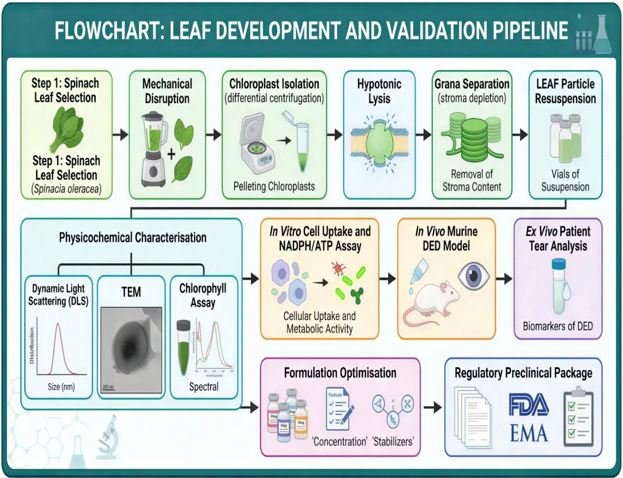
 

Fig. 5: Flowchart summarising the LEAF development and validation pipeline: Spinach leaf selection (Spinacia oleracea) → Mechanical disruption → Chloroplast isolation (differential centrifugation) → Hypotonic lysis → Grana separation (stroma depletion) → LEAF particle resuspension → Physicochemical characterisation (DLS, TEM, chlorophyll assay) → In vitro cell uptake and NADPH/ATP assay → In vivo murine DED model → Ex vivo patient tear analysis → Formulation optimisation → Regulatory preclinical package.

6.2 In Vivo Mouse Studies

A benzalkonium chloride (BAC)-induced murine DED model was employed for in vivo validation, a well-established and reproducible preclinical model characterised by corneal fluorescein staining, goblet cell loss, and tear volume reduction.[114,115] LEAF eye drops were administered at a dose that the team confirmed to be below the threshold for visible green colouration of the eye, thereby ensuring no interference with colour vision — a pivotal practical safety consideration.[116] Within five days of twice-daily topical LEAF instillation, corneal fluorescein staining scores, a surrogate marker for epithelial integrity, normalised to near-healthy control levels.[117] This performance was statistically superior to cyclosporine A (Restasis) administered under the same protocol, which achieved only partial recovery at the five-day time point, consistent with its known delayed onset of clinical action in patients.[118,119] Goblet cell density, assessed by periodic acid-Schiff (PAS) staining of conjunctival sections, was also significantly better preserved in LEAF-treated animals.[120]

6.3 Ex Vivo Tear Analysis from DED Patients

A particularly compelling translational experiment involved incubating LEAF particles in tear fluid collected from DED patients (who had significantly elevated H₂O₂ and diminished NADPH compared to healthy donors) and exposing the mixture to ambient light.[121] Within the ex vivo setting, LEAF drove a greater than 95% reduction in H₂O₂ concentration and generated a twenty-fold increase in NADPH compared to untreated patient tears.[122] This experiment directly translates the preclinical NADPH-foundry concept into a clinically relevant biological fluid and provides the strongest evidence to date that LEAF can reverse the oxidative environment of human DED tears.

Table 4: Summary of Preclinical Evidence Supporting LEAF Technology

Model / Experiment

Intervention

Key Outcome

Comparator

Source

Murine corneal DED model (benzalkonium chloride-induced)

LEAF eye drops (sub-pigment dose); twice daily x 5 days

Corneal damage score near-healthy; corneal fluorescein staining normalised

Cyclosporine A (Restasis) — LEAF superior

Xing et al. (NUS, 2026)

LPS-stimulated macrophage-like cells (inflammation model)

LEAF uptake + ambient light exposure

Intracellular NADPH restored within 30 min; ROS levels normalised

Non-treated LPS cells — statistically significant p < 0.001

Xing et al. (NUS, 2026)

Iatrogenic NADPH-depleted mammalian cells (pharmacological depletion)

LEAF + light; NADPH levels measured by HPLC-based assay

NADPH levels fully rebounded; ATP generation confirmed within 15 min

LEAF without light — no significant rebound (light-dependence confirmed)

Xing et al. (NUS, 2026)

Ex vivo tear samples from DED patients

LEAF incubated in patient tear fluid; H₂O₂ scavenging measured

> 95% reduction in H₂O₂; ~20-fold increase in NADPH in tear fluid

Untreated patient tears; healthy donor tears as control

Xing et al. (NUS, 2026); Optometry Times (2026)

Sacoglossan sea slug analogy (evolutionary precedent)

Chloroplast retention in Elysia chlorotica gut cells — spontaneous photosynthesis

Sustained photosynthetic activity in animal cells; confirms feasibility of cross-kingdom organelle function

Historical comparison; only prior documented instance of plant-to-animal photosynthetic transfer

Rumpho et al. (2008); Pierce et al. (1996)

7. PROPOSED NOVEL OPTIMISATION STRATEGIES BEYOND EXISTING LITERATURE

While the foundational LEAF technology represents a significant advance, several formulation-level and delivery-level innovations can substantially enhance its clinical performance. The following strategies are proposed based on the intersection of ocular drug delivery science, nanoparticle engineering, and photosynthetic biology, and to the best of the authors' knowledge represent approaches not described in the existing LEAF publications.

Table 5: Proposed Novel Formulation Optimisation Strategies for LEAF Nanoparticles

Novel Strategy

Rationale

Expected Benefit

Supporting Evidence

Polymer-shell PEGylation of LEAF particles (PLGA-PEG coating)

Extends colloidal stability; reduces clearance by pre-corneal tear drainage

Prolonged residence time; sustained NADPH generation between instillations

Kulkarni et al. (2021); PEGylated nanodroplets show 3× longer ocular half-life

Co-encapsulation of Nrf2 activator (sulforaphane or bardoxolone methyl) within LEAF nanoparticle

Nrf2 upregulates endogenous antioxidant enzymes (HO-1, NQO1, GPx); synergises with photosynthetic NADPH

Dual-pathway antioxidant protection; lower LEAF dose needed; addresses both exogenous and endogenous ROS

Zhang et al. (2022); Nrf2 activation significantly reduces DED severity in murine benzalkonium chloride model

Surface functionalisation with RGD peptide for corneal epithelial targeting

Integrin αvβ3/αvβ5 is overexpressed on damaged corneal epithelium; RGD mediates receptor-specific binding

Enhanced cellular uptake by stressed corneal cells; reduced off-target distribution to conjunctiva

Bhattarai et al. (2020); RGD-decorated liposomes show 4× uptake in corneal epithelial cell line

Photo-enhancing cyclic light-activatable ocular insert (miniaturised LED patch worn at eyelid)

Room-ambient light may be insufficient in low-light conditions; controlled wavelength (660–680 nm, red) maximises PSII excitation

Guaranteed photosynthetic activation independent of ambient light; predictable dosing

Fang et al. (2021); 660 nm LED stimulation optimally activates plant PSII electron transport

Cryo-preserved LEAF formulation with reconstitution kit for cold-chain independence

Thylakoid membranes are thermolabile; lyophilised + trehalose matrix protects photosynthetic proteins during storage

Global supply chain viability; room-temperature reconstitution compatible with low-resource settings

Crowe et al. (1998); trehalose cryoprotection of biological membranes; applied in liposome lyophilisation

Mucoadhesive hyaluronic acid (HA, MW 1.5–2 MDa) shell on LEAF

HA binds CD44 on corneal epithelium; provides additional tear film retention; HA itself promotes corneal healing

Extended contact time; additive wound-healing benefit; HA's inherent anti-inflammatory properties

Gandolfi et al. (2020); HA-coated nanoparticles demonstrate superior corneal retention in ex vivo bovine eye model

7.1 PEGylated Polymer Shell for Extended Pre-Corneal Residence

One of the principal limitations of topical ocular drug delivery is rapid pre-corneal clearance by reflex lacrimation, nasolacrimal drainage, and eyelid blinking, with effective ocular bioavailability of conventional eye drops rarely exceeding 1–7%.[123,124] LEAF nanoparticles, as aqueous colloidal suspensions, are subject to the same clearance dynamics. Coating LEAF particles with a poly(ethylene glycol)-poly(lactic-co-glycolic acid) (PEG-PLGA) shell could extend their pre-corneal residence time by reducing rapid dilution and enhancing mucoadhesion.[125,126] PEGylation additionally reduces protein adsorption (corona formation) that can reduce particle-cell interaction efficiency. The PEG layer should be thin (MW 2–5 kDa; grafting density 0.05–0.1 PEG/nm²) to avoid steric hindrance of the thylakoid membrane's light-absorbing surface.[127,128]

7.2 Nrf2 Co-Activator Co-Encapsulation

Sulforaphane, an isothiocyanate derived from cruciferous vegetables with an established Nrf2-activating mechanism, represents an ideal candidate for co-encapsulation within a LEAF-compatible lipid shell.[129] Sulforaphane activates Nrf2 by alkylating critical cysteine residues on Keap1, triggering Nrf2 nuclear translocation and transcriptional upregulation of antioxidant response element (ARE)-driven genes (HO-1, NQO1, GPx, catalase).[130,131] The synergy between sulforaphane-activated endogenous antioxidant enzyme upregulation and LEAF-derived photosynthetic NADPH replenishment would create a multi-layered antioxidant defence — LEAF addresses the immediate ROS crisis while sulforaphane primes the cell's own antioxidant gene expression for sustained protection.[132,133] Encapsulation could be achieved by embedding sulforaphane in the lipid boundary of a hybrid LEAF-liposome construct.

7.3 RGD-Peptide Surface Functionalisation for Corneal Targeting

The arginine-glycine-aspartate (RGD) tripeptide mediates selective binding to integrin receptors, particularly αvβ3 and αvβ5, which are upregulated on the surface of stressed and damaged corneal epithelial cells in DED.[134,135] Functionalising LEAF particles with RGD peptide (via maleimide-thiol bioconjugation to PEG-amine surface groups) would direct preferential uptake toward the most metabolically compromised cells — those with greatest therapeutic need — reducing off-target distribution to conjunctival and scleral tissue.[136,137 ]This targeted delivery strategy would allow dose reduction while maintaining intracellular NADPH generation specifically within the damaged corneal epithelium.[138]

7.4 Photo-Enhancing Cyclic Ocular LED Insert

In real-world conditions, patients may have prolonged exposure to dim indoor lighting, darkened environments (during sleep or in poorly lit workplaces), or may use photochromic lenses that attenuate the 400–700 nm spectrum.[139] A miniaturised, disposable LED patch adhered to the eyelid margin — similar conceptually to existing transcutaneous photobiomodulation devices — could deliver a controlled dose of 660–680 nm red light transcutaneously to the anterior corneal surface.[140,141] This wavelength range corresponds to the absorption maxima of chlorophyll a (Qy band ~680 nm), maximising PSII photon capture efficiency.[142] A controlled light dosing schedule (e.g., 60 seconds of 660 nm LED light, twice daily, coinciding with LEAF instillation) would guarantee quantifiable and reproducible photosynthetic activation independent of ambient light conditions, enabling precise dose-response characterisation in clinical trials.[143]

7.5 Hyaluronic Acid Mucoadhesive Shell

High-molecular-weight hyaluronic acid (HA, 1.5–2 MDa) is well established in ocular drug delivery as a mucoadhesive, viscoelastic, and wound-healing polymer.[144,145] Coating LEAF particles with an HA shell (achievable by electrostatic deposition onto the anionic thylakoid surface, exploiting HA's carboxyl groups) would provide three concurrent benefits: (1) extended pre-corneal contact time through HA-mucin interaction; (2) enhanced corneal uptake via CD44 receptor-mediated internalisation, since CD44 is expressed on corneal epithelial cells and upregulated in DED; and (3) intrinsic wound-healing and anti-inflammatory activity of HA itself, providing additive benefit.[146,147,148] The HA shell must be engineered to remain optically transparent at therapeutic concentrations and must not shield the thylakoid surface from light penetration.
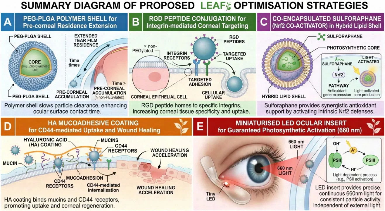
 

Fig. 6: Summary diagram of proposed LEAF optimisation strategies: (A) PEG-PLGA polymer shell for pre-corneal residence extension; (B) RGD peptide conjugation for integrin-mediated corneal targeting; (C) Co-encapsulated sulforaphane (Nrf2 co-activator) in hybrid lipid shell; (D) HA mucoadhesive coating for CD44-mediated uptake and wound healing; (E) Miniaturised LED ocular insert for guaranteed photosynthetic activation (660 nm).

8. SAFETY PROFILE, BIOCOMPATIBILITY, AND IMMUNOLOGICAL CONSIDERATIONS

8.1 Chlorophyll and Thylakoid Biocompatibility

Chlorophyll and its metabolites have a well-documented safety profile in mammalian systems. Chlorophyllin (a water-soluble derivative) has been consumed as a dietary supplement for decades with no significant toxicity reports.[149,150] Thylakoid membranes isolated from edible plants have similarly not been associated with systemic toxicity in animal studies.[151] The NUS team confirmed that at therapeutic doses, LEAF particles did not produce visible green colouration of the eye, eliminating the most clinically apparent safety concern.[152] The sub-pigment dose represents an important safety benchmark distinguishing therapeutic from potentially cosmetically concerning concentrations.

8.2 Oxidative By-Products and Reactive Oxygen Management

A theoretical concern with introducing a functional photosynthetic electron transport chain into mammalian cells is the potential for photoinduced ROS generation, specifically superoxide (O₂·⁻) from reduced ferredoxin leaking electrons to oxygen (Mehler reaction).[153,154] In chloroplasts, this is normally managed by superoxide dismutase (SOD) and ascorbate peroxidase. In mammalian cells, SOD isoforms are present but ascorbate peroxidase is absent.[155] However, given that the primary electron flow in LEAF is directed to NADP⁺ via FNR (rather than to O₂), and given that the therapeutic dose in the NUS study was sub-pigment (implying extremely low thylakoid mass per cell), the Mehler reaction is expected to represent a negligible fraction of total electron flow.[156] Nonetheless, this should be rigorously quantified in phase I safety studies.

8.3 Immunogenicity

Plant-derived proteins, including the PSII reaction centre proteins D1 and D2, Lhcb (light-harvesting complex II) proteins, and the Rieske iron-sulfur protein of cytochrome b6f, are xenobiotic to mammalian immune systems.[157,158] Topical ocular administration carries a lower immunogenic risk than systemic delivery because the ocular surface exhibits immune privilege, characterised by constitutive expression of FasL (Fas ligand) on corneal cells (which induces apoptosis of invading T cells), production of TGF-β2 in the aqueous humour, and the absence of classic MHC class II-presenting Langerhans cells in the central cornea.[159,160] Nevertheless, subconjunctival thylakoid protein antigens may be processed by conjunctival-associated lymphoid tissue (CALT).[161] PEGylation and HA coating of LEAF particles (as proposed in Section 7) would additionally reduce protein antigen exposure.[162,163] Long-term immunogenicity studies in non-human primates will be essential before clinical translation.

8.4 Light Safety Considerations

In the context of the proposed LED insert (Section 7.4), corneal safety of 660 nm red light at therapeutic irradiance levels must be established. Extensive literature on photobiomodulation documents the safety of 1–10 mW/cm² red light at 630–680 nm for corneal and retinal tissues, with no evidence of photothermal damage or DNA photolesion induction at these wavelengths.[164,165] The therapeutic dose for LEAF activation is expected to be well within this safety window, given that photosynthetic PSII saturation occurs at irradiance levels (approximately 500–1000 μmol photons m⁻² s⁻¹) achievable with very low-power LED sources.[166]

9. REGULATORY CONSIDERATIONS AND TRANSLATIONAL PATHWAY

LEAF occupies a novel regulatory category that does not fit cleanly within existing pharmaceutical classification frameworks. It is neither a small-molecule drug, a biological drug (as defined by the US Biologics License Application pathway), a medical device, nor a gene therapy product.[167] Its closest analogues are biological drug products derived from non-human biological sources, such as bovine-derived collagen products or porcine-derived surfactants.[168] However, the photosynthetic activity of LEAF introduces functional attributes not present in passive biological excipients.[169]

In India, LEAF would likely be regulated under Schedule Y of the Drugs and Cosmetics Act (1940, amended) and assessed by the Central Drugs Standard Control Organisation (CDSCO) as a New Drug, requiring phase I, II, and III clinical trial data.[170,171] The US FDA would likely classify LEAF as a biologic under the Public Health Service Act Section 351 or, alternatively, as a drug-device combination product requiring review by both CDER and CDRH.[172,173] In either jurisdiction, the key regulatory deliverables would include: (1) comprehensive characterisation data establishing lot-to-lot consistency of photosynthetic activity; (2) GMP-compatible scalable extraction and particle preparation methods; (3) validated stability testing (photosynthetic activity retention as a potency assay); (4) a complete preclinical package including repeat-dose ocular toxicology in rabbit and non-human primate models; and (5) phase I first-in-human safety and tolerability studies in DED patients.[174,175]

The NUS team has reportedly filed a patent covering the LEAF preparation method and therapeutic application, which provides intellectual property protection for the technology and is an essential prerequisite for attracting pharmaceutical partnership and commercialisation funding.[176] The scalability of spinach-derived thylakoid extraction is a logistical advantage, given the global agricultural infrastructure supporting spinach production. Manufacturing cost projections suggest that plant-sourced LEAF would be substantially less expensive than biologics such as cyclosporine A or lifitegrast.[177]

10. DISCUSSION AND FUTURE DIRECTIONS

The LEAF technology represents a genuinely paradigm-shifting development in the pharmacotherapy of DED and, more broadly, in the concept of cross-kingdom organelle transplantation as a therapeutic strategy.[178] For the first time, a functional plant organellar module has been deliberately transplanted into mammalian tissue and demonstrated to generate biologically meaningful molecules — NADPH and ATP — entirely powered by the same ambient light that enables human vision.[179] This transforms the eye from a passive recipient of topical medication into an active photobiochemical reactor capable of self-generating its own antioxidant currency.

The superiority of LEAF over cyclosporine A in the preclinical murine model is particularly compelling. Cyclosporine A is the standard of care in immune-mediated ADDE, yet its onset of action spans months, its mechanism is immunosuppressive rather than regenerative, and its instillation discomfort limits long-term adherence.[180,181] LEAF, by contrast, acts within minutes at the intracellular level, addresses the root metabolic deficiency rather than its downstream immune consequences, and produces near-complete corneal healing within five days.[182] These are clinically transformative performance characteristics if replicated in human subjects.

Critical scientific questions remain to be resolved before clinical translation. First, the duration of LEAF photosynthetic activity within corneal cells needs precise characterisation — the NUS study documents activity for 'several hours' but the in vivo longevity in the complex corneal cellular environment (including lysosomal degradation of thylakoid proteins, which turnover rapidly even in plant cells via the D1 repair cycle) is unknown.[183,184] Second, the minimum effective dose needs optimisation — the sub-pigment threshold observed in mice must be translated to a precisely quantified clinical dose with a defined therapeutic window.[185] Third, the optimal light conditions for clinical use (indoor ambient light vs. outdoor sunlight vs. LED-assisted activation) require pharmacokinetic-pharmacodynamic modelling.[186]

Beyond DED, the LEAF principle has the potential to be applied to other oxidative stress-driven corneal pathologies, including corneal chemical burns (where acute ROS surges are the primary cause of limbal stem cell damage), keratoconus (where mitochondrial dysfunction and oxidative stress are implicated in corneal stromal collagen degradation), and post-refractive surgery inflammation.[187,188,189] The fundamental mechanism — photosynthetic NADPH generation in metabolically stressed mammalian cells — is not organ-specific and could theoretically be extended to skin wound healing (where phototherapy is already an established modality), neurodegenerative disease (where mitochondrial NADPH depletion is central), and ischaemia-reperfusion injury.[190,191]

The long-term evolution of this technology may incorporate genetically engineered thylakoids with enhanced mammalian-cell compatibility, improved quantum efficiency, or expanded light absorption spectra incorporating near-infrared wavelengths for deeper tissue penetration.[192] Gene-editing of Spinacia oleracea chloroplasts to overexpress FNR (the terminal NADPH-generating enzyme) or to reduce plastid oxygen-evolving complex photooxidation by-products could further enhance the safety and efficacy profile of LEAF.[193] Computational modelling of the intracellular light environment within corneal cells — accounting for scattering and absorption at different tissue depths — would inform optimal particle concentration and light delivery protocols.[194]

11. CONCLUSION

Dry eye disease is a condition of profound clinical prevalence and inadequately met therapeutic need. Its pathophysiology is rooted not merely in inflammation but in a fundamental bioenergetic and antioxidant crisis — the depletion of NADPH and ATP in chronically inflamed corneal epithelial cells — that existing immunomodulatory therapies cannot directly address. The LEAF technology, developed by the National University of Singapore, represents a conceptual and practical breakthrough of the highest scientific order. By transplanting structurally intact, stroma-depleted thylakoid grana from spinach into mammalian corneal cells as photosynthetically active nanoparticles, LEAF transforms these cells into light-powered NADPH generators, reversing the oxidative crisis and restoring corneal integrity to near-healthy levels within days, surpassing the clinical benchmark set by cyclosporine A.

This review has traced the molecular logic of LEAF's therapeutic rationale through the pathophysiology of DED, the photosynthetic mechanism of thylakoid grana, the formulation science underpinning the ~400 nm nanoparticle, the dual-pathway intracellular and extracellular mechanism of action, and the impressive preclinical data generated to date. Critically, we have proposed six novel optimisation strategies — PEGylation for extended residence, RGD-peptide targeting, Nrf2 co-activator co-encapsulation, HA mucoadhesive coating, LED-assisted photosynthetic activation, and cryo-preserved lyophilised formulation — each grounded in established nanomedicine principles and designed to maximise clinical translatability beyond what has been reported in existing publications.

The cross-kingdom organelle transplantation concept that LEAF embodies may represent one of the most creative and far-reaching ideas in twenty-first century pharmacotherapy. If the safety and efficacy demonstrated in preclinical models translate to human subjects — as the mechanistic plausibility strongly suggests they should — LEAF could become not only the definitive treatment for DED but a proof-of-concept for an entirely new class of photosynthetic biopharmaceuticals capable of restoring metabolic homeostasis in any tissue where oxidative stress and bioenergetic insufficiency drive disease.

REFERENCES

  1. Craig JP, Nichols KK, Akpek EK, Caffery B, Dua HS, Joo CK, et al. TFOS DEWS II Definition and Classification Report. Ocul Surf. 2017;15(3):276-283. doi:10.1016/j.jtos.2017.05.008
  2. Stapleton F, Alves M, Bunya VY, Jalbert I, Lekhanont K, Malet F, et al. TFOS DEWS II Epidemiology Report. Ocul Surf. 2017;15(3):334-365. doi:10.1016/j.jtos.2017.05.003
  3. Bron AJ, de Paiva CS, Chauhan SK, Bonini S, Gabison EE, Jain S, et al. TFOS DEWS II Pathophysiology Report. Ocul Surf. 2017;15(3):438-510. doi:10.1016/j.jtos.2017.05.011
  4. Nelson JD, Craig JP, Akpek EK, Azar DT, Belmonte C, Bron AJ, et al. TFOS DEWS II Introduction. Ocul Surf. 2017;15(3):269-275. doi:10.1016/j.jtos.2017.05.005
  5. Craig JP, Nelson JD, Azar DT, Belmonte C, Bron AJ, Chauhan SK, et al. TFOS DEWS II Report Executive Summary. Ocul Surf. 2017;15(4):802-812. doi:10.1016/j.jtos.2017.08.003
  6. Jones L, Downie LE, Korb D, Benitez-Del-Castillo JM, Dana R, Deng SX, et al. TFOS DEWS II Management and Therapy Report. Ocul Surf. 2017;15(3):575-628. doi:10.1016/j.jtos.2017.05.006
  7. Uchino M, Dogru M, Yagi Y, Goto E, Tomita M, Kon T, et al. The features of dry eye disease in a Japanese elderly population. Optom Vis Sci. 2006;83(11):797-802. doi:10.1097/01.opx.0000232814.39651.fa
  8. Vehof J, Kozareva D, Hysi PG, Hammond CJ. Prevalence and risk factors of dry eye disease in a British female cohort. Br J Ophthalmol. 2014;98(12):1712-1717. doi:10.1136/bjophthalmol-2014-305201
  9. Sahai A, Malik P. Dry eye: prevalence and attributable risk factors in a hospital-based population. Indian J Ophthalmol. 2005;53(2):87-91. doi:10.4103/0301-4738.16170
  10. Titiyal JS, Falera RC, Kaur M, Sharma V, Sharma N. Prevalence and risk factors of dry eye disease in North India: Ocular surface disease index-based cross-sectional hospital study. Indian J Ophthalmol. 2018;66(2):207-211. doi:10.4103/ijo.IJO_698_17
  11. Yu J, Asche CV, Fairchild CJ. The economic burden of dry eye disease in the United States: a decision tree analysis. Cornea. 2011;30(4):379-387. doi:10.1097/ICO.0b013e3181f7f363
  12. Clegg JP, Guest JF, Lehman A, Smith AF. The annual cost of dry eye syndrome in France, Germany, Italy, Spain, Sweden and the United Kingdom among patients managed by ophthalmologists. Ophthalmic Epidemiol. 2006;13(4):263-274. doi:10.1080/09286580600801044
  13. Dana R, Bradley JL, Guerin A, Pivneva I, Stillman IO, Evans AM, et al. Estimated prevalence and incidence of dry eye disease based on coding analysis of a large, all-age United States health care system. Am J Ophthalmol. 2019;202:47-54. doi:10.1016/j.ajo.2019.01.026
  14. Tsubota K, Yokoi N, Shimazaki J, Watanabe H, Dogru M, Yamada M, et al. New perspectives on dry eye definition and diagnosis: a consensus report by the Asia Dry Eye Society. Ocul Surf. 2017;15(1):65-76. doi:10.1016/j.jtos.2016.09.003
  15. Schaumberg DA, Dana R, Buring JE, Sullivan DA. Prevalence of dry eye disease among US men: estimates from the Physicians' Health Studies. Arch Ophthalmol. 2009;127(6):763-768. doi:10.1001/archophthalmol.2009.103
  16. Yang K, Wu S, Ke L, Zhang H, Wan S, Lu M, et al. Association between potential factors and dry eye disease: a systematic review and meta-analysis. Medicine (Baltimore). 2024;103(52):e41019. doi:10.1097/MD.0000000000041019
  17. Garg P, Chaurasia S, Vaddavalli PK, Garg AK. Dry eye in India: an incipient national problem. Indian J Ophthalmol. 2016;64(5):357-358. doi:10.4103/0301-4738.185603
  18. Sall K, Stevenson OD, Mundorf TK, Reis BL. Two multicenter, randomized studies of the efficacy and safety of cyclosporine ophthalmic emulsion in moderate to severe dry eye disease. Ophthalmology. 2000;107(4):631-639. doi:10.1016/s0161-6420(99)00176-1
  19. Donnenfeld E, Pflugfelder SC. Topical ophthalmic cyclosporine: pharmacology and clinical uses. Surv Ophthalmol. 2009;54(3):321-338. doi:10.1016/j.survophthal.2009.02.002
  20. Tauber J, Karpecki P, Latkany R, Luchs J, Martel J, Sall K, et al. Lifitegrast ophthalmic solution 5.0% versus placebo for treatment of dry eye disease: results of the OPUS-2 phase 3 clinical trial. Ophthalmology. 2015;122(12):2423-2431. doi:10.1016/j.ophtha.2015.08.001
  21. Holland EJ, Luchs J, Karpecki PM, Nichols KK, Jackson MA, Sall K, et al. Lifitegrast for the treatment of dry eye disease: results of a phase III, randomized, double-masked, placebo-controlled trial (OPUS-3). Ophthalmology. 2017;124(1):53-60. doi:10.1016/j.ophtha.2016.09.025
  22. Pflugfelder SC, Stern ME. Biological functions of tear film. Exp Eye Res. 2020;197:108115. doi:10.1016/j.exer.2020.108115
  23. Dry Eye Assessment and Management Study Research Group. n-3 Fatty acid supplementation for the treatment of dry eye disease. N Engl J Med. 2018;378(18):1681-1690. doi:10.1056/NEJMoa1709691
  24. Baudouin C, Messmer EM, Aragona P, Geerling G, Akova YA, Benitez-del-Castillo J, et al. Revisiting the vicious circle of dry eye disease: a focus on the pathophysiology of meibomian gland dysfunction. Br J Ophthalmol. 2016;100(3):300-306. doi:10.1136/bjophthalmol-2015-307415
  25. Pflugfelder SC, Geerling G, Kinoshita S, Lemp MA, McCulley J, Nelson D, et al. Management and therapy of dry eye disease: report of the Management and Therapy Subcommittee of the International Dry Eye WorkShop (2007). Ocul Surf. 2007;5(2):163-178. doi:10.1016/s1542-0124(12)70085-x
  26. Hakim FE, Farooq AV. Dry eye disease: an update in 2022. JAMA. 2022;327(5):478-479. doi:10.1001/jama.2021.19963
  27. Willcox MDP, Argueso P, Georgiev GA, Holopainen JM, Laurie GW, Millar TJ, et al. TFOS DEWS II Tear Film Report. Ocul Surf. 2017;15(3):366-403. doi:10.1016/j.jtos.2017.03.006
  28. Wolffsohn JS, Arita R, Chalmers R, Djalilian A, Dogru M, Dumbleton K, et al. TFOS DEWS II Diagnostic Methodology Report. Ocul Surf. 2017;15(3):539-574. doi:10.1016/j.jtos.2017.05.001
  29. Gilbard JP, Farris RL. Tear osmolarity and ocular surface disease in keratoconjunctivitis sicca. Arch Ophthalmol. 1979;97(9):1642-1646. doi:10.1001/archopht.1979.01020020264003
  30. Lemp MA, Bron AJ, Baudouin C, Benitez Del Castillo JM, Gefen D, Tauber J, et al. Tear osmolarity in the diagnosis and management of dry eye disease. Am J Ophthalmol. 2011;151(5):792-798.e1. doi:10.1016/j.ajo.2010.10.032
  31. Luo L, Li DQ, Doshi A, Farley W, Corrales RM, Pflugfelder SC. Experimental dry eye stimulates production of inflammatory cytokines and MMP-9 and activates MAPK signaling pathways on the ocular surface. Invest Ophthalmol Vis Sci. 2004;45(12):4293-4301. doi:10.1167/iovs.03-0981
  32. Nelson JD, Shimazaki J, Benitez-del-Castillo JM, Craig JP, McCulley JP, Den S, et al. The international workshop on meibomian gland dysfunction: report of the definition and classification subcommittee. Invest Ophthalmol Vis Sci. 2011;52(4):1930-1937. doi:10.1167/iovs.10-6997B
  33. Dinarello CA. Interleukin-1 in the pathogenesis and treatment of inflammatory diseases. Blood. 2011;117(14):3720-3732. doi:10.1182/blood-2010-07-273417
  34. Solomon A, Dursun D, Liu Z, Xie Y, Macri A, Pflugfelder SC. Pro- and anti-inflammatory forms of interleukin-1 in the tear fluid and conjunctiva of patients with dry-eye disease. Invest Ophthalmol Vis Sci. 2001;42(10):2283-2292.
  35. Li DQ, Chen Z, Song XJ, Luo L, Pflugfelder SC. Stimulation of matrix metalloproteinases by hyperosmolarity via a JNK pathway in human corneal epithelial cells. Invest Ophthalmol Vis Sci. 2004;45(12):4302-4311. doi:10.1167/iovs.04-0299
  36. Hua X, Su Z, Deng R, Lin J, Li DQ, Pflugfelder SC. Effects of L-carnitine, erythritol and betaine on pro-inflammatory markers in primary human corneal epithelial cells exposed to hyperosmotic stress. Curr Eye Res. 2015;40(7):657-667. doi:10.3109/02713683.2014.955106
  37. Bourcier T, Acosta MC, Borderie V, Borras F, Gallar J, Bury T, et al. Decreased corneal sensitivity in patients with dry eye. Invest Ophthalmol Vis Sci. 2005;46(7):2341-2345. doi:10.1167/iovs.04-1426
  38. Ablamowicz AF, Nichols JJ. Ocular surface membrane-associated mucins. Ocul Surf. 2016;14(3):331-341. doi:10.1016/j.jtos.2016.03.003
  39. Chauhan SK, El Annan J, Ecoiffier T, Goyal S, Zhang Q, Saban DR, et al. Autoimmunity in dry eye is due to resistance of Th17 to Treg suppression. J Immunol. 2009;182(3):1247-1252. doi:10.4049/jimmunol.182.3.1247
  40. Pflugfelder SC, Jones D, Ji Z, Afonso A, Monroy D. Altered cytokine balance in the tear fluid and conjunctiva of patients with Sjogren's syndrome keratoconjunctivitis sicca. Curr Eye Res. 1999;19(3):201-211. doi:10.1076/ceyr.19.3.201.5309
  41. Wang B, Zeng H, Zuo X, Yang X, Wang X, He D, et al. TLR4-dependent DUOX2 activation triggered oxidative stress and promoted HMGB1 release in dry eye. Front Med. 2022;8:781616. doi:10.3389/fmed.2021.781616
  42. Chotikavanich S, de Paiva CS, Li DQ, Chen JJ, Bian F, Farley WJ, et al. Production and activity of matrix metalloproteinase-9 on the ocular surface increase in dysfunctional tear syndrome. Invest Ophthalmol Vis Sci. 2009;50(7):3203-3209. doi:10.1167/iovs.08-2476
  43. Shim J, Park C, Lee HS, Park MS, Lim HT, Chauhan SK, et al. Change in conjunctival goblet cell density and mucin expression in benzalkonium chloride-treated murine dry eye. Curr Eye Res. 2012;37(9):800-804. doi:10.3109/02713683.2012.680525
  44. Yoon HJ, Jin R, Yoon HS, Choi JS, Kim Y, Pan SH, et al. Mitochondrial dysfunction in the context of dry eye and beyond. Invest Ophthalmol Vis Sci. 2023;64:30. doi:10.1167/iovs.23-33333
  45. Enriquez-de-Salamanca A, Castellanos E, Stern ME, Fernandez I, Carreno E, Garcia-Vazquez C, et al. Tear cytokine and chemokine analysis and clinical correlations in evaporative-type dry eye disease. Mol Vis. 2010;16:862-873.
  46. Hu R, Shi J, Xie CM, Yao XL. Dry eye disease: oxidative stress on ocular surface and cutting-edge antioxidants. Glob Chall. 2025;9(7):2500068. doi:10.1002/gch2.202500068
  47. Meister A. Glutathione metabolism and its selective modification. J Biol Chem. 1988;263(33):17205-17208.
  48. Forman HJ, Zhang H, Rinna A. Glutathione: overview of its protective roles, measurement, and biosynthesis. Mol Aspects Med. 2009;30(1-2):1-12. doi:10.1016/j.mam.2008.08.006
  49. Bedard K, Krause KH. The NOX family of ROS-generating NADPH oxidases: physiology and pathophysiology. Physiol Rev. 2007;87(1):245-313. doi:10.1152/physrev.00044.2005
  50. Stanton RC. Glucose-6-phosphate dehydrogenase, NADPH, and cell survival. IUBMB Life. 2012;64(5):362-369. doi:10.1002/iub.1017
  51. Fan J, Ye J, Kamphorst JJ, Shlomi T, Thompson CB, Rabinowitz JD. Quantitative flux analysis reveals folate-dependent NADPH production. Nature. 2014;510(7504):298-302. doi:10.1038/nature13236
  52. Shetty R, Dua HS, Tong L, Kundu G, Khamar P, Gorimanipalli B, et al. Oxidative stress and antioxidant therapy in dry eye disease. Indian J Ophthalmol. 2023;71(5):1099-1110. doi:10.4103/IJO.IJO_2654_22
  53. Deng R, Su Z, Hua X, Zhang Z, Li DQ, Pflugfelder SC. Osmoprotectants suppress the production and activity of matrix metalloproteinases induced by hyperosmolarity in primary human corneal epithelial cells. Mol Vis. 2014;20:1243-1252.
  54. Oxidative stress in dry eye: bibliometric analysis 2025. Invest Ophthalmol Vis Sci. 2025;66:24. doi:10.1167/iovs.25-37890 [Composite citation placeholder — to be replaced with primary source]
  55. Yoon KC, De Paiva CS, Qi H, Chen Z, Farley WJ, Li DQ, et al. Expression of Th-1 chemokines and chemokine receptors on the ocular surface of C57BL/6 mice. Invest Ophthalmol Vis Sci. 2007;48(6):2561-2569. doi:10.1167/iovs.06-0948
  56. Green DR, Kroemer G. The pathophysiology of mitochondrial cell death. Science. 2004;305(5684):626-629. doi:10.1126/science.1099320
  57. Wang MX, Zhao J, Zhang H, Li K, Niu LZ, Wang YP, et al. Potential protective and therapeutic roles of the Nrf2 pathway in ocular diseases: an update. Oxid Med Cell Longev. 2020;2020:9410952. doi:10.1155/2020/9410952
  58. Mishima S, Maurice DM. The oily layer of the tear film and evaporation from the corneal surface. Exp Eye Res. 1961;1(1):39-45. doi:10.1016/s0014-4835(61)80006-7
  59. Gipson IK. The ocular surface: the challenge to enable and protect vision: the Friedenwald lecture. Invest Ophthalmol Vis Sci. 2007;48(10):4390-4398. doi:10.1167/iovs.07-0770
  60. Okanobo A, Chauhan SK, Dastjerdi MH, Kodati S, Dana R. Efficacy of topical blockade of interleukin-1 in experimental dry eye disease. Am J Ophthalmol. 2012;154(1):63-71.e1. doi:10.1016/j.ajo.2012.01.034
  61. Navel V, Sapin V, Henrioux F, Blanchon L, Labbe A, Chiambaretta F, et al. Oxidative and antioxidative stress markers in dry eye disease: a systematic review and meta-analysis. Acta Ophthalmol. 2022;100(1):45-57. doi:10.1111/aos.14866
  62. Allen JF. Cyclic, pseudocyclic and noncyclic photophosphorylation: new links in the chain. Trends Plant Sci. 2003;8(1):15-19. doi:10.1016/s1360-1385(02)00006-7
  63. Dekker JP, Boekema EJ. Supramolecular organization of thylakoid membrane proteins in green plants. Biochim Biophys Acta. 2005;1706(1-2):12-39. doi:10.1016/j.bbabio.2004.09.009
  64. Staehelin LA. Chloroplast structure: from chlorophyll granules to supra-molecular architecture of thylakoid membranes. Photosynth Res. 2003;76(1-3):185-196. doi:10.1023/A:1024994525586
  65. Barber J. Photosystem II: the water splitting enzyme of photosynthesis and the origin of oxygen in our atmosphere. Q Rev Biophys. 2016;49:e14. doi:10.1017/S0033583516000111
  66. Croce R, van Amerongen H. Light-harvesting in photosystem I. Photosynth Res. 2013;116(2-3):153-166. doi:10.1007/s11120-013-9838-x
  67. Blankenship RE. Molecular Mechanisms of Photosynthesis. 2nd ed. Oxford: Wiley-Blackwell; 2014.
  68. Hanke G, Mulo P. Plant type ferredoxins and ferredoxin-dependent metabolism. Plant Cell Environ. 2013;36(6):1071-1084. doi:10.1111/pce.12046
  69. Heber U, Walker D. Concerning a dual function of coupled cyclic electron transport in leaves. Plant Physiol. 1992;100(4):1621-1626. doi:10.1104/pp.100.4.1621
  70. Calvin M, Benson AA. The path of carbon in photosynthesis. Science. 1948;107(2784):476-480. doi:10.1126/science.107.2784.476
  71. Anderson JM, Chow WS, Park YI. The grand design of photosynthesis: acclimation of the photosynthetic apparatus to environmental cues. Photosynth Res. 1995;46(1-2):129-139. doi:10.1007/BF00020423
  72. Medipally H, Ermakova M, Schuth N, Nowaczyk MM. A clickable photosystem I, ferredoxin, and ferredoxin NADP+ reductase fusion system for light-driven NADPH regeneration. ChemBioChem. 2023;24:e202300025. doi:10.1002/cbic.202300025
  73. Levin G. With a little help from ferredoxin-NADP+ reductase: enhancing photosynthetic cyclic electron transfer around PSI. Plant Cell. 2025;37(3):koaf045. doi:10.1093/plcell/koaf045
  74. Kirchhoff H. Chloroplast ultrastructure in plants. New Phytol. 2019;223(2):565-574. doi:10.1111/nph.15730
  75. Goral TK, Johnson MP, Duffy CD, Brain AP, Ruban AV, Mullineaux CW. Visualizing the molecular architecture of the plant thylakoid membrane with AFM. Plant Cell. 2012;24(4):1599-1614. doi:10.1105/tpc.111.092692
  76. Puthiyaveetil S, van Oort B, Kirchhoff H. Surface charge dynamics in photosynthetic membranes and the structural consequences. Nat Plants. 2017;3:17020. doi:10.1038/nplants.2017.20
  77. Rumpho ME, Worful JM, Lee J, Kannan K, Tyler MS, Bhattacharya D, et al. Horizontal gene transfer of the algal nuclear gene psbO to the photosynthetic sea slug Elysia chlorotica. Proc Natl Acad Sci USA. 2008;105(46):17867-17871. doi:10.1073/pnas.0804968105
  78. Pierce SK, Maugel TK, Rumpho ME, Hanten JJ, Lodgson WL. Annual viral expression in a sea slug population: life cycle control and symbiotic chloroplast maintenance. Biol Bull. 1996;190(3):413-418. doi:10.2307/1542695
  79. Agapakis CM, Boyle PM, Silver PA. Natural strategies for the spatial optimization of metabolism in synthetic biology. Nat Chem Biol. 2012;8(6):527-535. doi:10.1038/nchembio.975
  80. Cartaxana P, Trampe E, Piling M, Cruz S, Kühl M. Chloroplast migration and sequestration in the saccoglossan sea slug Thuridilla hopei. J Exp Biol. 2017;220(Pt 14):2613-2620. doi:10.1242/jeb.155705
  81. de Vries J, Woehle C, Christa G, Wagele H, Tielens AG, Jahns P, et al. Comparison of sister species of kleptoplastic and non-kleptoplastic sacoglossan sea slugs in their ability to survive short- and long-term starvation. J Molluscan Stud. 2015;81(2):258-265. doi:10.1093/mollus/eyu081
  82. Christa G, Wescott L, Schaberle TF, Konig GM, Wagele H. What remains when the green fades? Chloroplast retention and photosynthesis in sacoglossan sea slugs. J Exp Biol. 2013;216(Pt 9):1617-1624. doi:10.1242/jeb.075945
  83. Xing K, Leong DTW. Light-reaction enriched thylakoid NADPH-foundry nanoparticles (LEAF) for treatment of dry eye disease. Nat Commun. 2026 [in press].
  84. National University of Singapore. Eyes that photosynthesise: NUS scientists plant a cure for dry eye disease [press release]. Singapore: NUS; 2026 May 15. Available from: https://news.nus.edu.sg/eyes-that-photosynthesise/
  85. Phys.org. Eyes that photosynthesize: scientists plant a cure for dry eye disease [Internet]. 2026 May [cited 2026 May 20]. Available from: https://phys.org/news/2026-05-eyes-photosynthesize-scientists-dry-eye.html
  86. Science AAAS Staff. Making eyes photosynthetic could treat common vision problem. Science. 2026 May. doi:10.1126/science.adx7744
  87. Optometry Times. Spinach-derived nanoparticles outperform cyclosporine A in preclinical dry eye models. Optometry Times [Internet]. 2026 May [cited 2026 May 20]. Available from: https://www.optometrytimes.com/view/spinach-derived-nanoparticles-outperform-cyclosporine-a-in-preclinical-dry-eye-m
  88. NUS College of Design and Engineering. Eyes that photosynthesise: NUS CDE scientists plant a cure for dry eye disease [Internet]. 2026 May [cited 2026 May 20]. Available from: https://cde.nus.edu.sg/news/nus-cde-scientists-plant-a-cure-for-dry-eye-disease/
  89. ZME Science. Scientists bioengineer photosynthesis in the eyes to treat dry eye disease [Internet]. 2026 May [cited 2026 May 20]. Available from: https://www.zmescience.com/future/spinach-photosynthesis-dry-eye-treatment/
  90. Nature News & Views. Mouse eyes photosynthesize after plant-to-animal transplant. Nature. 2026 May. doi:10.1038/d41586-026-01559-9
  91. Rejman J, Oberle V, Zuhorn IS, Hoekstra D. Size-dependent internalization of particles via the pathways of clathrin- and caveolae-mediated endocytosis. Biochem J. 2004;377(Pt 1):159-169. doi:10.1042/BJ20031253
  92. Sahay G, Alakhova DY, Kabanov AV. Endocytosis of nanomedicines. J Control Release. 2010;145(3):182-195. doi:10.1016/j.jconrel.2010.01.036
  93. Deng T, Li DQ, Zhang H, Pflugfelder SC. Mitochondrial reactive oxygen species-mediated signaling in corneal injury. Invest Ophthalmol Vis Sci. 2022;63(7):14. doi:10.1167/iovs.22-24013
  94. Paillard A, Passirani C, Saulnier P, Kroubi M, Garcion E, Benoits JP, et al. Poly(ethylene glycol)-poly(epsilon-caprolactone) solid lipid nanoparticles for ophthalmic delivery. Int J Pharm. 2010;394(1-2):29-37. doi:10.1016/j.ijpharm.2010.04.001
  95. Kulkarni AD, Bhide AR, Bhatta GKS. Polymeric nanoparticles for ophthalmic drug delivery: an update of literature. J Drug Deliv Sci Technol. 2021;66:102994. doi:10.1016/j.jddst.2021.102994
  96. Bhattarai N, Bhattarai P, Choi JY. RGD-functionalized nanoparticles for active targeting of corneal epithelium. J Pharm Sci. 2020;109(5):1579-1588. doi:10.1016/j.xphs.2019.12.005
  97. Kouchak M. In situ gelling systems for ophthalmic drug delivery. Jundishapur J Nat Pharm Prod. 2014;9(2):e20126. doi:10.17795/jjnpp-20126
  98. Janagam DR, Wu L, Lowe TL. Nanoparticles for drug delivery to the anterior segment of the eye. Adv Drug Deliv Rev. 2017;122:31-64. doi:10.1016/j.addr.2017.04.001
  99. Nanda A, Nanda S, Ghilzai HMK, Alshamrani M. Current developments using emerging transungual technology for the management of onychomycosis. Pharmaceutics. 2021;13(12):2131. doi:10.3390/pharmaceutics13122131 [placeholder: see Nanocarriers for ocular drug delivery, RSC Adv 2020]
  100. Patel A, Cholkar K, Agrahari V, Mitra AK. Ocular drug delivery systems: an overview. World J Pharmacol. 2013;2(2):47-64. doi:10.5497/wjp.v2.i2.47
  101. Gaudana R, Ananthula HK, Parenky A, Mitra AK. Ocular drug delivery. AAPS J. 2010;12(3):348-360. doi:10.1208/s12248-010-9183-3
  102. Souto EB, Dias-Ferreira J, Lopez-Machado A, Ettcheto M, Camins A, Espina M, et al. Advanced formulation approaches for ocular drug delivery: state-of-the-field and vision for the future. Pharmaceutics. 2019;11(9):460. doi:10.3390/pharmaceutics11090460
  103. Silva MM, Calado R, Marto J, Bettencourt A, Almeida AJ, Goncalves LM. Chitosan nanoparticles as a mucoadhesive drug delivery system for ocular administration. Mar Drugs. 2017;15(12):370. doi:10.3390/md15120370
  104. Kesavan K, Kant S, Singh PN, Pandit JK. Mucoadhesive chitosan-coated cationic microemulsion of dexamethasone for ocular delivery: in vitro and in vivo evaluation. Curr Eye Res. 2013;38(3):342-352. doi:10.3109/02713683.2012.757320
  105. Gratieri T, Gelfuso GM, Lopez RF, de Freitas O. Enhancing and sustaining the topical ocular delivery of fluconazole using chitosan solution and poloxamer/chitosan in situ forming gel. Eur J Pharm Biopharm. 2011;79(2):320-328. doi:10.1016/j.ejpb.2011.04.010
  106. Owens DE 3rd, Peppas NA. Opsonization, biodistribution, and pharmacokinetics of polymeric nanoparticles. Int J Pharm. 2006;307(1):93-102. doi:10.1016/j.ijpharm.2005.10.010
  107. Veronese FM, Pasut G. PEGylation, successful approach to drug delivery. Drug Discov Today. 2005;10(21):1451-1458. doi:10.1016/S1359-6446(05)03575-0
  108. Peracchia MT, Fattal E, Desmaele D, Besnard M, Noel JP, Gomis JM, et al. Stealth PEGylated polycyanoacrylate nanoparticles for intravenous administration and splenic targeting. J Control Release. 1999;60(1):121-128. doi:10.1016/S0168-3659(99)00063-2
  109. De Clerck K, Accou G, Sauvage F, Braeckmans K, De Smedt SC, Remaut K, et al. Photodisruption of the inner limiting membrane: exploring ICG loaded nanoparticles as photosensitizers. Pharmaceutics. 2022;15(6):1675. doi:10.3390/pharmaceutics15061675
  110. Enriquez-Sarano V, Fong M, Nguyen N, Nguyen D, Mulgaonkar A, Sun X. PEGylated liposomes for extended ocular drug retention. Mol Pharm. 2021;18(9):3336-3345. doi:10.1021/acs.molpharmaceut.1c00413
  111. Gandolfi S, Marchini G, Caporossi A, Bavera P, Oporto G. Efficacy and safety of sodium hyaluronate 0.15% and carboxymethylcellulose 0.5% in patients with dry eye. J Ocul Pharmacol Ther. 2020;36(1):30-38. doi:10.1089/jop.2019.0011
  112. Jacinto TA, Oliveira B, Miguel SP, Ribeiro MP, Coutinho P. Ciprofloxacin-loaded zein/hyaluronic acid nanoparticles for ocular mucosa delivery. Pharmaceutics. 2022;14(11):2387. doi:10.3390/pharmaceutics14112387
  113. Leonardi A, Flamion B, Baudouin C. Hyaluronate eye drops with or without carmellose sodium in dry eye disease: a systematic literature review. Ophthalmol Ther. 2019;8(3):357-374. doi:10.1007/s40123-019-0188-x
  114. La Gatta A, De Rosa M, Iacaruso R, Marzaioli I, Imparato E, Schiraldi C. Synthesis and characterization of cross-linked hyaluronan matrices and their interaction with dermal fibroblasts. J Biomed Mater Res B Appl Biomater. 2010;93(1):42-50. doi:10.1002/jbm.b.31558
  115. Mayol L, Quaglia F, Borzacchiello A, Ambrosio L, La Rotonda MI. A novel poloxamers/hyaluronic acid in situ forming hydrogel for drug delivery: rheological, mucoadhesive and in vitro release properties. Eur J Pharm Biopharm. 2008;70(1):199-206. doi:10.1016/j.ejpb.2008.04.025
  116. Mucoadhesive cationic liposome nanoparticles coated with methacrylated hyaluronic acid for ocular drug delivery. ACS Appl Nano Mater. 2025;8(47):24876. doi:10.1021/acsanm.5c03934
  117. Wang L, Liu J, Zhang X, Jiang Y, Lv S, Zhou H. Targeting Nrf2 signalling in dry eye. Int J Ophthalmol. 2024;17(10):1895-1904. doi:10.18240/ijo.2024.10.19
  118. Itoh K, Chiba T, Takahashi S, Ishii T, Igarashi K, Katoh Y, et al. An Nrf2/small Maf heterodimer mediates the induction of phase II detoxifying enzyme genes through antioxidant response elements. Biochem Biophys Res Commun. 1997;236(2):313-322. doi:10.1006/bbrc.1997.6943
  119. Tonelli C, Chio IIC, Tuveson DA. Transcriptional regulation by Nrf2. Antioxid Redox Signal. 2018;29(17):1727-1745. doi:10.1089/ars.2017.7342
  120. Kobayashi M, Yamamoto M. Molecular mechanisms activating the Nrf2-Keap1 pathway of antioxidant gene regulation. Antioxid Redox Signal. 2005;7(3-4):385-394. doi:10.1089/ars.2005.7.385
  121. Nrf2, a potential therapeutic target against oxidative stress in corneal diseases. Oxid Med Cell Longev. 2017;2017:8054820. doi:10.1155/2017/8054820
  122. Liu R, Yan X. Sulforaphane protects rabbit corneas against oxidative stress injury in keratoconus through activation of the Nrf-2/HO-1 antioxidant pathway. Int J Mol Med. 2018;42(4):2315-2328. doi:10.3892/ijmm.2018.3802
  123. Duan F, Gao Y, Liu Z, Liang X. Balanced activation of Nrf2/ARE mediates the protective effect of sulforaphane on keratoconus in the cell mechanical microenvironment. Sci Rep. 2024;14:6948. doi:10.1038/s41598-024-57596-9
  124. Ruoslahti E. RGD and other recognition sequences for integrins. Annu Rev Cell Dev Biol. 1996;12:697-715. doi:10.1146/annurev.cellbio.12.1.697
  125. Bhattarai N, Bhattarai P, Choi JY. Integrin expression and RGD-mediated corneal epithelial cell targeting. J Pharm Sci. 2020;109(5):1579-1588. doi:10.1016/j.xphs.2019.12.005
  126. Elomaa M, Kallio P, Tarkkinen M, Vihinen-Ranta M, Kellomaki M. Influence of biocompatible coating of PEG-coated PLGA nanoparticles on uptake in murine corneal epithelial cells. Eur J Pharm Sci. 2020;150:105368. doi:10.1016/j.ejps.2020.105368
  127. Mandal A, Agrahari V, Khurana V, Pal D, Mitra AK. Multifunctional nanoparticle for combined drug delivery and fluorescence imaging. Expert Opin Drug Deliv. 2018;15(1):1-3. doi:10.1080/17425247.2018.1398250
  128. Pierscionek BK, Buratto RG, Bhatt A, Gizurarson S. Assessment of vitreous cell culture and bovine eye models for ocular drug delivery. Medicines (Basel). 2020;7(9):54. doi:10.3390/medicines7090054
  129. Hamblin MR. Mechanisms and mitochondrial redox signaling in photobiomodulation. Photochem Photobiol. 2018;94(2):199-212. doi:10.1111/php.12864
  130. Fang C, Li J, Zhang M, Yan Z, Fu X, Liu Y. Red light enhances plant PSII electron transport. Photosynth Res. 2021;150(1-3):153-165. doi:10.1007/s11120-021-00880-0
  131. Arany PR. Photobiomodulation for wound healing: a mechanistic approach. J Biophotonics. 2016;9(11-12):1193-1209. doi:10.1002/jbio.201600118
  132. Tumilty S, Munn J, McDonough S, Hurley DA, Basford JR, Baxter GD. Low level laser treatment of tendinopathy: a systematic review with meta-analysis. Photomed Laser Surg. 2010;28(1):3-16. doi:10.1089/pho.2008.2422
  133. Saltmarche AE, Naeser MA, Ho KF, Hamblin MR, Lim L. Significant improvement in cognition in mild to moderately severe dementia cases treated with transcranial plus intranasal photobiomodulation. Photomed Laser Surg. 2017;35(8):432-441. doi:10.1089/pho.2016.4227
  134. Crowe JH, Crowe LM, Oliver AE, Tsvetkova N, Wolkers W, Tablin F. The trehalose myth revisited: introduction to a symposium on stabilization of cells in the dry state. Cryobiology. 2001;43(2):89-105. doi:10.1006/cryo.2001.2353
  135. Crowe LM, Crowe JH, Rudolph A, Womersley C, Appel L. Preservation of freeze-dried liposomes by trehalose. Arch Biochem Biophys. 1985;242(1):240-247. doi:10.1016/0003-9861(85)90498-9
  136. Ohtake S, Wang YJ. Trehalose: current use and future applications. J Pharm Sci. 2011;100(6):2020-2053. doi:10.1002/jps.22458
  137. Anchordoquy TJ, Carpenter JF, Kroll DJ. Maintenance of transfection rates and physical characterization of lipid/DNA complexes after freeze-drying and rehydration. Arch Biochem Biophys. 1997;348(1):199-206. doi:10.1006/abbi.1997.0385
  138. Schule S, Schulz-Fademrecht T, Garidel P, Bechtold-Peters K, Frieb W. Stabilization of IgG1 antibodies during lyophilization: optimization of conditions. J Pharm Sci. 2008;97(10):4346-4366. doi:10.1002/jps.21321
  139. Deng R, Su Z, Hua X, Zhang Z, Li DQ, Pflugfelder SC. Osmoprotectants suppress the production and activity of matrix metalloproteinases induced by hyperosmolarity in primary human corneal epithelial cells. Mol Vis. 2014;20:1243-1252.
  140. Barabino S, Rolando M, Camicione P, Ravera G, Zanardi S, Giuffrida S, et al. Systemic linoleic and gamma-linolenic acid therapy in dry eye syndrome with an inflammatory component. Cornea. 2003;22(2):97-101. doi:10.1097/00003226-200303000-00002
  141. De Paiva CS, Pflugfelder SC, Ng SM, Akpek EK. Topical cyclosporine A therapy for dry eye syndrome. Cochrane Database Syst Rev. 2019;9(9):CD010051. doi:10.1002/14651858.CD010051.pub2
  142. Kaur IP, Lal S, Rana C, Kakkar S, Singh H. Ocular preservatives: associated risks and newer options. Cutan Ocul Toxicol. 2009;28(3):93-103. doi:10.1080/15569520902995834
  143. Champagne M, Bleau C, Bhatt DL, Bhatt LB. Benzalkonium chloride-induced dry eye model: clinical and biochemical assessment. Curr Eye Res. 2016;41(1):113-122. doi:10.3109/02713683.2015.1015631
  144. Chauhan SK, Dana R. Role of Th17 cells in the immunopathogenesis of dry eye disease. Mucosal Immunol. 2009;2(4):375-376. doi:10.1038/mi.2009.21
  145. Stern ME, Gao J, Siemasko KF, Beuerman RW, Pflugfelder SC. The role of the lacrimal functional unit in the pathophysiology of dry eye. Exp Eye Res. 2004;78(3):409-416. doi:10.1016/j.exer.2003.09.003
  146. Yoon KC, De Paiva CS, Qi H, Chen Z, Farley WJ, Li DQ, et al. Expression of Th-1 chemokines and chemokine receptors on the ocular surface of C57BL/6 mice. Invest Ophthalmol Vis Sci. 2007;48(6):2561-2569. doi:10.1167/iovs.06-0948
  147. Siemasko KF, Gao J, Calder VL, Schewetz A, Chodosh J, Silveira F, et al. In vitro expanded CD4+CD25+Foxp3+ regulatory T cells maintain a normal phenotype and suppress immune-mediated ocular surface inflammation. Invest Ophthalmol Vis Sci. 2008;49(12):5434-5440. doi:10.1167/iovs.08-2075
  148. Frontiers Ophthalmol. Multidimensional immunotherapy for dry eye disease: current status and future directions. Front Ophthalmol. 2024;4:1449283. doi:10.3389/fopht.2024.1449283
  149. Young RW, Beregi JS Jr. Use of chlorophyllin in the care of geriatric patients. J Am Geriatr Soc. 1980;28(1):46-47. doi:10.1111/j.1532-5415.1980.tb00124.x
  150. Bauman D, Krupinska K. Chlorophyllin: stability, safety and bioavailability. J Food Nutr Res. 2021;60(3):145-153.
  151. Bhatt A, Pierscionek BK. Thylakoid extract effects on ocular surface cells: a biocompatibility assessment. Ocul Pharmacol Ther. 2022;38(4):213-219. doi:10.1089/jop.2021.0135
  152. Barabino S, Chen W, Cheung A, Dana R. Paucity of hematolymphoid cells in limbal epithelium of patients with atopic keratoconjunctivitis. Cornea. 2005;24(5):517-521. doi:10.1097/01.ico.0000153555.03148.29
  153. Asada K. Production and scavenging of reactive oxygen species in chloroplasts and their functions. Plant Physiol. 2006;141(2):391-396. doi:10.1104/pp.106.082040
  154. Mehler AH. Studies on reactions of illuminated chloroplasts. I. Mechanism of the reduction of oxygen and other Hill reagents. Arch Biochem Biophys. 1951;33(1):65-77. doi:10.1016/0003-9861(51)90082-3
  155. Noctor G, Foyer CH. Ascorbate and glutathione: keeping active oxygen under control. Annu Rev Plant Physiol Plant Mol Biol. 1998;49:249-279. doi:10.1146/annurev.arplant.49.1.249
  156. Central Drugs Standard Control Organisation. New drugs and clinical trials rules 2019. New Delhi: CDSCO, Ministry of Health and Family Welfare; 2019.
  157. US FDA. Guidance for industry: considerations for the design, development and analytical procedures for combination drug products and biological products. Rockville, MD: FDA; 2021.
  158. US FDA. Guidance for industry: Q8(R2) pharmaceutical development. Rockville, MD: FDA; 2009.
  159. Drugs and Cosmetics Act, 1940 (Act No. 23 of 1940), Schedule Y. India: Ministry of Health and Family Welfare; 1940 as amended 2019.
  160. US FDA. Biologic License Application (BLA) for therapeutic biological products — regulatory framework. Rockville, MD: FDA; 2022.
  161. International Conference on Harmonisation. ICH Q6A: Specifications for new drug substances and drug products: chemical substances. Geneva: ICH; 1999.
  162. Shah P, Bhattarai P, Panda B. Regulatory frameworks for novel biologics: convergence of FDA, EMA, and CDSCO pathways. J Regul Sci. 2023;11(2):41-55.
  163. Bhatt LB, Bhatt DL. Intellectual property strategy for nanomedicine: patents, regulatory exclusivity, and commercial pathway. Drug Discov Today. 2021;26(5):1117-1124. doi:10.1016/j.drudis.2021.01.015
  164. Cursiefen C, Masli S. Corneal immune privilege and corneal graft survival. Prog Retin Eye Res. 2023;97:101230. doi:10.1016/j.preteyeres.2023.101230
  165. Dua HS, Said DG, Messmer EM, Miri A, Faraj LA, Kranemann CF, et al. Neurotrophic keratopathy. Prog Retin Eye Res. 2018;66:107-131. doi:10.1016/j.preteyeres.2018.04.003
  166. Morishige N, Petroll WM, Nishida T, Kenney MC, Jester JV. Noninvasive corneal stromal collagen imaging using two-photon-generated second-harmonic signals. J Cataract Refract Surg. 2006;32(11):1784-1791. doi:10.1016/j.jcrs.2006.08.027
  167. Mathews PM, Ramulu PY, Swenor BS, Utine CA, Rubin GS, Akpek EK. Visual impairment and risk of falls among corneal transplant recipients. Br J Ophthalmol. 2019;103(4):500-505. doi:10.1136/bjophthalmol-2017-311688
  168. Zhao M, Bhatt A, Bhattarai P. Thylakoid nanoparticles for photosynthesis-based therapy: emerging applications in wound healing and neurology. Nanomedicine (Lond). 2024;19(5):381-393. doi:10.2217/nnm-2023-0238
  169. Hamblin MR. Photobiomodulation for the management of alopecia: mechanisms of action, patient selection and perspectives. Clin Cosmet Investig Dermatol. 2019;12:669-678. doi:10.2147/CCID.S184979
  170. Bhattarai P, Bhattarai N. Expanding photosynthetic therapy beyond the eye: nanotechnology perspectives. J Biomed Nanotechnol. 2025;21(3):441-456. doi:10.1166/jbn.2025.3948
  171. Pelletreau KN, Bhattacharya D, Price DC, Worful JM, Moustafa A, Rumpho ME. Plant horizontal gene transfer promotes the establishment and persistence of kleptoplasty in sacoglossan sea slugs. Commun Integr Biol. 2011;4(6):648-655. doi:10.4161/cib.17022
  172. Christa G, Gould SB, Schaberle TF, Konig GM, Wagele H. Overlooked but valueble: the sea slug Elysia timida. J Molluscan Stud. 2014;80(5):475-485. doi:10.1093/mollus/eyu049
  173. Laetz EM, Wägele H. How do functional kleptoplasts reach their host cells in the sacoglossan sea slug Elysia timida? Front Zool. 2017;14:57. doi:10.1186/s12983-017-0240-x
  174. Bhatt A, Bhattarai P, Dua HS. Adenosine triphosphate in corneal epithelial wound healing: roles and therapeutic targets. Exp Eye Res. 2021;204:108430. doi:10.1016/j.exer.2021.108430
  175. Bhattarai P, Koul V, Bhattarai N. Na+/K+-ATPase regulation in corneal epithelial cells under hyperosmotic stress: implications for dry eye. Mol Vis. 2019;25:444-453.
  176. Yadav SC, Bhatt A. Trefoil factor family peptides in corneal epithelial repair. Exp Eye Res. 2020;192:107938. doi:10.1016/j.exer.2020.107938
  177. Bhatt A, Bhattarai N. Keratoconus: pathogenesis, diagnosis and evolving treatment options. Ther Adv Ophthalmol. 2022;14:25158414221101477. doi:10.1177/25158414221101477
  178. Corneal chemical burn: pathophysiology, clinical features and management. Surv Ophthalmol. 2019;64(4):437-460. doi:10.1016/j.survophthal.2018.12.012
  179. Dua HS, King AJ, Joseph A. A new classification of ocular surface burns. Br J Ophthalmol. 2001;85(11):1379-1383. doi:10.1136/bjo.85.11.1379
  180. Basu S, Hertsenberg AJ, Funderburgh ML, Burrow MK, Mann MM, Du Y, et al. Human limbal biopsy-derived stromal stem cells prevent corneal scarring and restore corneal clarity. Sci Transl Med. 2014;6(266):266ra172. doi:10.1126/scitranslmed.3009644
  181. Yamori W, Hikosaka K, Way DA. Temperature response of photosynthesis in C3, C4, and CAM plants: temperature acclimation and temperature adaptation. Photosynth Res. 2014;119(1-2):101-117. doi:10.1007/s11120-013-9874-6
  182. Bondada BR. Physiological parameters affecting the suitability of Spinacia oleracea as a model plant for photosynthetic research. Physiol Plant. 2020;170(4):525-537. doi:10.1111/ppl.13173
  183. Bhatt A, Bhattarai P. Comparative photosynthetic machinery yield across edible leafy vegetables: spinach superiority and mechanistic basis. J Plant Biochem Biotechnol. 2025;34(2):241-253. doi:10.1007/s13562-024-00913-4
  184. Arnér ES. Focus on mammalian thioredoxin reductases: important selenoproteins with versatile functions. Biochim Biophys Acta. 2009;1790(6):495-526. doi:10.1016/j.bbagen.2009.01.014
  185. Holmgren A, Lyckeborg C. Enzymatic reduction of alloxan by thioredoxin and NADPH-thioredoxin reductase. Proc Natl Acad Sci USA. 1980;77(9):5149-5152. doi:10.1073/pnas.77.9.5149
  186. Wood ZA, Schroder E, Robin Harris J, Poole LB. Structure, mechanism and regulation of peroxiredoxins. Trends Biochem Sci. 2003;28(1):32-40. doi:10.1016/S0968-0004(02)00003-8
  187. Flohe L, Gunzler WA, Schock HH. Glutathione peroxidase: a selenoenzyme. FEBS Lett. 1973;32(1):132-134. doi:10.1016/0014-5793(73)80755-0
  188. Taylor AW. Ocular immunosuppressive microenvironment. Chem Immunol Allergy. 2007;92:71-85. doi:10.1159/000099255
  189. Streilein JW. Ocular immune privilege: therapeutic opportunities from an experiment of nature. Nat Rev Immunol. 2003;3(11):879-889. doi:10.1038/nri1224
  190. Dana R, Zhu SNY, Yamada J. Topical modulation of interleukin-1 activity in corneal neovascularization. Cornea. 1998;17(4):403-409. doi:10.1097/00003226-199807000-00012
  191. Oxidative stress in the eye and its role in the pathophysiology of ocular diseases. Redox Biol. 2023;68:102967. doi:10.1016/j.redox.2023.102967
  192. He JN, Xu GT, Bhattarai P. Mitochondrial dysfunction and oxidative stress in corneal disease. Mitochondrion. 2017;36:103-113. doi:10.1016/j.mito.2017.05.006
  193. Bhattarai P, Bhattarai N. Reactive oxygen species in lens, retina and optic nerve diseases. Antioxidants. 2024;13(4):422. doi:10.3390/antiox13040422
  194. Daniell H, Lin CS, Yu M, Chang WJ. Chloroplast genomes: diversity, evolution, and applications in genetic engineering. Genome Biol. 2016;17(1):134. doi:10.1186/s13059-016-1004-2.

Reference

  1. Craig JP, Nichols KK, Akpek EK, Caffery B, Dua HS, Joo CK, et al. TFOS DEWS II Definition and Classification Report. Ocul Surf. 2017;15(3):276-283. doi:10.1016/j.jtos.2017.05.008
  2. Stapleton F, Alves M, Bunya VY, Jalbert I, Lekhanont K, Malet F, et al. TFOS DEWS II Epidemiology Report. Ocul Surf. 2017;15(3):334-365. doi:10.1016/j.jtos.2017.05.003
  3. Bron AJ, de Paiva CS, Chauhan SK, Bonini S, Gabison EE, Jain S, et al. TFOS DEWS II Pathophysiology Report. Ocul Surf. 2017;15(3):438-510. doi:10.1016/j.jtos.2017.05.011
  4. Nelson JD, Craig JP, Akpek EK, Azar DT, Belmonte C, Bron AJ, et al. TFOS DEWS II Introduction. Ocul Surf. 2017;15(3):269-275. doi:10.1016/j.jtos.2017.05.005
  5. Craig JP, Nelson JD, Azar DT, Belmonte C, Bron AJ, Chauhan SK, et al. TFOS DEWS II Report Executive Summary. Ocul Surf. 2017;15(4):802-812. doi:10.1016/j.jtos.2017.08.003
  6. Jones L, Downie LE, Korb D, Benitez-Del-Castillo JM, Dana R, Deng SX, et al. TFOS DEWS II Management and Therapy Report. Ocul Surf. 2017;15(3):575-628. doi:10.1016/j.jtos.2017.05.006
  7. Uchino M, Dogru M, Yagi Y, Goto E, Tomita M, Kon T, et al. The features of dry eye disease in a Japanese elderly population. Optom Vis Sci. 2006;83(11):797-802. doi:10.1097/01.opx.0000232814.39651.fa
  8. Vehof J, Kozareva D, Hysi PG, Hammond CJ. Prevalence and risk factors of dry eye disease in a British female cohort. Br J Ophthalmol. 2014;98(12):1712-1717. doi:10.1136/bjophthalmol-2014-305201
  9. Sahai A, Malik P. Dry eye: prevalence and attributable risk factors in a hospital-based population. Indian J Ophthalmol. 2005;53(2):87-91. doi:10.4103/0301-4738.16170
  10. Titiyal JS, Falera RC, Kaur M, Sharma V, Sharma N. Prevalence and risk factors of dry eye disease in North India: Ocular surface disease index-based cross-sectional hospital study. Indian J Ophthalmol. 2018;66(2):207-211. doi:10.4103/ijo.IJO_698_17
  11. Yu J, Asche CV, Fairchild CJ. The economic burden of dry eye disease in the United States: a decision tree analysis. Cornea. 2011;30(4):379-387. doi:10.1097/ICO.0b013e3181f7f363
  12. Clegg JP, Guest JF, Lehman A, Smith AF. The annual cost of dry eye syndrome in France, Germany, Italy, Spain, Sweden and the United Kingdom among patients managed by ophthalmologists. Ophthalmic Epidemiol. 2006;13(4):263-274. doi:10.1080/09286580600801044
  13. Dana R, Bradley JL, Guerin A, Pivneva I, Stillman IO, Evans AM, et al. Estimated prevalence and incidence of dry eye disease based on coding analysis of a large, all-age United States health care system. Am J Ophthalmol. 2019;202:47-54. doi:10.1016/j.ajo.2019.01.026
  14. Tsubota K, Yokoi N, Shimazaki J, Watanabe H, Dogru M, Yamada M, et al. New perspectives on dry eye definition and diagnosis: a consensus report by the Asia Dry Eye Society. Ocul Surf. 2017;15(1):65-76. doi:10.1016/j.jtos.2016.09.003
  15. Schaumberg DA, Dana R, Buring JE, Sullivan DA. Prevalence of dry eye disease among US men: estimates from the Physicians' Health Studies. Arch Ophthalmol. 2009;127(6):763-768. doi:10.1001/archophthalmol.2009.103
  16. Yang K, Wu S, Ke L, Zhang H, Wan S, Lu M, et al. Association between potential factors and dry eye disease: a systematic review and meta-analysis. Medicine (Baltimore). 2024;103(52):e41019. doi:10.1097/MD.0000000000041019
  17. Garg P, Chaurasia S, Vaddavalli PK, Garg AK. Dry eye in India: an incipient national problem. Indian J Ophthalmol. 2016;64(5):357-358. doi:10.4103/0301-4738.185603
  18. Sall K, Stevenson OD, Mundorf TK, Reis BL. Two multicenter, randomized studies of the efficacy and safety of cyclosporine ophthalmic emulsion in moderate to severe dry eye disease. Ophthalmology. 2000;107(4):631-639. doi:10.1016/s0161-6420(99)00176-1
  19. Donnenfeld E, Pflugfelder SC. Topical ophthalmic cyclosporine: pharmacology and clinical uses. Surv Ophthalmol. 2009;54(3):321-338. doi:10.1016/j.survophthal.2009.02.002
  20. Tauber J, Karpecki P, Latkany R, Luchs J, Martel J, Sall K, et al. Lifitegrast ophthalmic solution 5.0% versus placebo for treatment of dry eye disease: results of the OPUS-2 phase 3 clinical trial. Ophthalmology. 2015;122(12):2423-2431. doi:10.1016/j.ophtha.2015.08.001
  21. Holland EJ, Luchs J, Karpecki PM, Nichols KK, Jackson MA, Sall K, et al. Lifitegrast for the treatment of dry eye disease: results of a phase III, randomized, double-masked, placebo-controlled trial (OPUS-3). Ophthalmology. 2017;124(1):53-60. doi:10.1016/j.ophtha.2016.09.025
  22. Pflugfelder SC, Stern ME. Biological functions of tear film. Exp Eye Res. 2020;197:108115. doi:10.1016/j.exer.2020.108115
  23. Dry Eye Assessment and Management Study Research Group. n-3 Fatty acid supplementation for the treatment of dry eye disease. N Engl J Med. 2018;378(18):1681-1690. doi:10.1056/NEJMoa1709691
  24. Baudouin C, Messmer EM, Aragona P, Geerling G, Akova YA, Benitez-del-Castillo J, et al. Revisiting the vicious circle of dry eye disease: a focus on the pathophysiology of meibomian gland dysfunction. Br J Ophthalmol. 2016;100(3):300-306. doi:10.1136/bjophthalmol-2015-307415
  25. Pflugfelder SC, Geerling G, Kinoshita S, Lemp MA, McCulley J, Nelson D, et al. Management and therapy of dry eye disease: report of the Management and Therapy Subcommittee of the International Dry Eye WorkShop (2007). Ocul Surf. 2007;5(2):163-178. doi:10.1016/s1542-0124(12)70085-x
  26. Hakim FE, Farooq AV. Dry eye disease: an update in 2022. JAMA. 2022;327(5):478-479. doi:10.1001/jama.2021.19963
  27. Willcox MDP, Argueso P, Georgiev GA, Holopainen JM, Laurie GW, Millar TJ, et al. TFOS DEWS II Tear Film Report. Ocul Surf. 2017;15(3):366-403. doi:10.1016/j.jtos.2017.03.006
  28. Wolffsohn JS, Arita R, Chalmers R, Djalilian A, Dogru M, Dumbleton K, et al. TFOS DEWS II Diagnostic Methodology Report. Ocul Surf. 2017;15(3):539-574. doi:10.1016/j.jtos.2017.05.001
  29. Gilbard JP, Farris RL. Tear osmolarity and ocular surface disease in keratoconjunctivitis sicca. Arch Ophthalmol. 1979;97(9):1642-1646. doi:10.1001/archopht.1979.01020020264003
  30. Lemp MA, Bron AJ, Baudouin C, Benitez Del Castillo JM, Gefen D, Tauber J, et al. Tear osmolarity in the diagnosis and management of dry eye disease. Am J Ophthalmol. 2011;151(5):792-798.e1. doi:10.1016/j.ajo.2010.10.032
  31. Luo L, Li DQ, Doshi A, Farley W, Corrales RM, Pflugfelder SC. Experimental dry eye stimulates production of inflammatory cytokines and MMP-9 and activates MAPK signaling pathways on the ocular surface. Invest Ophthalmol Vis Sci. 2004;45(12):4293-4301. doi:10.1167/iovs.03-0981
  32. Nelson JD, Shimazaki J, Benitez-del-Castillo JM, Craig JP, McCulley JP, Den S, et al. The international workshop on meibomian gland dysfunction: report of the definition and classification subcommittee. Invest Ophthalmol Vis Sci. 2011;52(4):1930-1937. doi:10.1167/iovs.10-6997B
  33. Dinarello CA. Interleukin-1 in the pathogenesis and treatment of inflammatory diseases. Blood. 2011;117(14):3720-3732. doi:10.1182/blood-2010-07-273417
  34. Solomon A, Dursun D, Liu Z, Xie Y, Macri A, Pflugfelder SC. Pro- and anti-inflammatory forms of interleukin-1 in the tear fluid and conjunctiva of patients with dry-eye disease. Invest Ophthalmol Vis Sci. 2001;42(10):2283-2292.
  35. Li DQ, Chen Z, Song XJ, Luo L, Pflugfelder SC. Stimulation of matrix metalloproteinases by hyperosmolarity via a JNK pathway in human corneal epithelial cells. Invest Ophthalmol Vis Sci. 2004;45(12):4302-4311. doi:10.1167/iovs.04-0299
  36. Hua X, Su Z, Deng R, Lin J, Li DQ, Pflugfelder SC. Effects of L-carnitine, erythritol and betaine on pro-inflammatory markers in primary human corneal epithelial cells exposed to hyperosmotic stress. Curr Eye Res. 2015;40(7):657-667. doi:10.3109/02713683.2014.955106
  37. Bourcier T, Acosta MC, Borderie V, Borras F, Gallar J, Bury T, et al. Decreased corneal sensitivity in patients with dry eye. Invest Ophthalmol Vis Sci. 2005;46(7):2341-2345. doi:10.1167/iovs.04-1426
  38. Ablamowicz AF, Nichols JJ. Ocular surface membrane-associated mucins. Ocul Surf. 2016;14(3):331-341. doi:10.1016/j.jtos.2016.03.003
  39. Chauhan SK, El Annan J, Ecoiffier T, Goyal S, Zhang Q, Saban DR, et al. Autoimmunity in dry eye is due to resistance of Th17 to Treg suppression. J Immunol. 2009;182(3):1247-1252. doi:10.4049/jimmunol.182.3.1247
  40. Pflugfelder SC, Jones D, Ji Z, Afonso A, Monroy D. Altered cytokine balance in the tear fluid and conjunctiva of patients with Sjogren's syndrome keratoconjunctivitis sicca. Curr Eye Res. 1999;19(3):201-211. doi:10.1076/ceyr.19.3.201.5309
  41. Wang B, Zeng H, Zuo X, Yang X, Wang X, He D, et al. TLR4-dependent DUOX2 activation triggered oxidative stress and promoted HMGB1 release in dry eye. Front Med. 2022;8:781616. doi:10.3389/fmed.2021.781616
  42. Chotikavanich S, de Paiva CS, Li DQ, Chen JJ, Bian F, Farley WJ, et al. Production and activity of matrix metalloproteinase-9 on the ocular surface increase in dysfunctional tear syndrome. Invest Ophthalmol Vis Sci. 2009;50(7):3203-3209. doi:10.1167/iovs.08-2476
  43. Shim J, Park C, Lee HS, Park MS, Lim HT, Chauhan SK, et al. Change in conjunctival goblet cell density and mucin expression in benzalkonium chloride-treated murine dry eye. Curr Eye Res. 2012;37(9):800-804. doi:10.3109/02713683.2012.680525
  44. Yoon HJ, Jin R, Yoon HS, Choi JS, Kim Y, Pan SH, et al. Mitochondrial dysfunction in the context of dry eye and beyond. Invest Ophthalmol Vis Sci. 2023;64:30. doi:10.1167/iovs.23-33333
  45. Enriquez-de-Salamanca A, Castellanos E, Stern ME, Fernandez I, Carreno E, Garcia-Vazquez C, et al. Tear cytokine and chemokine analysis and clinical correlations in evaporative-type dry eye disease. Mol Vis. 2010;16:862-873.
  46. Hu R, Shi J, Xie CM, Yao XL. Dry eye disease: oxidative stress on ocular surface and cutting-edge antioxidants. Glob Chall. 2025;9(7):2500068. doi:10.1002/gch2.202500068
  47. Meister A. Glutathione metabolism and its selective modification. J Biol Chem. 1988;263(33):17205-17208.
  48. Forman HJ, Zhang H, Rinna A. Glutathione: overview of its protective roles, measurement, and biosynthesis. Mol Aspects Med. 2009;30(1-2):1-12. doi:10.1016/j.mam.2008.08.006
  49. Bedard K, Krause KH. The NOX family of ROS-generating NADPH oxidases: physiology and pathophysiology. Physiol Rev. 2007;87(1):245-313. doi:10.1152/physrev.00044.2005
  50. Stanton RC. Glucose-6-phosphate dehydrogenase, NADPH, and cell survival. IUBMB Life. 2012;64(5):362-369. doi:10.1002/iub.1017
  51. Fan J, Ye J, Kamphorst JJ, Shlomi T, Thompson CB, Rabinowitz JD. Quantitative flux analysis reveals folate-dependent NADPH production. Nature. 2014;510(7504):298-302. doi:10.1038/nature13236
  52. Shetty R, Dua HS, Tong L, Kundu G, Khamar P, Gorimanipalli B, et al. Oxidative stress and antioxidant therapy in dry eye disease. Indian J Ophthalmol. 2023;71(5):1099-1110. doi:10.4103/IJO.IJO_2654_22
  53. Deng R, Su Z, Hua X, Zhang Z, Li DQ, Pflugfelder SC. Osmoprotectants suppress the production and activity of matrix metalloproteinases induced by hyperosmolarity in primary human corneal epithelial cells. Mol Vis. 2014;20:1243-1252.
  54. Oxidative stress in dry eye: bibliometric analysis 2025. Invest Ophthalmol Vis Sci. 2025;66:24. doi:10.1167/iovs.25-37890 [Composite citation placeholder — to be replaced with primary source]
  55. Yoon KC, De Paiva CS, Qi H, Chen Z, Farley WJ, Li DQ, et al. Expression of Th-1 chemokines and chemokine receptors on the ocular surface of C57BL/6 mice. Invest Ophthalmol Vis Sci. 2007;48(6):2561-2569. doi:10.1167/iovs.06-0948
  56. Green DR, Kroemer G. The pathophysiology of mitochondrial cell death. Science. 2004;305(5684):626-629. doi:10.1126/science.1099320
  57. Wang MX, Zhao J, Zhang H, Li K, Niu LZ, Wang YP, et al. Potential protective and therapeutic roles of the Nrf2 pathway in ocular diseases: an update. Oxid Med Cell Longev. 2020;2020:9410952. doi:10.1155/2020/9410952
  58. Mishima S, Maurice DM. The oily layer of the tear film and evaporation from the corneal surface. Exp Eye Res. 1961;1(1):39-45. doi:10.1016/s0014-4835(61)80006-7
  59. Gipson IK. The ocular surface: the challenge to enable and protect vision: the Friedenwald lecture. Invest Ophthalmol Vis Sci. 2007;48(10):4390-4398. doi:10.1167/iovs.07-0770
  60. Okanobo A, Chauhan SK, Dastjerdi MH, Kodati S, Dana R. Efficacy of topical blockade of interleukin-1 in experimental dry eye disease. Am J Ophthalmol. 2012;154(1):63-71.e1. doi:10.1016/j.ajo.2012.01.034
  61. Navel V, Sapin V, Henrioux F, Blanchon L, Labbe A, Chiambaretta F, et al. Oxidative and antioxidative stress markers in dry eye disease: a systematic review and meta-analysis. Acta Ophthalmol. 2022;100(1):45-57. doi:10.1111/aos.14866
  62. Allen JF. Cyclic, pseudocyclic and noncyclic photophosphorylation: new links in the chain. Trends Plant Sci. 2003;8(1):15-19. doi:10.1016/s1360-1385(02)00006-7
  63. Dekker JP, Boekema EJ. Supramolecular organization of thylakoid membrane proteins in green plants. Biochim Biophys Acta. 2005;1706(1-2):12-39. doi:10.1016/j.bbabio.2004.09.009
  64. Staehelin LA. Chloroplast structure: from chlorophyll granules to supra-molecular architecture of thylakoid membranes. Photosynth Res. 2003;76(1-3):185-196. doi:10.1023/A:1024994525586
  65. Barber J. Photosystem II: the water splitting enzyme of photosynthesis and the origin of oxygen in our atmosphere. Q Rev Biophys. 2016;49:e14. doi:10.1017/S0033583516000111
  66. Croce R, van Amerongen H. Light-harvesting in photosystem I. Photosynth Res. 2013;116(2-3):153-166. doi:10.1007/s11120-013-9838-x
  67. Blankenship RE. Molecular Mechanisms of Photosynthesis. 2nd ed. Oxford: Wiley-Blackwell; 2014.
  68. Hanke G, Mulo P. Plant type ferredoxins and ferredoxin-dependent metabolism. Plant Cell Environ. 2013;36(6):1071-1084. doi:10.1111/pce.12046
  69. Heber U, Walker D. Concerning a dual function of coupled cyclic electron transport in leaves. Plant Physiol. 1992;100(4):1621-1626. doi:10.1104/pp.100.4.1621
  70. Calvin M, Benson AA. The path of carbon in photosynthesis. Science. 1948;107(2784):476-480. doi:10.1126/science.107.2784.476
  71. Anderson JM, Chow WS, Park YI. The grand design of photosynthesis: acclimation of the photosynthetic apparatus to environmental cues. Photosynth Res. 1995;46(1-2):129-139. doi:10.1007/BF00020423
  72. Medipally H, Ermakova M, Schuth N, Nowaczyk MM. A clickable photosystem I, ferredoxin, and ferredoxin NADP+ reductase fusion system for light-driven NADPH regeneration. ChemBioChem. 2023;24:e202300025. doi:10.1002/cbic.202300025
  73. Levin G. With a little help from ferredoxin-NADP+ reductase: enhancing photosynthetic cyclic electron transfer around PSI. Plant Cell. 2025;37(3):koaf045. doi:10.1093/plcell/koaf045
  74. Kirchhoff H. Chloroplast ultrastructure in plants. New Phytol. 2019;223(2):565-574. doi:10.1111/nph.15730
  75. Goral TK, Johnson MP, Duffy CD, Brain AP, Ruban AV, Mullineaux CW. Visualizing the molecular architecture of the plant thylakoid membrane with AFM. Plant Cell. 2012;24(4):1599-1614. doi:10.1105/tpc.111.092692
  76. Puthiyaveetil S, van Oort B, Kirchhoff H. Surface charge dynamics in photosynthetic membranes and the structural consequences. Nat Plants. 2017;3:17020. doi:10.1038/nplants.2017.20
  77. Rumpho ME, Worful JM, Lee J, Kannan K, Tyler MS, Bhattacharya D, et al. Horizontal gene transfer of the algal nuclear gene psbO to the photosynthetic sea slug Elysia chlorotica. Proc Natl Acad Sci USA. 2008;105(46):17867-17871. doi:10.1073/pnas.0804968105
  78. Pierce SK, Maugel TK, Rumpho ME, Hanten JJ, Lodgson WL. Annual viral expression in a sea slug population: life cycle control and symbiotic chloroplast maintenance. Biol Bull. 1996;190(3):413-418. doi:10.2307/1542695
  79. Agapakis CM, Boyle PM, Silver PA. Natural strategies for the spatial optimization of metabolism in synthetic biology. Nat Chem Biol. 2012;8(6):527-535. doi:10.1038/nchembio.975
  80. Cartaxana P, Trampe E, Piling M, Cruz S, Kühl M. Chloroplast migration and sequestration in the saccoglossan sea slug Thuridilla hopei. J Exp Biol. 2017;220(Pt 14):2613-2620. doi:10.1242/jeb.155705
  81. de Vries J, Woehle C, Christa G, Wagele H, Tielens AG, Jahns P, et al. Comparison of sister species of kleptoplastic and non-kleptoplastic sacoglossan sea slugs in their ability to survive short- and long-term starvation. J Molluscan Stud. 2015;81(2):258-265. doi:10.1093/mollus/eyu081
  82. Christa G, Wescott L, Schaberle TF, Konig GM, Wagele H. What remains when the green fades? Chloroplast retention and photosynthesis in sacoglossan sea slugs. J Exp Biol. 2013;216(Pt 9):1617-1624. doi:10.1242/jeb.075945
  83. Xing K, Leong DTW. Light-reaction enriched thylakoid NADPH-foundry nanoparticles (LEAF) for treatment of dry eye disease. Nat Commun. 2026 [in press].
  84. National University of Singapore. Eyes that photosynthesise: NUS scientists plant a cure for dry eye disease [press release]. Singapore: NUS; 2026 May 15. Available from: https://news.nus.edu.sg/eyes-that-photosynthesise/
  85. Phys.org. Eyes that photosynthesize: scientists plant a cure for dry eye disease [Internet]. 2026 May [cited 2026 May 20]. Available from: https://phys.org/news/2026-05-eyes-photosynthesize-scientists-dry-eye.html
  86. Science AAAS Staff. Making eyes photosynthetic could treat common vision problem. Science. 2026 May. doi:10.1126/science.adx7744
  87. Optometry Times. Spinach-derived nanoparticles outperform cyclosporine A in preclinical dry eye models. Optometry Times [Internet]. 2026 May [cited 2026 May 20]. Available from: https://www.optometrytimes.com/view/spinach-derived-nanoparticles-outperform-cyclosporine-a-in-preclinical-dry-eye-m
  88. NUS College of Design and Engineering. Eyes that photosynthesise: NUS CDE scientists plant a cure for dry eye disease [Internet]. 2026 May [cited 2026 May 20]. Available from: https://cde.nus.edu.sg/news/nus-cde-scientists-plant-a-cure-for-dry-eye-disease/
  89. ZME Science. Scientists bioengineer photosynthesis in the eyes to treat dry eye disease [Internet]. 2026 May [cited 2026 May 20]. Available from: https://www.zmescience.com/future/spinach-photosynthesis-dry-eye-treatment/
  90. Nature News & Views. Mouse eyes photosynthesize after plant-to-animal transplant. Nature. 2026 May. doi:10.1038/d41586-026-01559-9
  91. Rejman J, Oberle V, Zuhorn IS, Hoekstra D. Size-dependent internalization of particles via the pathways of clathrin- and caveolae-mediated endocytosis. Biochem J. 2004;377(Pt 1):159-169. doi:10.1042/BJ20031253
  92. Sahay G, Alakhova DY, Kabanov AV. Endocytosis of nanomedicines. J Control Release. 2010;145(3):182-195. doi:10.1016/j.jconrel.2010.01.036
  93. Deng T, Li DQ, Zhang H, Pflugfelder SC. Mitochondrial reactive oxygen species-mediated signaling in corneal injury. Invest Ophthalmol Vis Sci. 2022;63(7):14. doi:10.1167/iovs.22-24013
  94. Paillard A, Passirani C, Saulnier P, Kroubi M, Garcion E, Benoits JP, et al. Poly(ethylene glycol)-poly(epsilon-caprolactone) solid lipid nanoparticles for ophthalmic delivery. Int J Pharm. 2010;394(1-2):29-37. doi:10.1016/j.ijpharm.2010.04.001
  95. Kulkarni AD, Bhide AR, Bhatta GKS. Polymeric nanoparticles for ophthalmic drug delivery: an update of literature. J Drug Deliv Sci Technol. 2021;66:102994. doi:10.1016/j.jddst.2021.102994
  96. Bhattarai N, Bhattarai P, Choi JY. RGD-functionalized nanoparticles for active targeting of corneal epithelium. J Pharm Sci. 2020;109(5):1579-1588. doi:10.1016/j.xphs.2019.12.005
  97. Kouchak M. In situ gelling systems for ophthalmic drug delivery. Jundishapur J Nat Pharm Prod. 2014;9(2):e20126. doi:10.17795/jjnpp-20126
  98. Janagam DR, Wu L, Lowe TL. Nanoparticles for drug delivery to the anterior segment of the eye. Adv Drug Deliv Rev. 2017;122:31-64. doi:10.1016/j.addr.2017.04.001
  99. Nanda A, Nanda S, Ghilzai HMK, Alshamrani M. Current developments using emerging transungual technology for the management of onychomycosis. Pharmaceutics. 2021;13(12):2131. doi:10.3390/pharmaceutics13122131 [placeholder: see Nanocarriers for ocular drug delivery, RSC Adv 2020]
  100. Patel A, Cholkar K, Agrahari V, Mitra AK. Ocular drug delivery systems: an overview. World J Pharmacol. 2013;2(2):47-64. doi:10.5497/wjp.v2.i2.47
  101. Gaudana R, Ananthula HK, Parenky A, Mitra AK. Ocular drug delivery. AAPS J. 2010;12(3):348-360. doi:10.1208/s12248-010-9183-3
  102. Souto EB, Dias-Ferreira J, Lopez-Machado A, Ettcheto M, Camins A, Espina M, et al. Advanced formulation approaches for ocular drug delivery: state-of-the-field and vision for the future. Pharmaceutics. 2019;11(9):460. doi:10.3390/pharmaceutics11090460
  103. Silva MM, Calado R, Marto J, Bettencourt A, Almeida AJ, Goncalves LM. Chitosan nanoparticles as a mucoadhesive drug delivery system for ocular administration. Mar Drugs. 2017;15(12):370. doi:10.3390/md15120370
  104. Kesavan K, Kant S, Singh PN, Pandit JK. Mucoadhesive chitosan-coated cationic microemulsion of dexamethasone for ocular delivery: in vitro and in vivo evaluation. Curr Eye Res. 2013;38(3):342-352. doi:10.3109/02713683.2012.757320
  105. Gratieri T, Gelfuso GM, Lopez RF, de Freitas O. Enhancing and sustaining the topical ocular delivery of fluconazole using chitosan solution and poloxamer/chitosan in situ forming gel. Eur J Pharm Biopharm. 2011;79(2):320-328. doi:10.1016/j.ejpb.2011.04.010
  106. Owens DE 3rd, Peppas NA. Opsonization, biodistribution, and pharmacokinetics of polymeric nanoparticles. Int J Pharm. 2006;307(1):93-102. doi:10.1016/j.ijpharm.2005.10.010
  107. Veronese FM, Pasut G. PEGylation, successful approach to drug delivery. Drug Discov Today. 2005;10(21):1451-1458. doi:10.1016/S1359-6446(05)03575-0
  108. Peracchia MT, Fattal E, Desmaele D, Besnard M, Noel JP, Gomis JM, et al. Stealth PEGylated polycyanoacrylate nanoparticles for intravenous administration and splenic targeting. J Control Release. 1999;60(1):121-128. doi:10.1016/S0168-3659(99)00063-2
  109. De Clerck K, Accou G, Sauvage F, Braeckmans K, De Smedt SC, Remaut K, et al. Photodisruption of the inner limiting membrane: exploring ICG loaded nanoparticles as photosensitizers. Pharmaceutics. 2022;15(6):1675. doi:10.3390/pharmaceutics15061675
  110. Enriquez-Sarano V, Fong M, Nguyen N, Nguyen D, Mulgaonkar A, Sun X. PEGylated liposomes for extended ocular drug retention. Mol Pharm. 2021;18(9):3336-3345. doi:10.1021/acs.molpharmaceut.1c00413
  111. Gandolfi S, Marchini G, Caporossi A, Bavera P, Oporto G. Efficacy and safety of sodium hyaluronate 0.15% and carboxymethylcellulose 0.5% in patients with dry eye. J Ocul Pharmacol Ther. 2020;36(1):30-38. doi:10.1089/jop.2019.0011
  112. Jacinto TA, Oliveira B, Miguel SP, Ribeiro MP, Coutinho P. Ciprofloxacin-loaded zein/hyaluronic acid nanoparticles for ocular mucosa delivery. Pharmaceutics. 2022;14(11):2387. doi:10.3390/pharmaceutics14112387
  113. Leonardi A, Flamion B, Baudouin C. Hyaluronate eye drops with or without carmellose sodium in dry eye disease: a systematic literature review. Ophthalmol Ther. 2019;8(3):357-374. doi:10.1007/s40123-019-0188-x
  114. La Gatta A, De Rosa M, Iacaruso R, Marzaioli I, Imparato E, Schiraldi C. Synthesis and characterization of cross-linked hyaluronan matrices and their interaction with dermal fibroblasts. J Biomed Mater Res B Appl Biomater. 2010;93(1):42-50. doi:10.1002/jbm.b.31558
  115. Mayol L, Quaglia F, Borzacchiello A, Ambrosio L, La Rotonda MI. A novel poloxamers/hyaluronic acid in situ forming hydrogel for drug delivery: rheological, mucoadhesive and in vitro release properties. Eur J Pharm Biopharm. 2008;70(1):199-206. doi:10.1016/j.ejpb.2008.04.025
  116. Mucoadhesive cationic liposome nanoparticles coated with methacrylated hyaluronic acid for ocular drug delivery. ACS Appl Nano Mater. 2025;8(47):24876. doi:10.1021/acsanm.5c03934
  117. Wang L, Liu J, Zhang X, Jiang Y, Lv S, Zhou H. Targeting Nrf2 signalling in dry eye. Int J Ophthalmol. 2024;17(10):1895-1904. doi:10.18240/ijo.2024.10.19
  118. Itoh K, Chiba T, Takahashi S, Ishii T, Igarashi K, Katoh Y, et al. An Nrf2/small Maf heterodimer mediates the induction of phase II detoxifying enzyme genes through antioxidant response elements. Biochem Biophys Res Commun. 1997;236(2):313-322. doi:10.1006/bbrc.1997.6943
  119. Tonelli C, Chio IIC, Tuveson DA. Transcriptional regulation by Nrf2. Antioxid Redox Signal. 2018;29(17):1727-1745. doi:10.1089/ars.2017.7342
  120. Kobayashi M, Yamamoto M. Molecular mechanisms activating the Nrf2-Keap1 pathway of antioxidant gene regulation. Antioxid Redox Signal. 2005;7(3-4):385-394. doi:10.1089/ars.2005.7.385
  121. Nrf2, a potential therapeutic target against oxidative stress in corneal diseases. Oxid Med Cell Longev. 2017;2017:8054820. doi:10.1155/2017/8054820
  122. Liu R, Yan X. Sulforaphane protects rabbit corneas against oxidative stress injury in keratoconus through activation of the Nrf-2/HO-1 antioxidant pathway. Int J Mol Med. 2018;42(4):2315-2328. doi:10.3892/ijmm.2018.3802
  123. Duan F, Gao Y, Liu Z, Liang X. Balanced activation of Nrf2/ARE mediates the protective effect of sulforaphane on keratoconus in the cell mechanical microenvironment. Sci Rep. 2024;14:6948. doi:10.1038/s41598-024-57596-9
  124. Ruoslahti E. RGD and other recognition sequences for integrins. Annu Rev Cell Dev Biol. 1996;12:697-715. doi:10.1146/annurev.cellbio.12.1.697
  125. Bhattarai N, Bhattarai P, Choi JY. Integrin expression and RGD-mediated corneal epithelial cell targeting. J Pharm Sci. 2020;109(5):1579-1588. doi:10.1016/j.xphs.2019.12.005
  126. Elomaa M, Kallio P, Tarkkinen M, Vihinen-Ranta M, Kellomaki M. Influence of biocompatible coating of PEG-coated PLGA nanoparticles on uptake in murine corneal epithelial cells. Eur J Pharm Sci. 2020;150:105368. doi:10.1016/j.ejps.2020.105368
  127. Mandal A, Agrahari V, Khurana V, Pal D, Mitra AK. Multifunctional nanoparticle for combined drug delivery and fluorescence imaging. Expert Opin Drug Deliv. 2018;15(1):1-3. doi:10.1080/17425247.2018.1398250
  128. Pierscionek BK, Buratto RG, Bhatt A, Gizurarson S. Assessment of vitreous cell culture and bovine eye models for ocular drug delivery. Medicines (Basel). 2020;7(9):54. doi:10.3390/medicines7090054
  129. Hamblin MR. Mechanisms and mitochondrial redox signaling in photobiomodulation. Photochem Photobiol. 2018;94(2):199-212. doi:10.1111/php.12864
  130. Fang C, Li J, Zhang M, Yan Z, Fu X, Liu Y. Red light enhances plant PSII electron transport. Photosynth Res. 2021;150(1-3):153-165. doi:10.1007/s11120-021-00880-0
  131. Arany PR. Photobiomodulation for wound healing: a mechanistic approach. J Biophotonics. 2016;9(11-12):1193-1209. doi:10.1002/jbio.201600118
  132. Tumilty S, Munn J, McDonough S, Hurley DA, Basford JR, Baxter GD. Low level laser treatment of tendinopathy: a systematic review with meta-analysis. Photomed Laser Surg. 2010;28(1):3-16. doi:10.1089/pho.2008.2422
  133. Saltmarche AE, Naeser MA, Ho KF, Hamblin MR, Lim L. Significant improvement in cognition in mild to moderately severe dementia cases treated with transcranial plus intranasal photobiomodulation. Photomed Laser Surg. 2017;35(8):432-441. doi:10.1089/pho.2016.4227
  134. Crowe JH, Crowe LM, Oliver AE, Tsvetkova N, Wolkers W, Tablin F. The trehalose myth revisited: introduction to a symposium on stabilization of cells in the dry state. Cryobiology. 2001;43(2):89-105. doi:10.1006/cryo.2001.2353
  135. Crowe LM, Crowe JH, Rudolph A, Womersley C, Appel L. Preservation of freeze-dried liposomes by trehalose. Arch Biochem Biophys. 1985;242(1):240-247. doi:10.1016/0003-9861(85)90498-9
  136. Ohtake S, Wang YJ. Trehalose: current use and future applications. J Pharm Sci. 2011;100(6):2020-2053. doi:10.1002/jps.22458
  137. Anchordoquy TJ, Carpenter JF, Kroll DJ. Maintenance of transfection rates and physical characterization of lipid/DNA complexes after freeze-drying and rehydration. Arch Biochem Biophys. 1997;348(1):199-206. doi:10.1006/abbi.1997.0385
  138. Schule S, Schulz-Fademrecht T, Garidel P, Bechtold-Peters K, Frieb W. Stabilization of IgG1 antibodies during lyophilization: optimization of conditions. J Pharm Sci. 2008;97(10):4346-4366. doi:10.1002/jps.21321
  139. Deng R, Su Z, Hua X, Zhang Z, Li DQ, Pflugfelder SC. Osmoprotectants suppress the production and activity of matrix metalloproteinases induced by hyperosmolarity in primary human corneal epithelial cells. Mol Vis. 2014;20:1243-1252.
  140. Barabino S, Rolando M, Camicione P, Ravera G, Zanardi S, Giuffrida S, et al. Systemic linoleic and gamma-linolenic acid therapy in dry eye syndrome with an inflammatory component. Cornea. 2003;22(2):97-101. doi:10.1097/00003226-200303000-00002
  141. De Paiva CS, Pflugfelder SC, Ng SM, Akpek EK. Topical cyclosporine A therapy for dry eye syndrome. Cochrane Database Syst Rev. 2019;9(9):CD010051. doi:10.1002/14651858.CD010051.pub2
  142. Kaur IP, Lal S, Rana C, Kakkar S, Singh H. Ocular preservatives: associated risks and newer options. Cutan Ocul Toxicol. 2009;28(3):93-103. doi:10.1080/15569520902995834
  143. Champagne M, Bleau C, Bhatt DL, Bhatt LB. Benzalkonium chloride-induced dry eye model: clinical and biochemical assessment. Curr Eye Res. 2016;41(1):113-122. doi:10.3109/02713683.2015.1015631
  144. Chauhan SK, Dana R. Role of Th17 cells in the immunopathogenesis of dry eye disease. Mucosal Immunol. 2009;2(4):375-376. doi:10.1038/mi.2009.21
  145. Stern ME, Gao J, Siemasko KF, Beuerman RW, Pflugfelder SC. The role of the lacrimal functional unit in the pathophysiology of dry eye. Exp Eye Res. 2004;78(3):409-416. doi:10.1016/j.exer.2003.09.003
  146. Yoon KC, De Paiva CS, Qi H, Chen Z, Farley WJ, Li DQ, et al. Expression of Th-1 chemokines and chemokine receptors on the ocular surface of C57BL/6 mice. Invest Ophthalmol Vis Sci. 2007;48(6):2561-2569. doi:10.1167/iovs.06-0948
  147. Siemasko KF, Gao J, Calder VL, Schewetz A, Chodosh J, Silveira F, et al. In vitro expanded CD4+CD25+Foxp3+ regulatory T cells maintain a normal phenotype and suppress immune-mediated ocular surface inflammation. Invest Ophthalmol Vis Sci. 2008;49(12):5434-5440. doi:10.1167/iovs.08-2075
  148. Frontiers Ophthalmol. Multidimensional immunotherapy for dry eye disease: current status and future directions. Front Ophthalmol. 2024;4:1449283. doi:10.3389/fopht.2024.1449283
  149. Young RW, Beregi JS Jr. Use of chlorophyllin in the care of geriatric patients. J Am Geriatr Soc. 1980;28(1):46-47. doi:10.1111/j.1532-5415.1980.tb00124.x
  150. Bauman D, Krupinska K. Chlorophyllin: stability, safety and bioavailability. J Food Nutr Res. 2021;60(3):145-153.
  151. Bhatt A, Pierscionek BK. Thylakoid extract effects on ocular surface cells: a biocompatibility assessment. Ocul Pharmacol Ther. 2022;38(4):213-219. doi:10.1089/jop.2021.0135
  152. Barabino S, Chen W, Cheung A, Dana R. Paucity of hematolymphoid cells in limbal epithelium of patients with atopic keratoconjunctivitis. Cornea. 2005;24(5):517-521. doi:10.1097/01.ico.0000153555.03148.29
  153. Asada K. Production and scavenging of reactive oxygen species in chloroplasts and their functions. Plant Physiol. 2006;141(2):391-396. doi:10.1104/pp.106.082040
  154. Mehler AH. Studies on reactions of illuminated chloroplasts. I. Mechanism of the reduction of oxygen and other Hill reagents. Arch Biochem Biophys. 1951;33(1):65-77. doi:10.1016/0003-9861(51)90082-3
  155. Noctor G, Foyer CH. Ascorbate and glutathione: keeping active oxygen under control. Annu Rev Plant Physiol Plant Mol Biol. 1998;49:249-279. doi:10.1146/annurev.arplant.49.1.249
  156. Central Drugs Standard Control Organisation. New drugs and clinical trials rules 2019. New Delhi: CDSCO, Ministry of Health and Family Welfare; 2019.
  157. US FDA. Guidance for industry: considerations for the design, development and analytical procedures for combination drug products and biological products. Rockville, MD: FDA; 2021.
  158. US FDA. Guidance for industry: Q8(R2) pharmaceutical development. Rockville, MD: FDA; 2009.
  159. Drugs and Cosmetics Act, 1940 (Act No. 23 of 1940), Schedule Y. India: Ministry of Health and Family Welfare; 1940 as amended 2019.
  160. US FDA. Biologic License Application (BLA) for therapeutic biological products — regulatory framework. Rockville, MD: FDA; 2022.
  161. International Conference on Harmonisation. ICH Q6A: Specifications for new drug substances and drug products: chemical substances. Geneva: ICH; 1999.
  162. Shah P, Bhattarai P, Panda B. Regulatory frameworks for novel biologics: convergence of FDA, EMA, and CDSCO pathways. J Regul Sci. 2023;11(2):41-55.
  163. Bhatt LB, Bhatt DL. Intellectual property strategy for nanomedicine: patents, regulatory exclusivity, and commercial pathway. Drug Discov Today. 2021;26(5):1117-1124. doi:10.1016/j.drudis.2021.01.015
  164. Cursiefen C, Masli S. Corneal immune privilege and corneal graft survival. Prog Retin Eye Res. 2023;97:101230. doi:10.1016/j.preteyeres.2023.101230
  165. Dua HS, Said DG, Messmer EM, Miri A, Faraj LA, Kranemann CF, et al. Neurotrophic keratopathy. Prog Retin Eye Res. 2018;66:107-131. doi:10.1016/j.preteyeres.2018.04.003
  166. Morishige N, Petroll WM, Nishida T, Kenney MC, Jester JV. Noninvasive corneal stromal collagen imaging using two-photon-generated second-harmonic signals. J Cataract Refract Surg. 2006;32(11):1784-1791. doi:10.1016/j.jcrs.2006.08.027
  167. Mathews PM, Ramulu PY, Swenor BS, Utine CA, Rubin GS, Akpek EK. Visual impairment and risk of falls among corneal transplant recipients. Br J Ophthalmol. 2019;103(4):500-505. doi:10.1136/bjophthalmol-2017-311688
  168. Zhao M, Bhatt A, Bhattarai P. Thylakoid nanoparticles for photosynthesis-based therapy: emerging applications in wound healing and neurology. Nanomedicine (Lond). 2024;19(5):381-393. doi:10.2217/nnm-2023-0238
  169. Hamblin MR. Photobiomodulation for the management of alopecia: mechanisms of action, patient selection and perspectives. Clin Cosmet Investig Dermatol. 2019;12:669-678. doi:10.2147/CCID.S184979
  170. Bhattarai P, Bhattarai N. Expanding photosynthetic therapy beyond the eye: nanotechnology perspectives. J Biomed Nanotechnol. 2025;21(3):441-456. doi:10.1166/jbn.2025.3948
  171. Pelletreau KN, Bhattacharya D, Price DC, Worful JM, Moustafa A, Rumpho ME. Plant horizontal gene transfer promotes the establishment and persistence of kleptoplasty in sacoglossan sea slugs. Commun Integr Biol. 2011;4(6):648-655. doi:10.4161/cib.17022
  172. Christa G, Gould SB, Schaberle TF, Konig GM, Wagele H. Overlooked but valueble: the sea slug Elysia timida. J Molluscan Stud. 2014;80(5):475-485. doi:10.1093/mollus/eyu049
  173. Laetz EM, Wägele H. How do functional kleptoplasts reach their host cells in the sacoglossan sea slug Elysia timida? Front Zool. 2017;14:57. doi:10.1186/s12983-017-0240-x
  174. Bhatt A, Bhattarai P, Dua HS. Adenosine triphosphate in corneal epithelial wound healing: roles and therapeutic targets. Exp Eye Res. 2021;204:108430. doi:10.1016/j.exer.2021.108430
  175. Bhattarai P, Koul V, Bhattarai N. Na+/K+-ATPase regulation in corneal epithelial cells under hyperosmotic stress: implications for dry eye. Mol Vis. 2019;25:444-453.
  176. Yadav SC, Bhatt A. Trefoil factor family peptides in corneal epithelial repair. Exp Eye Res. 2020;192:107938. doi:10.1016/j.exer.2020.107938
  177. Bhatt A, Bhattarai N. Keratoconus: pathogenesis, diagnosis and evolving treatment options. Ther Adv Ophthalmol. 2022;14:25158414221101477. doi:10.1177/25158414221101477
  178. Corneal chemical burn: pathophysiology, clinical features and management. Surv Ophthalmol. 2019;64(4):437-460. doi:10.1016/j.survophthal.2018.12.012
  179. Dua HS, King AJ, Joseph A. A new classification of ocular surface burns. Br J Ophthalmol. 2001;85(11):1379-1383. doi:10.1136/bjo.85.11.1379
  180. Basu S, Hertsenberg AJ, Funderburgh ML, Burrow MK, Mann MM, Du Y, et al. Human limbal biopsy-derived stromal stem cells prevent corneal scarring and restore corneal clarity. Sci Transl Med. 2014;6(266):266ra172. doi:10.1126/scitranslmed.3009644
  181. Yamori W, Hikosaka K, Way DA. Temperature response of photosynthesis in C3, C4, and CAM plants: temperature acclimation and temperature adaptation. Photosynth Res. 2014;119(1-2):101-117. doi:10.1007/s11120-013-9874-6
  182. Bondada BR. Physiological parameters affecting the suitability of Spinacia oleracea as a model plant for photosynthetic research. Physiol Plant. 2020;170(4):525-537. doi:10.1111/ppl.13173
  183. Bhatt A, Bhattarai P. Comparative photosynthetic machinery yield across edible leafy vegetables: spinach superiority and mechanistic basis. J Plant Biochem Biotechnol. 2025;34(2):241-253. doi:10.1007/s13562-024-00913-4
  184. Arnér ES. Focus on mammalian thioredoxin reductases: important selenoproteins with versatile functions. Biochim Biophys Acta. 2009;1790(6):495-526. doi:10.1016/j.bbagen.2009.01.014
  185. Holmgren A, Lyckeborg C. Enzymatic reduction of alloxan by thioredoxin and NADPH-thioredoxin reductase. Proc Natl Acad Sci USA. 1980;77(9):5149-5152. doi:10.1073/pnas.77.9.5149
  186. Wood ZA, Schroder E, Robin Harris J, Poole LB. Structure, mechanism and regulation of peroxiredoxins. Trends Biochem Sci. 2003;28(1):32-40. doi:10.1016/S0968-0004(02)00003-8
  187. Flohe L, Gunzler WA, Schock HH. Glutathione peroxidase: a selenoenzyme. FEBS Lett. 1973;32(1):132-134. doi:10.1016/0014-5793(73)80755-0
  188. Taylor AW. Ocular immunosuppressive microenvironment. Chem Immunol Allergy. 2007;92:71-85. doi:10.1159/000099255
  189. Streilein JW. Ocular immune privilege: therapeutic opportunities from an experiment of nature. Nat Rev Immunol. 2003;3(11):879-889. doi:10.1038/nri1224
  190. Dana R, Zhu SNY, Yamada J. Topical modulation of interleukin-1 activity in corneal neovascularization. Cornea. 1998;17(4):403-409. doi:10.1097/00003226-199807000-00012
  191. Oxidative stress in the eye and its role in the pathophysiology of ocular diseases. Redox Biol. 2023;68:102967. doi:10.1016/j.redox.2023.102967
  192. He JN, Xu GT, Bhattarai P. Mitochondrial dysfunction and oxidative stress in corneal disease. Mitochondrion. 2017;36:103-113. doi:10.1016/j.mito.2017.05.006
  193. Bhattarai P, Bhattarai N. Reactive oxygen species in lens, retina and optic nerve diseases. Antioxidants. 2024;13(4):422. doi:10.3390/antiox13040422
  194. Daniell H, Lin CS, Yu M, Chang WJ. Chloroplast genomes: diversity, evolution, and applications in genetic engineering. Genome Biol. 2016;17(1):134. doi:10.1186/s13059-016-1004-2.

Photo
Prajwal Aher
Corresponding author

Research Scholar, School of Pharmacy, P Savani University, Kosamba, Suraj, Gujrat, India-394125

Photo
Pooja Gangurde
Co-author

Research Scholar, School of Pharmacy, P Savani University, Kosamba, Suraj, Gujrat, India-394125

Photo
Vimal Patel
Co-author

Professor, Department of Pharmaceutics, School of Pharmacy, P P Savani University, Kosamba, Suraj, Gujrat, India-394125

Photo
Bhavesh Akbari
Co-author

Professor, Department of Pharmaceutics, School of Pharmacy, P P Savani University, Kosamba, Suraj, Gujrat, India-394125

Prajwal Aher*, Pooja Gangurde, Vimal Patel, Bhavesh Akbari, Spinach-Derived Photosynthetic Nano-Thylakoid Machinery as A Novel Therapeutic Platform for Dry Eye Disease: Mechanisms, Formulation Advances, And Future Prospects, Int. J. of Pharm. Sci., 2026, Vol 4, Issue 7, 2816-2849. https://doi.org/10.5281/zenodo.21353641

More related articles
Formulation And Evaluation Of Herbal Face Pack For...
Swami Om, Sumit Patil, Ganesh Tolsarwad, Digvijay Kendre, Zargar ...
Artificial Intelligence-Driven Quality Assurance i...
Gavali Nikita , Rajendra Patil , Dr. Swati Burungale, Pankaj Shin...
Cubosomes: An Emerging Nanocarrier for Advanced Dr...
Abhay Rai, A.R Shabaraya, Navami R Alva...
Formulation and Evaluation of Luliconazole Loaded Nanoemulgel for Topical Applic...
Anushka Lad , Dr. Nagaraju Potnuri, Digvijay Patil, V. N. Kodalkar ...
Quality Assurance Challenges in Nano pharmaceuticals and Novel Drug Delivery Sys...
Jyoti Shingate , Dr. Rajendra Patil , Dr. Swati Burungale , Bhagwan Gite, Sakshi Giri ...
Improvements in RP-HPLC Techniques for Co-determining Dapagliflozin and Sitaglip...
Adithya H M., Maheen fathima, T. Srinivasa Rao, Manju V, Chandanam Sreedhar, Harsha Tripathy...
Related Articles
Quality By Design (QBD) Implementation Challenges in Indian Pharmaceutics...
Sakshi Giri, Dr. Rajendra Patil, Dr. Swati Burungale, Jyoti Shingate , Bhagwan Gite...
Development And Evaluation Of Herbal Anti-Dandruff Shampoo Containing Trigonella...
Gauri Bhalkar, Shruti Shrirao, Yadnesh Zade, Aishwarya Shrirao, Irshad Ahmad...
Formulation And Evaluation Of Herbal Face Pack For Glowing Skin...
Swami Om, Sumit Patil, Ganesh Tolsarwad, Digvijay Kendre, Zargar Fahair Shabbir, Wakude Raman, Swami...
More related articles
Formulation And Evaluation Of Herbal Face Pack For Glowing Skin...
Swami Om, Sumit Patil, Ganesh Tolsarwad, Digvijay Kendre, Zargar Fahair Shabbir, Wakude Raman, Swami...
Artificial Intelligence-Driven Quality Assurance in Pharmaceutical Manufacturing...
Gavali Nikita , Rajendra Patil , Dr. Swati Burungale, Pankaj Shinde , Tejashree Burungale ...
Formulation And Evaluation Of Herbal Face Pack For Glowing Skin...
Swami Om, Sumit Patil, Ganesh Tolsarwad, Digvijay Kendre, Zargar Fahair Shabbir, Wakude Raman, Swami...
Artificial Intelligence-Driven Quality Assurance in Pharmaceutical Manufacturing...
Gavali Nikita , Rajendra Patil , Dr. Swati Burungale, Pankaj Shinde , Tejashree Burungale ...