

We use cookies to ensure our website works properly and to personalise your experience. Cookies policy
Department of Pharmaceutics, Poona District Education. Associations Seth Govind Raghunath Sable College of Pharmacy, Saswad.
Microneedle-assisted transdermal drug delivery has emerged as an innovative platform for the administration of biologics and vaccines, overcoming many limitations associated with traditional injectable methods.[1,2,3,20,26] These systems consist of microscopic needle arrays engineered to create transient microchannels across the stratum corneum without stimulating deeper pain receptors, thereby enabling painless and efficient delivery. By facilit and conventional parenteral administration. This approach enhances patient comfort, increases adherence to therapy, and minimizes dependence on skilled healthcare professionals.Technological advancements have led to diverse microneedle designs, including solid, drug-coated, dissolving, and hydrogel-forming systems, each developed to achieve precise control over drug loading, release behavior, and immune activation. Progress in biomaterials research—particularly the development of biocompatible polymers and stimuli-responsive hydrogels—has improved formulation stability and enabled sustained or programmable release of sensitive proteins and vaccine antigens.[12,13,17,25] Furthermore, integration with wearable technologies and smart monitoring tools offers opportunities for real-time feedback and individualized dosing.Preclinical investigations and early-phase clinical trials indicate that this vaccines can elicit strong and sometimes superior immune responses compared with conventional injections.[4,10,11,21] The possibility of self-administration also presents a significant advantage for expanding access to essential biologics, especially in resource-limited regions. With ongoing refinement in regulatory frameworks and scalable manufacturing strategies,it is a positioned to transform pain-free therapeutic administration and enhance the global reach of biologic and vaccine interventions.
Biologic therapeutics and vaccines have significantly advanced contemporary medicine by providing targeted approaches for disease prevention and management. Despite their clinical importance, administration of these agents still primarily relies on conventional hypodermic injections.[1,2,20] Such methods are frequently associated with discomfort, needle anxiety, accidental needle-stick injuries, strict cold-chain requirements, and dependence on skilled healthcare providers. In addition, the considerable molecular weight and structural sensitivity of proteins, peptides, and nucleic acid–based vaccines restrict their delivery through traditional transderal routes, as the stratum corneum acts as an effective barrier against macromolecular penetration.[5,18,26] These limitations have driven the exploration of alternative, minimally invasive delivery technologies that enhance patient convenience and safety. Microneedle-mediated transdermal systems have emerged as an innovative strategy to address these challenges. By forming transient microchannels within the outer skin layer without activating deeper nociceptors, microneedles enable painless and efficient deposition of biologics into the viable epidermis and dermis. [1,3,9]These skin layers are abundant in antigen-presenting cells, which can amplify immune activation when vaccines are administered via this route.[7,18,19] Based on structural design and material selection, microneedles can be engineered as solid, coated, dissolvable, hollow, or hydrogel-forming configurations, allowing precise modulation of dose delivery and release profiles.[3,6,17,23,24] Advancements in polymer science, microfabrication technologies, and formulation optimization have further enhanced the stability, loading capacity, and therapeutic performance of delicate biologic molecules. Consequently, microneedle-based platforms are gaining recognition as transformative systems capable of facilitating pain-free, self-administered, and widely accessible treatments, particularly advantageous for mass immunization campaigns and long-term management of chronic disorders.[2,10,21,26]
2. Skin Anatomy and Barrier Function
The skin is a complex, multilayered organ that serves as the primary protective interface between the body and the external environment. Structurally, it is composed of three principal layers: the epidermis, dermis, and hypodermis. The outermost epidermis consists of stratified keratinized epithelium and is further subdivided into the stratum basale, spinosum, granulosum, lucidum (in thick skin), and stratum corneum. Among these, the stratum corneum plays the most critical role in transdermal drug delivery. It is formed by terminally differentiated keratinocytes embedded within a highly organized lipid matrix, often described as a “brick-and-mortar” arrangement. This dense lipid architecture severely restricts the diffusion of hydrophilic molecules and macromolecules such as proteins, peptides, and vaccine antigens. Beneath the epidermis lies the dermis, a connective tissue layer rich in collagen, elastin fibers, blood vessels, lymphatics, and immune cells including Langerhans cells and dermal dendritic cells. These immune-active components are particularly relevant for vaccine delivery, as they facilitate antigen recognition and immune activation. The hypodermis, composed mainly of adipose tissue, provides structural support and thermal insulation. The barrier function of the skin is primarily attributed to the stratum corneum’s low permeability and tight lipid organization, which prevent water loss and block pathogen entry. While this protective role is essential for physiological homeostasis, it also limits the passive transport of large biologics. Microneedle systems overcome this constraint by creating transient microchannels[1,8,9] that bypass the stratum corneum, enabling efficient and minimally invasive delivery of therapeutic macromolecules into immunologically responsive skin layers.[5,18,20,26] Molecules larger than 500 Daltons face significant resistance due to lipid bilayer organization in corneocytes.[20,26] Biologics such as peptides, proteins, monoclonal antibodies, and nucleic acid vaccines require mechanical enhancement strategies for delivery.
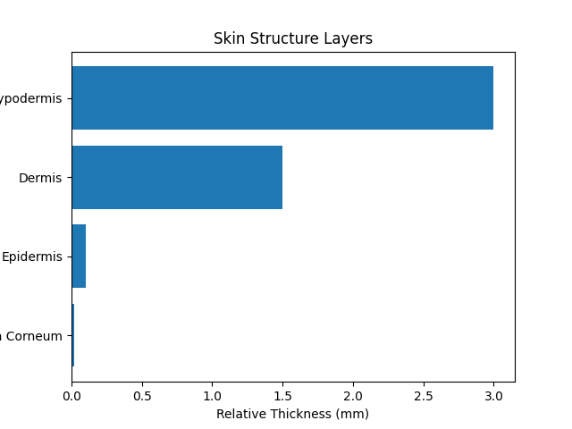
Figure 1: Structural organization of human skin layers
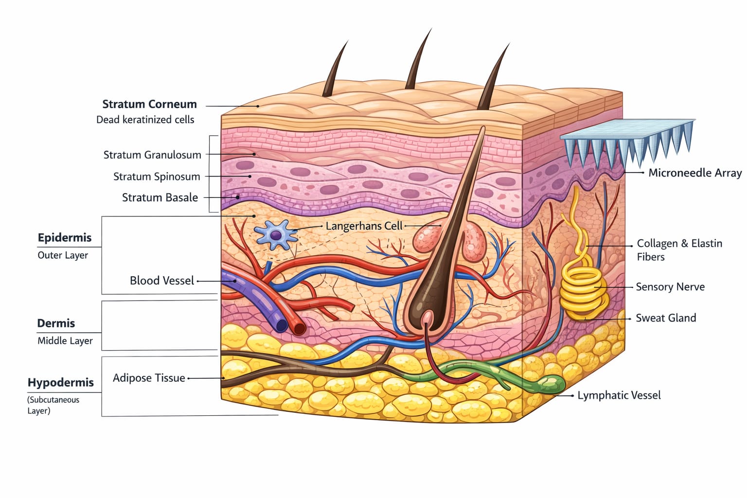
3. Types of Microneedles
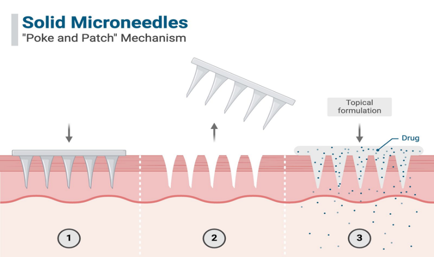
Concept- Solid microneedles are compact,non-hollow projections fabricated from materials similar as silicon, stainless steel, titanium, or polymers. They're primarily used in the “ poke- and- patch ” approach. First, the microneedle array is pressed into the skin to produce flash microchannels. After discarding, a topical medicine formulation( gel, cream, or patch) is applied over the treated point, allowing the medicine to diffuse through the created pores.
Mechanism
Insertion of Solid Microneedle Array
↓
Creation of TemporaryMicro-pores
↓
Removal of Microneedle Device
↓
use of medicine Formulation Over Treated Area
↓
medicine Diffuses Through Microchannels
↓
medicine Reaches feasible Epidermis/ Dermis
↓
Systemic or Original
remedial Action[1,3,8,9]
Crucial Features
Advantages
Limitations
Indications
2. Coated Microneedles
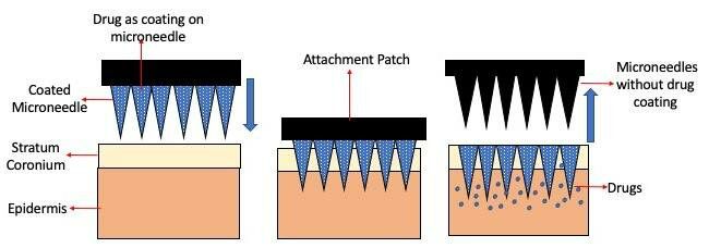
Concept- Coated microneedles contains of solid microneedles whose shells are concentrated with a thin film of medicine formulation. Upon insertion into the skin, the coating dissolves quickly, releasing the medicine directly into epidermal or dermal layers.
Mechanism
Insertion of medicine- covered Microneedle
↓
Contact with Interstitial Fluid
↓
Rapid Dissolution of Drug Coating
↓
Direct Deposit of medicine into Skin Layers
↓
medicine Diffusion into Capillary Network
↓
Pharmacological Response
Crucial Features
Advantages
Limitations
Indication
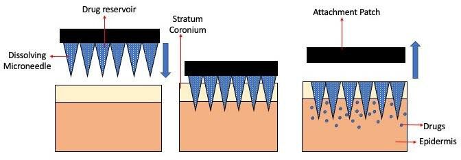
3. Dissolving (Biodegradable) Microneedles
Concept- Dissolving microneedles are fabricated entirely from water- solvable or biodegradable polymers( e.g., PVP, PVA, hyaluronic acid) that encases the medicine within the needle matrix. After insertion, the microneedles dissolve in interstitial fluid, releasing the medicament in a controlled manner.
Mechanism
Insertion of Polymer- Depended Microneedle
↓
immersion of Interstitial Fluid by Polymer Matrix
↓
Gradual Dissolution of Microneedle Structure
↓
Controlled Release of Encapsulated Drug
↓
medicine Diffusion in Dermal Tissue
↓
Sustained remedial Effect
Crucial Features
Advantages
Limitations
Indications
4. Hollow Microneedles
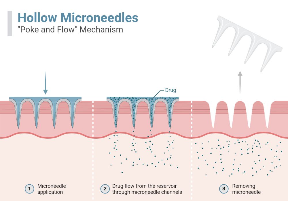
Concept- Hollow microneedles function also to atomic hypodermic needles. They contain a central drag that allows liquid medicine phrasings to flow directly into the dermal microcirculation, either through unresistant prolixity or controlled infusion.
Mechanism
Insertion of Hollow Microneedle into Skin
↓
Connection to Drug Reservoir or Syringe
↓
Controlled Infusion of Liquid Drug
↓
Direct Delivery into Dermal Microcirculation
↓
Rapid Systemic Distribution
↓
Immediate Pharmacological Effect
crucial Features
Advantages
Limitations
Applications
5. Hydrogel-Forming Microneedles
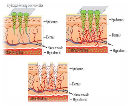
Concept- Hydrogel- forming microneedles are composed ofcross-linked polymer networks. Upon insertion into the skin, they absorb interstitial fluid and swell, forming nonstop channels between a medicine- containing patch force and the dermal microcirculation. The medicine itself is n't bedded in the needles but diffuses through the blown hydrogel matrix.[17]
Mechanism
Insertion ofCross-Linked Hydrogel Microneedle
↓
immersion of Interstitial Fluid
↓
lump of Polymer Network
↓
Formation of nonstop Diffusion Pathway
↓
Drug Movement from Attached Reservoir
↓
Sustained medicine Transport into Dermis
↓
extended remedial Activity
Crucial Features
Advantages
Limitations
Applications
4. Materials and Fabrication Techniques
Common fabrication materials include stainless steel, silicon, polylactic-co-glycolic acid (PLGA), polyvinylpyrrolidone (PVP), and carbohydrate matrices such as trehalose.[9,13,17,24,26] Micro-molding remains the most scalable fabrication method for polymeric microneedles. Photolithography and etching are used for silicon microneedles, while 3D printing enables customizable geometries.
5. Mechanism of Action
Apply of Microneedle Patch
↓
Microneedle Penetrates Stratum Corneum
↓
Formation of Controlled Microchannels
↓
Bypass of Skin Barrier ( Stratum Corneum)
↓
medicine Deposit in Epidermis/ Dermis
↓
Diffusion into Interstitial Fluid
↓
Absorbed into Original Tissue or Systemic blood
↓
Therapeutic Effect.[1,8,20,26]
6. Delivery of Biologics
Microneedle systems enhance bioavailability by bypassing gastrointestinal degradation and hepatic first-pass metabolism.[2,3,20,26]
Protein stabilization strategies[12,25] include incorporation of disaccharides and lyophilization-compatible polymers.
Sustained release[13,17] kinetics can be engineered through biodegradable matrices.
7. Delivery of Vaccines
Intradermal vaccination improves antigen presentation and immune activation.[7,18,19]
Microneedle patches can induce both humoral and cellular immune responses.[4,10,21]
Emerging research focuses on mRNA vaccines[16] and thermostable formulations.
8. Pharmacokinetics and Immunological Response
Drug absorption depends on microneedle geometry, insertion force, and dissolution kinetics.[1,8,9] Immune activation involves antigen processing, MHC presentation, and T-cell proliferation.[7,18,19]
9. Safety and Patient Compliance
Microneedle systems are considered a safe and patient-friendly alternative to conventional hypodermic injections because they are specifically engineered to penetrate only the outermost layer of the skin without reaching deeper pain receptors or major blood vessels. Due to their microscopic length, microneedles create controlled and superficial microchannels in the stratum corneum, which significantly reduces pain, bleeding, and tissue trauma. Most users report only mild pressure or a slight tingling sensation during application. The microchannels formed during insertion naturally close within a short period, which lowers the risk of infection when proper hygiene is maintained. Furthermore, dissolving and hydrogel-forming microneedles eliminate the generation of sharp medical waste, thereby reducing accidental needle-stick injuries and improving overall procedural safety. The materials used in fabrication—such as medical-grade metals, biodegradable polymers, and biocompatible hydrogels—are selected to ensure minimal toxicity, low irritation potential, and safe degradation within the skin.[8,11,21,27] From a patient compliance perspective, microneedles offer substantial advantages that improve treatment adherence. Their painless and minimally invasive nature reduces needle phobia, a common barrier to vaccination and chronic injectable therapies. The patch-based format allows simple and convenient self-administration without the need for trained healthcare professionals, decreasing hospital visits and healthcare costs. Because microneedle patches are discreet and easy to use, patients can maintain their daily activities during treatment, enhancing comfort and convenience. In vaccination and long-term therapies such as insulin or hormone delivery, the reduced discomfort and simplified application process lead to greater patient satisfaction and higher adherence rates. Overall, the combination of enhanced safety, reduced anxiety, ease of use, and improved comfort makes microneedle technology a highly promising strategy for increasing both therapeutic effectiveness and patient compliance in modern drug delivery systems.[10,11,21,27]
10. Challenges and Future Perspectives
Challenges include limited drug loading capacity, large-scale manufacturing, and long-term biologic stability.[9,25,26] Microneedle-assisted transdermal delivery of biologics and vaccines is expected to advance through the development of smart, stimuli-responsive systems capable of controlled and sustained release[13,15]. Future platforms may integrate biosensors for real-time monitoring of therapeutic response, enabling personalized dosing strategies.[16] Innovations in biodegradable and mechanically robust materials will expand the range of deliverable macromolecules while maintaining safety. Scalable manufacturing techniques such as microfabrication and 3D printing will improve affordability and global accessibility.[9,26] Additionally, combination delivery approaches incorporating adjuvants or immunomodulators may enhance immune responses, positioning microneedles as a transformative technology for painless, patient-centered therapeutics.
CONCLUSION
Microneedle-assisted transdermal systems represent a paradigm shift in painless biologic and vaccine delivery. Continued advancements in materials science, immunology, and pharmaceutical engineering will accelerate clinical translation.
REFERENCES





Sanskruti Abnave, Pooja Choudhari, Rushikesh Bhagat, Pranav Bhosle, Ankita Javalkar, Microneedle-Assisted Transdermal Delivery of Biologics and Vaccines: Emerging Technologies Enabling Painless and Patient-Centric Therapeutics, Int. J. of Pharm. Sci., 2026, Vol 4, Issue 2, 3033-3043. https://doi.org/10.5281/zenodo.18697953
 10.5281/zenodo.18697953