

We use cookies to ensure our website works properly and to personalise your experience. Cookies policy
1,3,4,5,6Research Scholar Department of Pharmacy, Siddhi's Institute of Pharmacy, Nandgaon, Maharashtra, India.
2Assistant Professor, Siddhi's Institute of Pharmacy, Nandgaon, Maharashtra, India.
Spectroscopic techniques play an important role in pharmaceutical analysis by providing information about the identity, structure, purity, and concentration of pharmaceutical substances. This review aims to discuss the principles, instrumentation, and applications of major spectroscopic techniques used in pharmaceutical analysis. These include ultraviolet-visible (UV-Visible) spectroscopy, infrared (IR) and Fourier transform infrared (FTIR) spectroscopy, Raman spectroscopy, fluorescence spectroscopy, nuclear magnetic resonance (NMR) spectroscopy, and mass spectrometry. The review also highlights the role of spectroscopic methods in drug identification, quantitative analysis, impurity detection, structural characterization, stability studies, and quality control. Each technique offers specific advantages depending on the nature of the sample and the analytical requirements. The use of modern spectroscopic instruments has improved the sensitivity, accuracy, and efficiency of pharmaceutical testing. Furthermore, the combination of spectroscopic techniques with other analytical methods can provide complementary information about drug substances and formulations. Overall, spectroscopic techniques are valuable tools in pharmaceutical research, drug development, formulation analysis, and quality assurance, supporting the production of pharmaceutical products that meet established quality standards.
Pharmaceutical analysis is an important branch of pharmaceutical science concerned with the analysis and quality control of substances used in pharmaceutical preparations. It involves various processes such as identification, quantitative determination, separation, purification, and structural characterization of chemical compounds. The major substances investigated include active pharmaceutical ingredients (APIs) and excipients such as binders, disintegrants, surfactants, suspending agents, viscosity enhancers, polymers, adhesives, and lubricants. Pharmaceutical analysis also helps in detecting impurities and contaminants, thereby contributing to the quality,purity, safety, and efficacy of pharmaceutical products [1].
Pharmaceutical products are assessed by a range of chemical, physical and instrumental analytical techniques. The quality of analytical procedures directly affects the quality and safety of pharmaceutical products and, therefore, precise and reproducible analytical results are important. Therefore appropriate analytical methods and quality control procedures are required during the development, manufacturing and testing of medicines. Among modern instrumental techniques, spectroscopic methods are widely used because they provide rapid, sensitive, and reliable information for the identification, characterization, and quantitative analysis of pharmaceutical substances [2].
It involves the use of different analytical techniques to obtain information about the identity, quality, purity, strength, stability, and other important properties of pharmaceutical substances. Analytical methods are essential throughout drug development, from the characterization of the active pharmaceutical ingredient (API) to the development, manufacture, and quality evaluation of the final dosage form.In pharmaceutical analysis, different analytical techniques are employed such as chromatographic, spectroscopic and other instrumental methods. These techniques are applied for the detection and quantification of active ingredients and related substances. Appropriate accuracy, precision and reliability are needed for quantitative analysis, because analytical results are important for manufacturing control, stability assessment and quality evaluation of pharmaceutical products.Pharmaceutical analysis plays an important role in ensuring that pharmaceutical products meet the required quality standards and are safe and suitable for their intended use [3,4].
CLASSIFICATION OF ANALYTICAL TECHNIQUES
Classical Analytical Techniques
Classical analytical methods are the traditional methods of qualitative and quantitative analysis of substances. They mostly deal with chemical reactions and measurement of mass or volume [5]
A .Volumetric analysis (Titrimetric)
:-The quantity of an analyte is determined by the volume of a standard solution that completely reacts with it. It is frequently used for assay and concentration determination [5]
B. Analysis :-
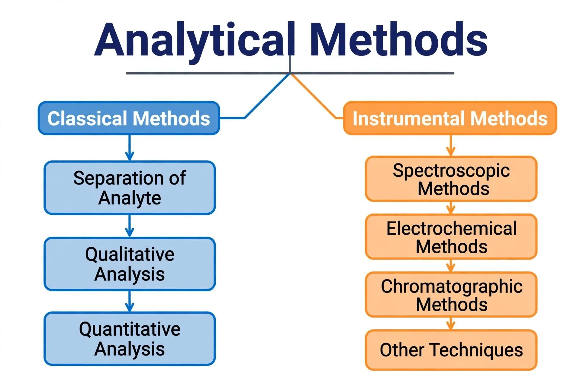
Gravimetric analysis is a traditional analytical technique where the analyte is isolated, precisely weighed, and transformed into a pure and stable molecule. The measured mass of the analyte is then used to compute its quantity [6].Analytical equipment is used in instrumental analytical techniques to quantify the physical or physicochemical characteristics of substances. Because they offer sensitive, precise, and quick analysis, they are frequently employed in pharmaceutical analysis [7].
1. Spectroscopic Techniques:
Spectroscopic techniques are analytical methods that rely on the interaction of electromagnetic radiation with matter. They are used in pharmaceutical analysis for identification, structural characterization and quantitative determination of drugs. These are UV-Visible, IR, fluorescence, Raman and NMR spectroscopy [8].
2. Chromatography Methods:
Chromatographic techniques are analytical methods used to separate the components of a mixture based on their different interactions with a stationary phase and a mobile phase. They are widely used in pharmaceutical analysis for drug identification, assay and impurity profiling. Examples are thin-layer chromatography (TLC),
high-performance liquid chromatography (HPLC), and gas chromatography (GC) [9].
3. Electroanalytical Techniques:
Electroanalytical methods Analytical methods based on the measurement of electrical properties (potential, current or conductivity) to determine concentration. They are used in pharmaceutical analysis for drug estimation and for pH measurement. Examples are potentiometry, conductometry and voltammetry [10].
4. Physical analytical techniques:
Techniques of Physical AnalysisPhysical analytical techniques are the determination of physical properties of substances for their identification and characterization. These properties are the melting point, refractive index, optical rotation and density. Useful in the determination of identity and purity of pharmaceutical substances [2].
INTRODUCTION OF SPECTROSCOPY
Spectroscopy is extensively used in pharmaceutical analysis for the identification of drug substances, determination of their concentration, study of molecular frameworks, and quality control of pharmaceutical products. It is also useful for the detection of impurities, the study of interactions between drugs and excipients or the evaluation of the stability of pharmaceutical formulations [11,12].
The selection of spectroscopic techniques depends on the nature of the sample and the analytical needs. UV-Visible spectroscopy is often used for the determination of drug concentrations, and infrared spectroscopy is useful for the identification of functional groups. Sensitive detection of substances is possible using fluorescence spectroscopy. Detailed information about the molecular framework is provided by nuclear magnetic resonance (NMR) spectroscopy. These techniques are useful for pharmaceutical research, formulation development and quality assurance [13].The choice of spectroscopic techniques depends on the chemical properties of the sample and the aim of the analysis. Different techniques are used to obtain specific information about the structure, composition and characteristics of pharmaceutical substances. These approaches are important for pharmaceutical research, drug characterization, analytical testing and quality control . Therefore, spectroscopy serves as an essential tool for ensuring the identity, purity, and quality of pharmaceutical products [11,13].
Classification of spectroscopy techniques
Spectroscopic techniques can be classified according to the interaction of electromagnetic radiation with matter and the type of information obtained. The choice of technique will depend on the chemical properties of the sample and the purpose of the analysis. These techniques are used in pharmaceutical analysis for drug identification, structural characterization, quantitative estimation and quality control [7].
The major spectroscopic techniques include
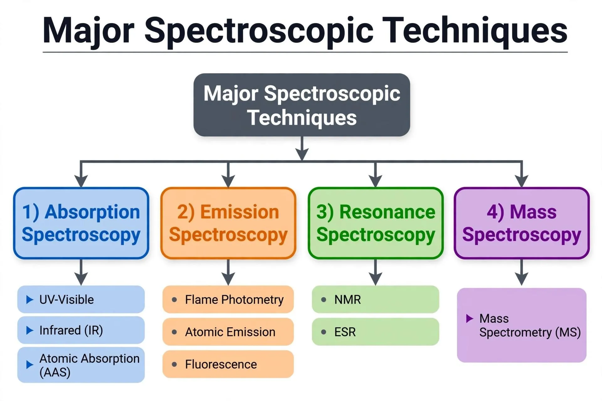
Figer 1:major spectroscopic technique
1. UV-VIS SPECTROSCOPY:
Ultraviolet–Visible (UV–Vis) spectroscopy is an analytical technique that employs the radiation in the ultraviolet and visible regions of the electromagnetic spectrum, usually from 200 to 800 nm (50,000–12,500 cm−1) . In some applications the technique is extended into the near infrared (NIR) region, to wavelengths of about 2000 nm, and is then referred to as UV–Vis–NIR spectroscopy. The UV region normally ranges from 200 to 400 nm, the visible region from 400 to 800 nm, and the NIR region starts above 800 nm. This technique involves measuring the absorption, scattering or reflection of a sample to UV, visible and NIR radiation. The visible region is related to the colors that the human eye is capable of detecting, and therefore, the color of a sample can offer a first indication of its interaction with light and it can help to determine if UV–Vis–NIR spectroscopy is appropriate for its analysis [14]. UV-Vis spectroscopy is based on the principle that molecules of the pollutant in the water are able to absorb UV-Vis light at a particular wavelength. Light of a particular wavelength is absorbed as electrons are moved from the ground state to an excited state and this decreases the amount of light transmitted. The absorption spectrum and the concentration of the substance are strongly correlated according to Lambert–Beer law, which is the principle of UV-Vis spectroscopy [15].
Lambert Beer’s Law:
The quantitative analysis of water quality parameters by UV-Vis spectroscopy is based on the Lambert-Beer law . The measurement principle of Lambert-Beer law is based on the monochromatic parallel light beam irradiating the surface of the medium under test [12].After passing through the medium with a certain thickness, the medium absorbs part of the light energy, weakening the intensity of the transmitted light passing through the medium. The absorbance of the absorption medium is directly proportional to its thickness
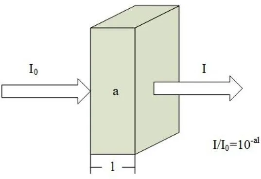
Figure 3: Schematic Diagram of the Lambert-Beer Law
Mathematical expression of the Lambert Beer law is as follows:
A = ε × c × l
Where:
A = Absorbance
ε = Molar absorptivity
c = Concentration of the solution l = Path length of the sample cell
Absorbance can also be expressed as:
A = log₁₀ (I₀/I) = −log₁₀ T A = log (1/T) = Kal. (1)
In Equation (1): A is the absorbance; T is the transmittance that the ratio of the intensity of the outgoing light (I) to the intensity of the incident light (I0); K is the molar absorption coefficient related to the nature of the absorbing substance and the wavelength λ of the incident light; a is the concentration of the absorbing substance in mol/L; and l is the thickness of the absorbing layer in cm [15].
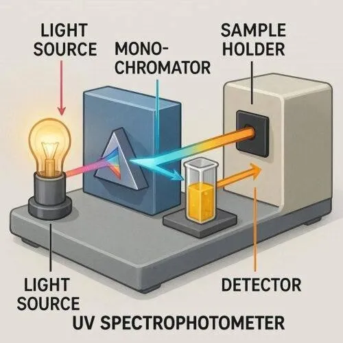
Instrumentation of uv - visible spectroscophy
UV–Visible spectroscopy comprises light source, wavelength selector (monochromator), sample holder and detector. These parts work together to measure the transmission or absorption of a sample’s UV and visible radiation. A spectrum is taken, and the substance is assayed from the measurements recorded. [16]
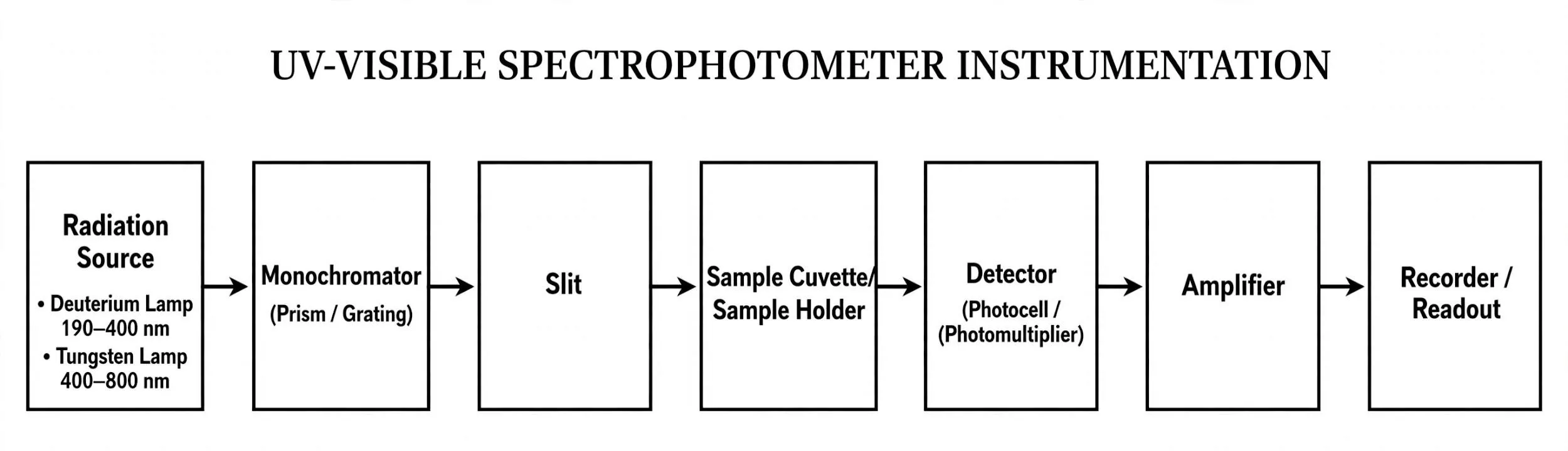
Flow diagram:
Figer 4:I Instrumentation of uv visible spectroscophy
Radiation sources :
1.DEUTERIUM LAMP :
In the ultraviolet region, a deuterium lamp is typically used as the radiation source. It is filled with deuterium gas . When an electrical discharge excites the gas , light is generated . It provides a continuous spectrum of UV radiation, typically between 160-400 nm, depending on the lamp and instrument design.
Working :When a high voltage is applied an electric discharge occurs in the deuterium gas. The excited species emit ultraviolet radiation. The radiation then
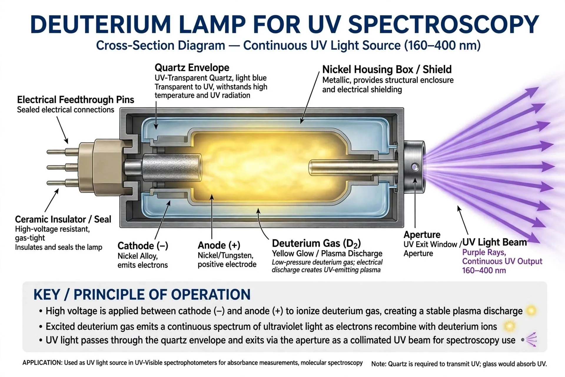
Figure 5: Deuterium Lamp
passes through the wavelength selector which selects the wavelength required for the analysis of the sample. [16,17]
2.TUNGSTEN-HALOGEN LAMP:
A common source of radiation in the visible region is a tungsten-halogen lamp. This can also be used into the near infrared region. It has a tungsten filament in a bulb filled with a halogen gas. Current is passed through the filament, which gets hot and emits radiation.
Working: Electrical energy heats the tungsten filament to a high temperature. The hot filament radiates a broad band of radiation. The instrument selects the required wavelength using a wavelength selector [17, 18].
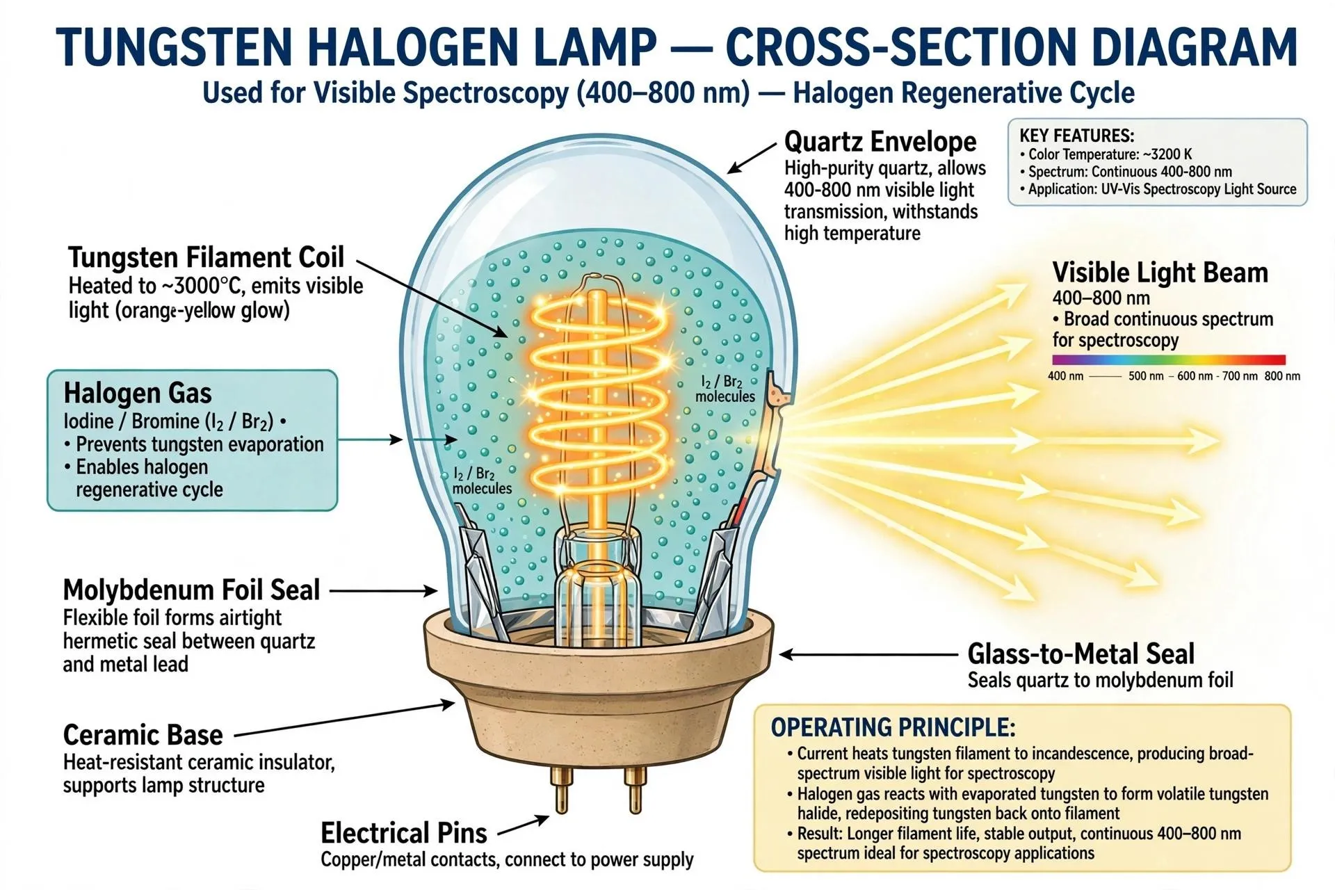
Figer 6: Tungsten Halogen Lamp
3. LAMP XENON :
Another radiation source used in some of the UV-Visible spectrophotometers is the xenon arc lamp. It emits strongly over a wide range of wavelengths, including the ultraviolet and visible regions. The exact wavelength range is determined by the design of the lamp and the optical materials used in the instrument.
working : An electrical current through xenon gas produces intense light. The emitted radiation passes through the optical system, where the desired wavelength for analysis is selected [18,20].
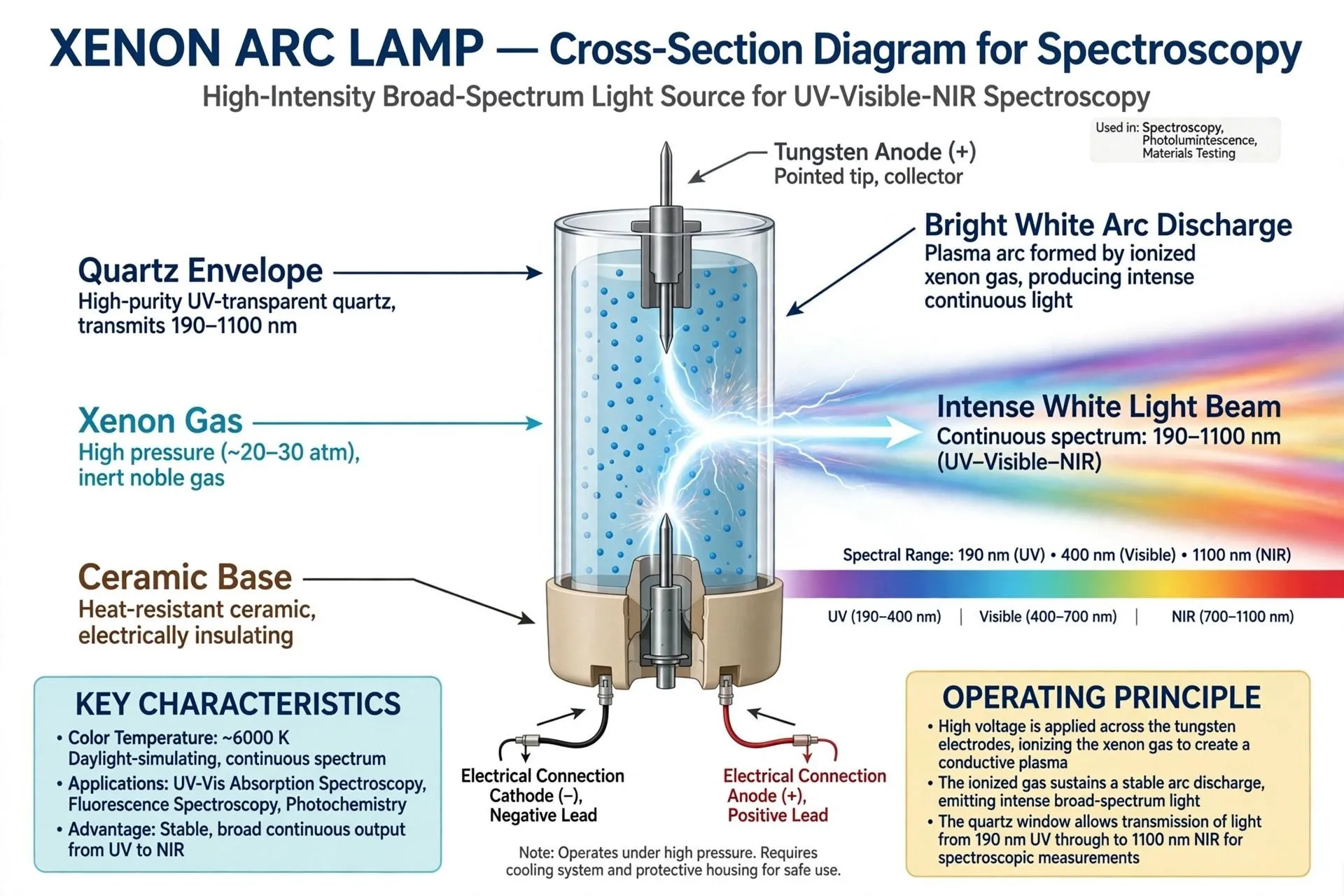
Figer 7:Xenon Arc Lamp
Monochromator are used in uv visible spectroscopy
1. Prism Monochromator:
A prism monochromator is an optical device that separates white light into its component wavelengths based on the concept of "refraction and dispersion". It has a small entrance slit, a dispersing prism, collimating lens or mirror and an exit slit.The collimated beam of polychromatic light then passes through the monochromator and into the prism . Different wavelengths bend differently because of their different indices of refraction. Blue, shorter wavelengths bend more than red, longer wavelengths. The prism can be rotated or the exit slit changed to select one wavelength. Prism monochromators are widely used in spectrophotometers, fluorimeters and in optical analysis equipment, due to their high throughput and stable wavelength output [21].
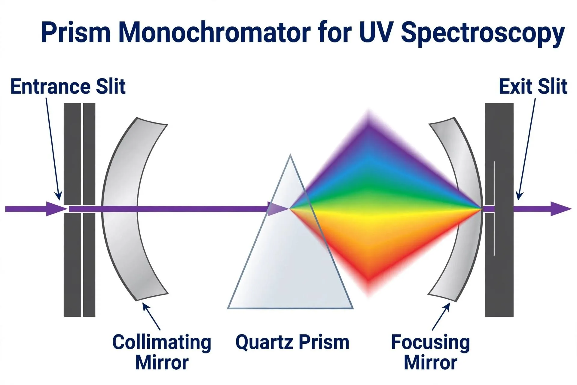
Figer 8:Prism Monocromator for UV spectroscophy
2. Grating Monochromator:
A grating monochromator is an optical instrument that separates out specific wavelengths of light using a "diffraction grating" as the dispersive element. It comprises an input slit, collimating optics, a reflecting or transmissive diffraction grating and an output slit. Because the refractive indices of polychromatic light vary at different wavelengths, the closely spaced parallel line curve on the grating's beam of polychromatic light bends differently at different wavelengths. Collimated polychromatic light can diffract at wavelength-dependent angles. Blue, shorter wavelengths bend more than red, longer wavelengths. The prism can be rotated or the exit slit changed to select one wavelength. Prism monochromators have a high throughput and constant wavelength output, and are widely used in spectrophotometers, fluorimeters and optical analysis equipment [22].
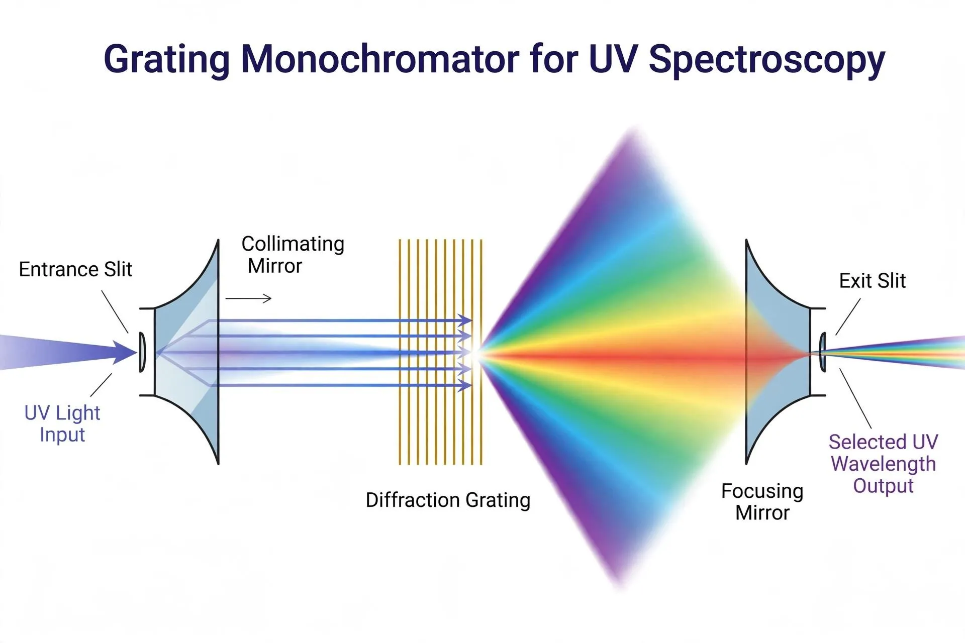
Figer 9:Grating Monochromator For UV Spectroscophy
C .sample holder in uv-visible spectrophotometer
1. Sample holder (cuvette holder):
The sample holder is an important part of a UV-Visible spectrophotometer which holds the sample cuvette in the correct position in the light path.In order to accurately measure absorbance or transmittance, it guarantees that light passes through the sample. Depending on the wavelength range and sample requirements, sample holders are typically made to fit quartz, glass, or plastic cuvettes. While glass or plastic cuvettes can be used for appropriate visible-region measurements, quartz cuvettes are recommended for UV measurements due to their efficient transmission of ultraviolet radiation. To reduce measurement errors, the sample holder should maintain the cuvette's stability and proper alignment. [23, 24].
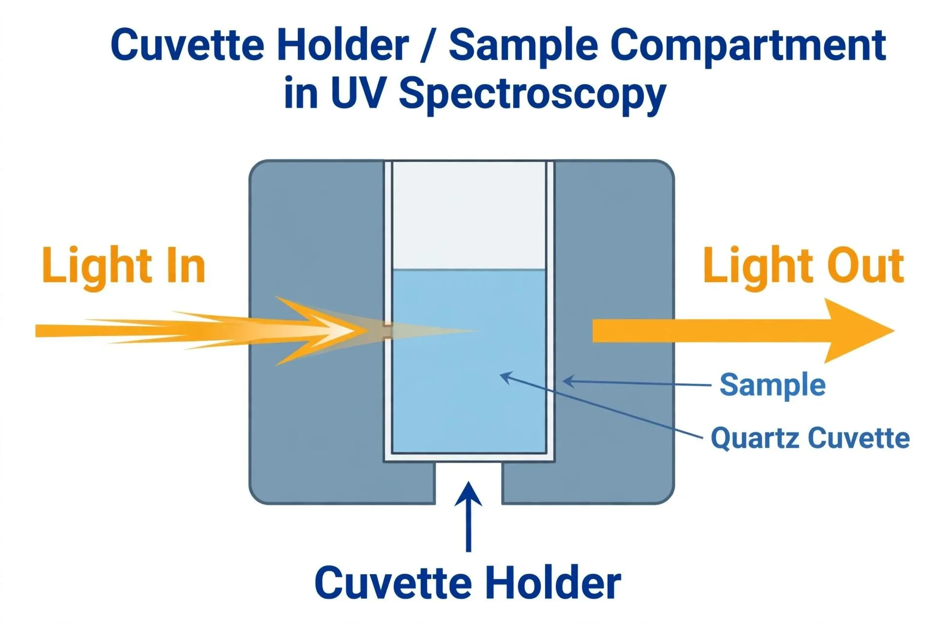
Figer 9: Cuvette Holder in UV Spectroscophy
D. Detector use in uv visible spectroscopy
Defination:
An essential part of a UV-visible spectrophotometer is a detector, which picks up light passing through the sample and transforms it into an electrical signal. After processing, the signal is shown as either transmittance or absorbance. For quantitative analysis of pharmaceutical substances, the detector aids in determining the amount of light absorbed by a sample.
1. PHOTOTUBE SENSOR:
A vacuum phototube includes a curved cathode. constructed from (or covered with) a light-emitting substance that releases electrons when exposed to photons. It seems like your text got cut off. Please provide the complete text you want paraphrased, and I'll be happy to help!electrons from the cathode are negatively charged by the photons traverse the vacuum to the positive anode and into the amplifier. The count of electrons emitted is directly proportional to the intensity of the incoming radiation. The responsiveness of a photoemissive cathode to different wavelengths is determined by the makeup of the source emitting facade. A surface that is sensitive to red light is covered with a series of metals usually Na/K/Cs/Sb, while
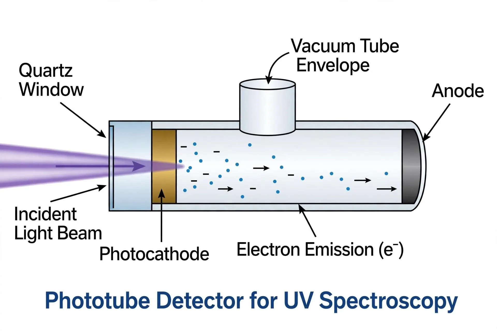
NIRSensitivity is derived from coatings produced with Ga/In/As. Typically, phototubes exhibit a minimal dark current occurring [26].
Figer 10 :Phototube detector for UV Spectroscophy
2. Photovoltaic Cell Detector:
A solar cell sensor, commonly known as asolar cell sensor or photovoltaic diode, is alight-detecting device that generates an electriccurrent generated from incoming light without requiringfor an extra energy source. The "solar energy""Effect" refers to the operation, in which photons strike a "p–njunction" generates electron-hole pairs, and the inherent electric field in the depletion region separates these charges.carriers, leading to a voltage and current that arerelative to the brightness of the light. While photovoltaic cell sensors exhibit "excellent stability, strong "linearity and minimal noise," they react at a slower pace.than photodiodes operating in photoconductive mode. Solar radiation detectors, mobile instruments, and additional applications needing low-level, self-sustaining light sensing often utilizes them [27].
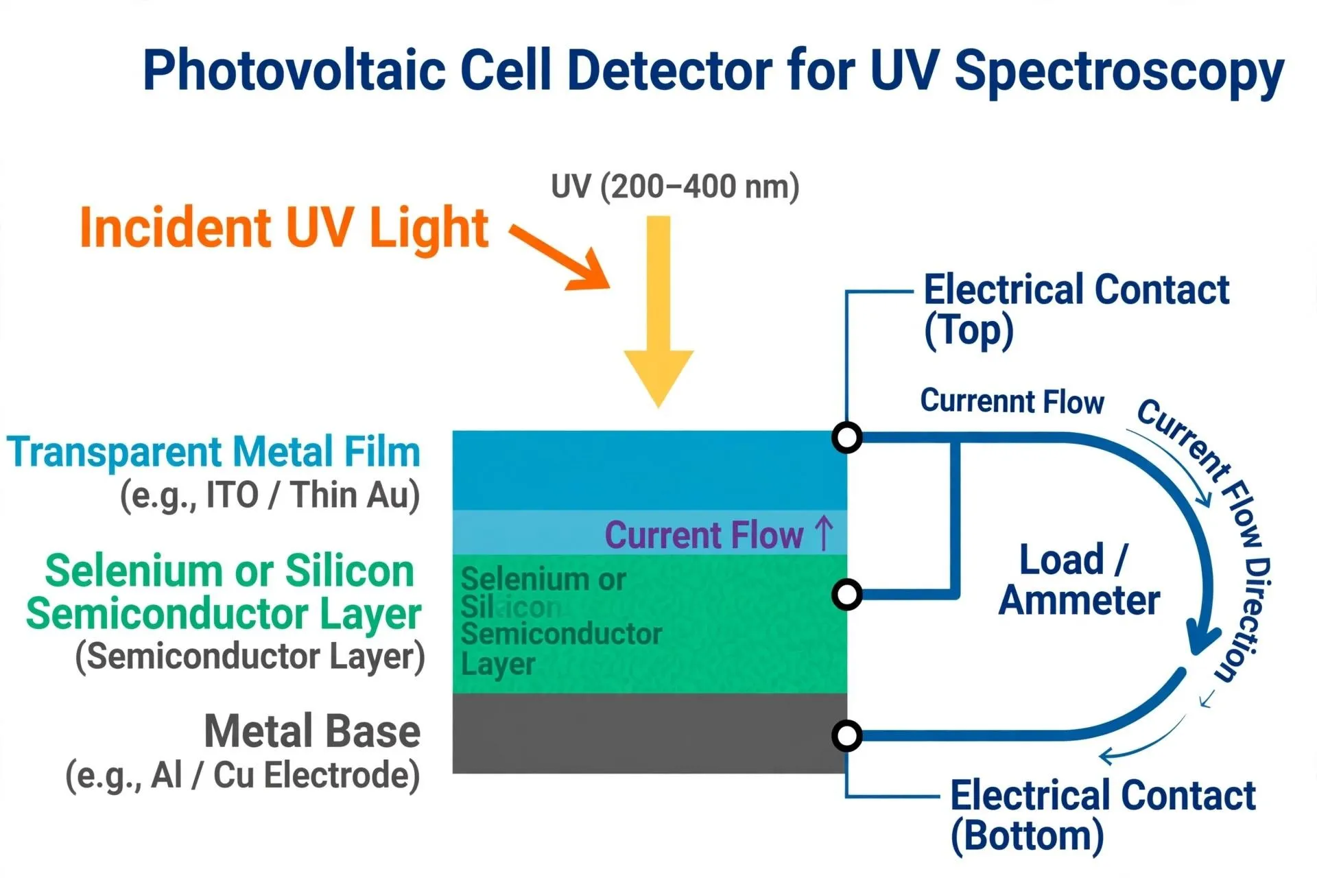
Figer 11: Photovoltaic Cell Detector For UV Spectroscophy
3. Silicon photodetector:
Photovoltaic or photoconductive phenomenon” is the foundation for the functioning of a silicon photodiode, alight sensor based on semiconductors. It consists of a silicon “p–n or p–i–n junction” which, upon exposure to light photons with sufficient energy strike the junction region, generate electron–hole pairs. The electric field within the depletion layer divides these charge carriers, resulting in a current that is proportional to the strength of the incoming light. For a wide variety of wavelengths, especially in the visible and near-infrared ranges, silicon photodiodes offer “excellent stability, swift reaction, minimal noise, and high linearity. Due to their compact dimensions, low cost, and reliability, they are frequently employed in spectrophotometric devices, light communication networks, medical instruments, barcode readers, and various light-detecting applications [28].
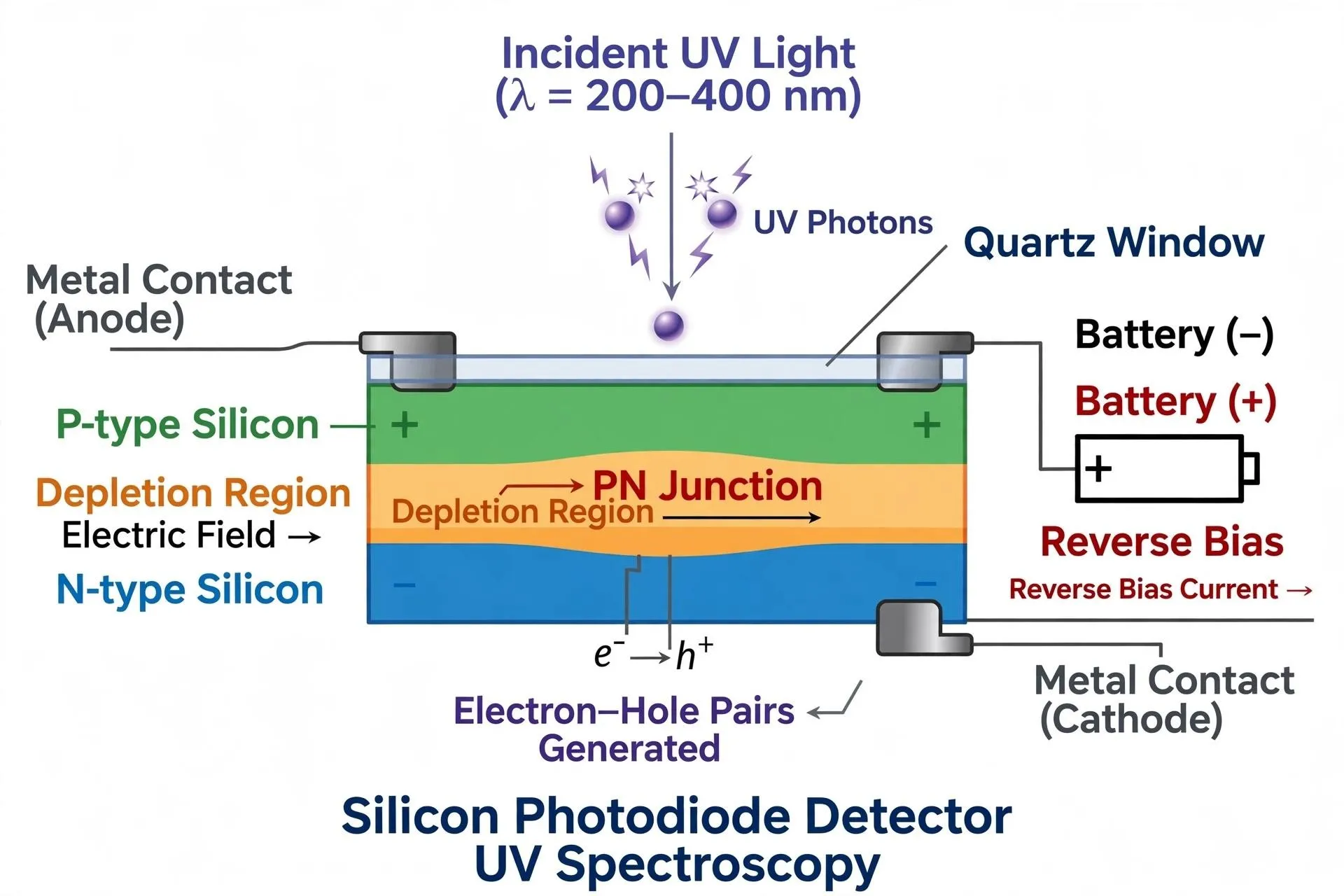
Figer 12 :Silicon photodetector:
4. Photomultiplier tube (PMT) application in UV visible spectroscopy
A photomultiplier tube (PMT) is an extremely sensitive light-sensing device that depends on secondary emission of electrons and the outside photoelectric impact. When photons of light hit the photocathode, primary electrons are emitted, resulting in a strong negative voltage is utilized to propel them toward numerous dynodes. Every incoming electron emits numerous secondary electrons since every dynode is kept at an increasingly higher voltage. This multiplication leads to a significantly high overall increase, that advances through various stages. The PMT is capable of perceive extremely dim light conditions with high sensitivity and velocity since the resulting multiplied electrons are collected at the anode to generate a powerful electrical signal [29].
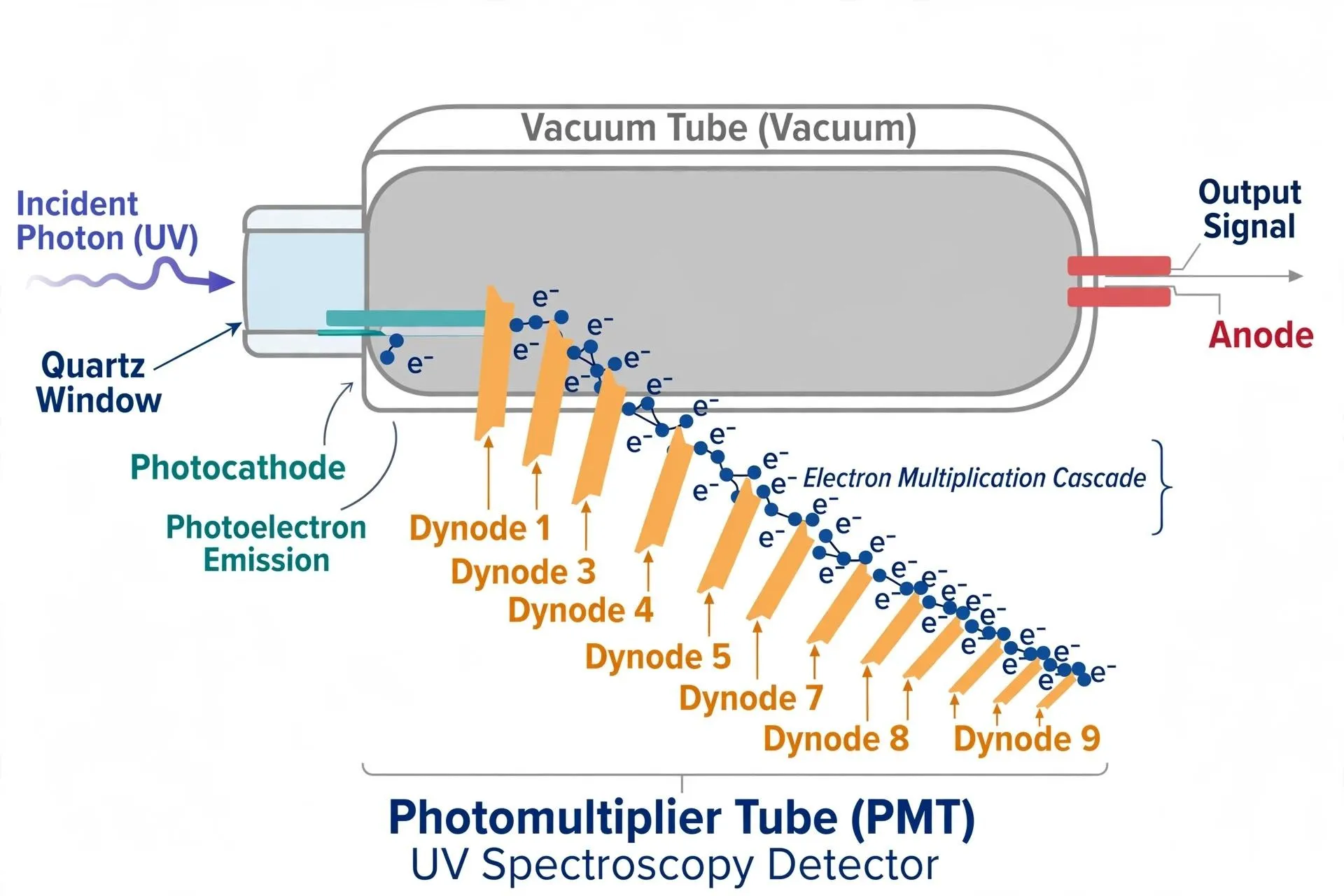
Figer 13:photomultiplier tube
2. INFRARED SPECTROMETRY:
Infrared spectroscopy is undoubtedly among the most significant analytical techniques. accessible to contemporary researchers. A significant benefit of infrared spectroscopy allows for the examination of nearly any sample in almost any condition.Liquids, solutions, pastes, powders, films, fibers, gases, and surfaces can all be analyzed using a careful selection of sampling method. Due to the enhanced instrumentation, a range of newly developed sensitive methods have now has been created to analyze previously unmanageable samples[30]. Over the last few years, there has been an increasing interest in the use of IR techniques in catalysis inquiries. One of the reasons, along with others, is their extensive prevalence (currently, IR and/or FT-IR Spectrometers are a common component of standard equipment. every scientific lab) and the comparatively low expenses (compared to other contemporary physico-chemical methods for surface characterization of the substrate device [31].
2.1 Types IR Spectroscopy
Near-infrared spectroscopy functions within a wavelength range of roughly 0.78–2.5 µm. It primarily comprises overtones and combination bands of molecular
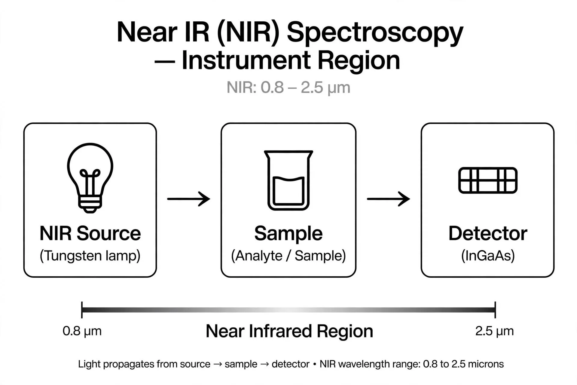
oscillations. In pharmaceutical analysis, NIR serves to identify raw materials, ascertain moisture levels, and facilitate quick quality control [32].
Figer 14 :IR Spectroscopy
2.2 Mid-Infrared (MIR) Spectroscopy:
The mid-infrared spectral range spans roughly 2.5–15 µm (4000–667 cm⁻¹). It is extensively utilized to detect functional groups and describe chemical compounds via their distinct absorption bands. In pharmaceutical analysis, it aids in drug identification, solid-state characterization, and studies on drug-excipient compatibility. Sure[3].
2.3 Far-Infrared (FIR) Spectroscopy
Far-infrared spectroscopy typically spans about 15–200 µm (667–50 cm⁻¹). This area offers insights into low-frequency molecular vibrations, lattice oscillations, and specific metal–ligand interactions. It is beneficial for exploring molecular compositions and characteristics of solid-state materials.[34]
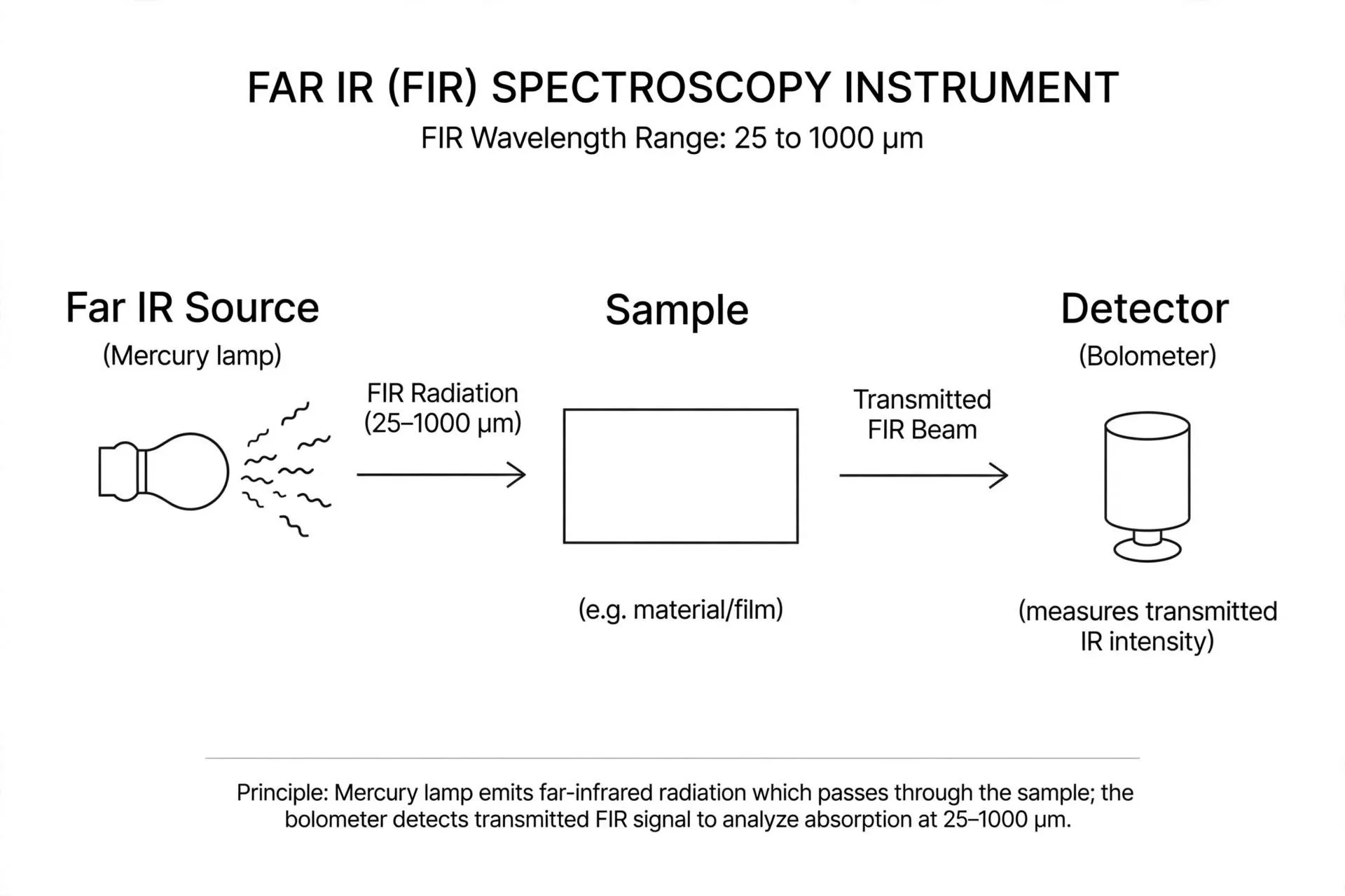
Figer 15 : Instrumentation of IR spectroscopy. Applications of IR Spectroscopy in Pharmaceutical Analysis:
Infrared (IR) spectroscopy is extensively employed in pharmaceutical analysis for the identification, characterization, and quality assessment of pharmaceutical compounds. It offers details on functional groups and molecular structure through the measurement of infrared radiation absorption. FTIR spectroscopy serves as a valuable tool for both qualitative and quantitative evaluations of pharmaceutical products. [33,35]
1. Drug Identification: IR spectroscopy identifies drugs by comparing their characteristic absorption spectra with reference spectra. [35]
2. Functional Group Identification: It helps identify functional groups such as O–H, N–H, C=O, and C–O in pharmaceutical compounds. [33]
3. Purity Testing: It can help assess drug purity and detect certain impurities when suitable analytical methods are used. [35]
4. Drug–Excipient Compatibility: IR spectroscopy is used to investigate possible interactions between active pharmaceutical ingredients and excipients. [36]
5. Quantitative Analysis: FTIR methods can be developed to determine the concentration of pharmaceutical compounds in suitable formulations. [35]
3. Nuclear Magnetic Resonance (NMR) Spectroscopy
Nuclear Magnetic Resonance (NMR) spectroscopy is a crucial analytical method employed to ascertain the molecular architecture and chemical makeup of pharmaceutical substances. It relies on the interaction of specific atomic nuclei with radiofrequency radiation under a strong magnetic field. The nuclei that are most frequently investigated are hydrogen (1H) and carbon-13. NMR spectroscopy offers insights into the chemical surroundings, connectivity, and atom arrangement in a molecule, proving valuable for identifying compounds and elucidating structures [37,38].NMR spectroscopy is applied in pharmaceutical analysis to identify drug compounds, characterize impurities and degradation products, assess drug purity, and conduct quantitative analyses. Solution-state and solid-state NMR methods are valuable for examining pharmaceutical substances, such as their molecular configuration, polymorphic varieties, and interactions between drugs and excipients. NMR spectroscopy is crucial in pharmaceutical research, drug development, and quality control because it offers detailed structural information [38,39].
PRINCIPLE-
Nuclear Magnetic Resonance (NMR) spectroscopy relies on the absorption of radiofrequency energy by specific atomic nuclei situated in a powerful external magnetic field. Nuclei like hydrogen-1 and carbon-13 have nuclear spin and act as small magnets. In a magnetic field, these nuclei reside in various energy states. When radiofrequency energy at the correct frequency is applied, the nuclei take in energy and transition between these states. The signals obtained are documented and examined to gather data regarding the atomic molecular structure and chemical surroundings [40,41].
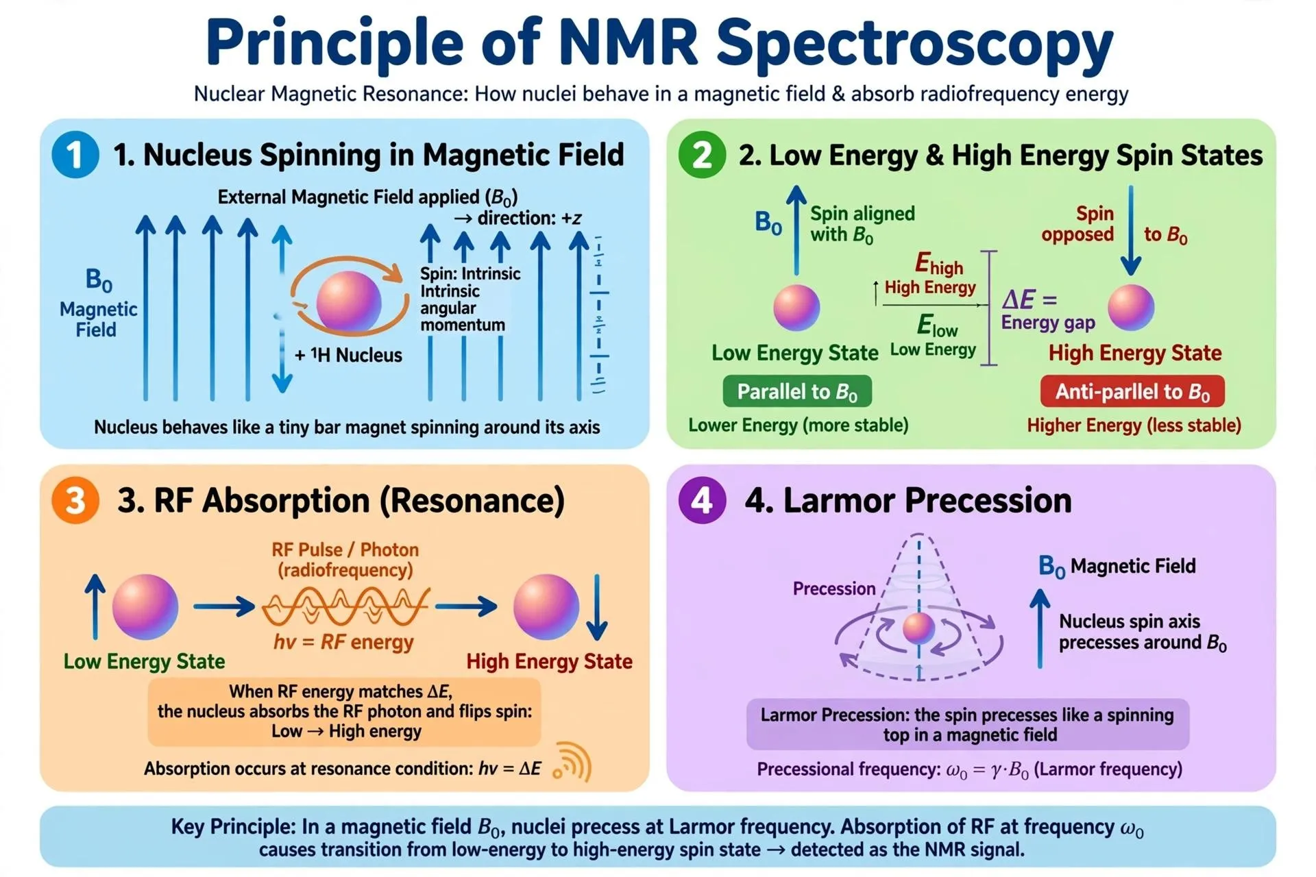
Figer 16:NMR SPECTROSCOPY TYPES OF NMR SPECTROSCOPY
1. Proton NMR (Nuclear Magnetic Resonance):
Proton NMR examines hydrogen atoms within a molecule. It offers insights into the quantity, chemical surroundings, and configuration of hydrogen atoms. It is commonly utilized for identifying drugs and clarifying structures [40].
2. Carbon-13 nuclear magnetic resonance (NMR):
This method examines carbon-13 nuclei and aids in identifying the carbon framework of a molecule [40].
3. Solution-State NMR:
In this technique, the sample is dissolved in a suitable solvent. It is commonly used to investigate molecular structures, drug purity, and impurities [38].
4. Solid-State NMR:
This technique analyzes solid samples without requiring them to dissolve. It is useful for studying pharmaceutical polymorphs, crystal structures, and drug–excipient interactions [39].
Applications of NMR Spectroscopy in Pharmaceutical Analysis
Nuclear Magnetic Resonance (NMR) spectroscopy is a vital analytical method employed for identifying, structurally characterizing, assessing purity, and quantitatively analyzing pharmaceutical compounds. It offers comprehensive insights into molecular structure and assists in assessing the quality of drugs and pharmaceutical products. [42,43]
1. Drug Identification: NMR spectroscopy identifies pharmaceutical substances by analyzing their characteristic spectra and comparing them with reference data. [42]
2. Structural Elucidation: It determines the arrangement and chemical environment of atoms in a molecule, supporting the characterization of drug compounds. [43]
3. Purity Determination: Quantitative NMR (qNMR) is used to assess the purity and content of pharmaceutical substances. [44]
4. Impurity Profiling: NMR helps identify and quantify impurities formed during drug synthesis, manufacturing, or degradation. [43]
5. Quantitative Analysis: qNMR determines the amount of active pharmaceutical ingredients (APIs) in suitable samples and formulations. [42,44]
4. MASS SPECTROMETRY:
Mass spectrometry (MS) is an analytical method employed to ascertain the molecular weight, molecular makeup, and structural properties of chemical substances. It operates by transforming molecules into charged entities known as ions and segregating these ions based on their mass-to-charge ratio. The produced mass spectrum delivers insights into the molecular weight and fragmentation pattern of a substance [45,46].Mass spectrometry is extensively utilized in pharmaceutical analysis for identifying drugs, profiling impurities, identifying metabolites, and quantitatively determining active pharmaceutical ingredients. When used alongside chromatographic methods like liquid chromatography (LC-MS) or gas chromatography (GC-MS), it offers excellent sensitivity and selectivity for analyzing intricate pharmaceutical samples [45,47].
TYPE OF MASS SPECTROSCOPY
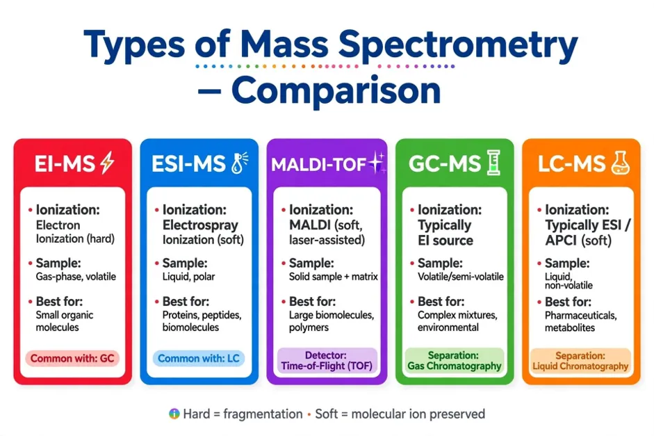
Figer 17:Type of Mass spectroscopy
1. Electron Ionization Mass Spectrometry (EI-MS)
Electron ionization is a rigorous ionization method where high-energy electrons engage with gaseous molecules to generate positively charged ions. These ions can split into smaller pieces, creating a distinct mass spectrum. The pattern of fragmentation aids in recognizing compounds and offers insights into their molecular composition. EI-MS is often combined with gas chromatography (GC-MS) for analyzing volatile drugs, organic substances, and pharmaceutical contaminants [46].
2. Electron Ionization Mass Spectrometry (EI-MS)
Electron ionization is a rigorous ionization method where high-energy electrons engage with gaseous molecules to generate positively charged ions. These ions can split into smaller pieces, creating a distinct mass spectrum. The pattern of fragmentation aids in recognizing compounds and offers insights into their molecular composition. EI-MS is often combined with gas chromatography (GC-MS) for analyzing volatile drugs, organic substances, and pharmaceutical contaminants [46].
3. Electron Ionization Mass Spectrometry (EI-MS)
Electron ionization is a rigorous ionization method where high-energy electrons engage with gaseous molecules to generate positively charged ions. These ions can split into smaller pieces, creating a distinct mass spectrum. The pattern of fragmentation aids in recognizing compounds and offers insights into their molecular composition. EI-MS is often combined with gas chromatography (GC-MS) for analyzing volatile drugs, organic substances, and pharmaceutical contaminants [46].
4. Electron Ionization Mass Spectrometry (EI-MS)
Electron ionization is a rigorous ionization method where high-energy electrons engage with gaseous molecules to generate positively charged ions. These ions can split into smaller pieces, creating a distinct mass spectrum. The pattern of fragmentation aids in recognizing compounds and offers insights into their molecular composition. EI-MS is often combined with gas chromatography (GC-MS) for analyzing volatile drugs, organic substances, and pharmaceutical contaminants [46].
5. Electrospray Ionization Mass Spectrometry (ESI-MS):
Electrospray ionization is a gentle ionization method employed to transform molecules in a liquid solution into ions in the gas phase. A strong electric voltage creates charged droplets, and the evaporation of the solvent frees the ions for mass analysis. ESI-MS is especially ideal for polar, large, and thermally sensitive compounds, such as peptides, proteins, and drug molecules. It is commonly employed with liquid chromatography (LC-MS) for analyzing drugs, identifying metabolites, and determining quantities [48].
6. Matrix-Assisted Laser Desorption/Ionization Mass Spectrometry (MALDI-MS):
MALDI-MS is a gentle ionization method where the sample is combined with an appropriate matrix that captures laser energy. The laser helps in moving sample
molecules into the gas phase and creating ions. This method usually results in minimal fragmentation, which makes it effective for studying large molecules like peptides, proteins, and polymers. In pharmaceutical research, MALDI-MS aids in the characterization of biomolecules and the evaluation of intricate drug-related compounds [49].
7. Tandem Mass Spectrometry (MS/MS):
Tandem mass spectrometry consists of multiple stages of mass analysis to gather in-depth information regarding a compound. Initially, a precursor ion is chosen and broken down into product ions, which are subsequently examined based on their mass-to-charge ratios. The resulting fragmentation pattern aids in identifying compounds and differentiating substances that have similar molecular weights. LC-MS/MS is extensively employed in pharmaceutical investigations for quantifying drugs, profiling impurities, identifying metabolites, and analyzing drugs in biological samples [47].
5. Raman spectroscopy:
Raman spectroscopy is a technique in vibrational spectroscopy employed to recognize and describe chemical substances. It relies on the interplay between monochromatic light, typically emitted by a laser, and molecules present in a sample. A tiny fraction of the dispersed light experiences a frequency shift, offering insights into molecular vibrations and chemical connections. Raman spectroscopy is applied in pharmaceutical analysis for identifying drugs, characterizing solid forms, and conducting quality control [50,51].
PRINCIPLE-
Raman spectroscopy is based on the principle of inelastic light scattering, referred to as the Raman effect. When monochromatic light strikes a sample, nearly all the light is scattered without any alteration in frequency; this phenomenon is known as Rayleigh scattering. A tiny fraction of the dispersed light possesses a different frequency due to energy transfer between the incoming light and the molecular vibrations. This phenomenon is referred to as Raman scattering.
The variance between the frequencies of incident light and scattered light is termed the Raman shift. It offers details regarding the vibrational energy states and chemical connections of molecules. Due to the unique Raman shifts of various molecules, their spectra can serve for identification and structural analysis [50,51].
TYPES OF RAMAN SPECTROSCOPY
1. Standard Raman Spectroscopy:
Standard Raman spectroscopy is a method that relies on the inelastic scattering of monochromatic light, typically emitted from a laser. When light interacts with a sample, the majority is scattered without any change in energy, while a small fraction experiences a change in energy. This energy variation reveals details regarding the vibrational modes of molecules. In pharmaceutical analysis, conventional Raman spectroscopy is utilized for identifying drugs, examining molecular structure, and assessing the quality of pharmaceutical compounds [50].
2. Resonance Raman Spectroscopy:
Resonance Raman spectroscopy is an adapted Raman method where the wavelength of the excitation laser is chosen near an electronic absorption band of the sample. This enhances the Raman signal of certain molecular groups and aids in identifying specific substances at low concentrations. It is beneficial for analyzing molecules with chromophores, biological materials, and specific pharmaceutical agents [52].
3. Raman Spectroscopy Enhanced by Surface (SERS):
Surface-enhanced Raman spectroscopy enhances the Raman signal when molecules are located on or close to specifically designed metal nanostructures, typically composed of gold or silver. These surfaces amplify the electromagnetic field surrounding the molecules, enabling faint Raman signals to significantly increase in strength. SERS is effective for identifying minimal amounts of pharmaceutical components, recognizing trace contaminants, and examining medications in intricate samples [53].
4. Tip-Enhanced Raman Spectroscopy (TERS)
Tip-enhanced Raman spectroscopy merges Raman spectroscopy with a scanning probe microscope equipped with a sharp, metal-coated tip. The tip amplifies the local electromagnetic field, enabling the collection of Raman signals from an extremely small region. This method offers intricate chemical details at the nanoscale. It holds possible uses in examining pharmaceutical particles, surface characteristics, and the arrangement of chemical elements in intricate materials [54].
CONCLUSION. :
Spectroscopic techniques are essential analytical tools in pharmaceutical analysis because they provide valuable information about the identity, structure, purity, and concentration of pharmaceutical substances. Techniques such as UV-Visible, IR/FTIR, Raman, fluorescence, nuclear magnetic resonance (NMR), and mass spectrometry have wide applications in drug identification, quantitative analysis, impurity detection, structural characterization, and quality control. Each technique
has its own advantages and limitations, and the selection of a suitable method depends on the nature of the sample and the purpose of the analysis. Advances in spectroscopic instrumentation have improved the sensitivity, accuracy, speed, and reliability of pharmaceutical testing. Furthermore, combining different spectroscopic techniques with other analytical methods can provide more comprehensive information about drug substances and formulations. Therefore, spectroscopic techniques play a significant role in pharmaceutical research, drug development, stability studies, and quality assurance, helping to ensure the quality, safety, and efficacy of pharmaceutical products.
DISCUSSION:
Spectroscopic techniques play an important role in pharmaceutical analysis because they provide valuable information about the identity, concentration, molecular structure, purity, and stability of pharmaceutical substances. This review discusses the principles, instrumentation, and applications of major spectroscopic techniques, including UV–Visible, infrared (IR/FTIR), Raman, fluorescence, nuclear magnetic resonance (NMR), and mass spectrometry. Each technique provides different types of analytical information and is selected according to the properties of the sample and the objective of the analysis.
UV–Visible spectroscopy is widely used for the quantitative estimation of drugs that absorb ultraviolet or visible radiation. Its relatively simple instrumentation, rapid analysis, and suitability for routine testing make it useful in pharmaceutical quality control. The Beer–Lambert law establishes the relationship between absorbance and concentration under appropriate experimental conditions. Infrared and FTIR spectroscopy are useful for identifying functional groups, characterizing drug substances, and investigating possible interactions between active pharmaceutical ingredients and excipients. Raman spectroscopy provides complementary molecular information and can be particularly useful for analysing certain pharmaceutical materials with minimal sample preparation.
Fluorescence spectroscopy offers high sensitivity for substances that emit fluorescence and is useful for detecting drugs at low concentrations. NMR spectroscopy provides detailed information about the chemical environment of atomic nuclei and helps determine molecular structures and assess the composition of pharmaceutical compounds. Mass spectrometry measures the mass-to-charge ratio of ions and is valuable for molecular-mass determination, impurity profiling, and structural characterization, particularly when combined with separation techniques such as liquid chromatography.
Although these techniques offer several advantages, their performance depends on factors such as sample composition, instrument calibration, analytical conditions,
and potential interference from excipients or other substances. UV–Visible methods may have limited selectivity when different compounds absorb at similar wavelengths, while fluorescence analysis is applicable only to naturally fluorescent substances or compounds that can be suitably derivatized. IR and Raman spectra may contain overlapping signals, and NMR and mass spectrometry generally require more specialized instrumentation and interpretation.
The combined application of spectroscopic techniques can provide complementary information and improve the reliability of pharmaceutical analysis. Appropriate method validation, instrument calibration, reference standards, and quality-control procedures are necessary to obtain accurate and reproducible results. Overall, spectroscopic techniques are important tools in pharmaceutical research, drug development, formulation evaluation, stability studies, and quality assurance. Their appropriate selection and combined use support the identification, characterization, and quality assessment of pharmaceutical products.
REFERENCES






Renuka Rothe, Girish Kokate, Vaishali Javare, Pragati Tekade, Namrata Isame, Supriya Tekade, A Comprehensive Review of Spectroscopic Techniques in Pharmaceutical Analysis, Int. J. of Pharm. Sci., 2026, Vol 4, Issue 10, 3715-3733. https://doi.org/10.5281/zenodo.23640477
 10.5281/zenodo.23640477