

We use cookies to ensure our website works properly and to personalise your experience. Cookies policy
Modern Institute of Pharmaceutical Sciences, Indore.
Atherosclerosis is a chronic cardiovascular disorder associated with dyslipidemia, oxidative stress, and inflammation. The present study aimed to evaluate the potential anti-atherogenic activity of the hydroalcoholic leaf extract of Mussaenda philippica in an experimental model. The extract was prepared from 100 g of powdered leaves, yielding 13 g of dried extract (13% w/w). Preliminary phytochemical screening revealed flavonoids, phenolic compounds, tannins, saponins, and terpenoids. Thirty male Wistar rats were divided into five groups (n = 6): normal control, disease control, atorvastatin-treated (10 mg/kg), and extract-treated groups receiving 200 or 400 mg/kg. The extract improved the lipid profile, with the 400 mg/kg dose reducing total cholesterol by 35.75%, triglycerides by 35.58%, LDL cholesterol by 53.24%, and VLDL cholesterol by 35.44% compared with the disease control group. HDL cholesterol increased by 56.45%. The same dose reduced malondialdehyde (MDA) levels by 45.36% and increased superoxide dismutase (SOD), catalase (CAT), and glutathione (GSH) levels by 90.74%, 94.31%, and 115.63%, respectively. Inflammatory biomarkers, including TNF-?, IL-6, and IL-1?, decreased by approximately 45–46%. Histopathological observations also indicated comparatively improved vascular architecture and reduced lipid deposition.
1.1 Atherosclerosis
Atherosclerosis is a chronic disease characterised by the accumulation of lipids and inflammatory cells within the arterial wall. The process involves endothelial dysfunction, retention of low-density lipoproteins (LDL), foam-cell formation and gradual development of atherosclerotic plaques. These plaques may narrow the arteries and restrict blood flow, increasing the risk of coronary artery disease and stroke. Lipid accumulation, inflammation and vascular injury collectively contribute to disease progression (Libby et al., 2019).
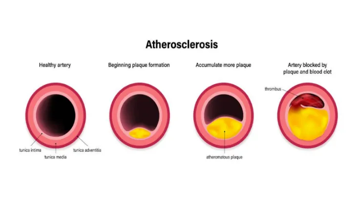
Figure 1: Atherosclerosis
1.2 Role of Dyslipidemia in Atherosclerosis
Dyslipidemia is an important risk factor for atherosclerosis, particularly when circulating levels of LDL and other atherogenic lipoproteins are elevated. These particles can become retained within the arterial wall, promoting lipid accumulation and inflammatory responses. High triglyceride levels and reduced HDL-cholesterol may also indicate an unfavourable lipid profile. Therefore, controlling abnormal lipid levels is an important approach to reducing atherosclerotic cardiovascular risk (Ference et al., 2017).
1.3 Role of Oxidative Stress and Inflammation
Oxidative stress and inflammation contribute to the development and progression of atherosclerosis. Excessive production of reactive oxygen species can damage vascular cells and promote LDL oxidation. Modified LDL is taken up by macrophages, leading to foam-cell formation. Inflammatory mediators, including TNF-α, IL-6 and IL-1β, further influence vascular inflammation and plaque development. These processes provide important targets for investigating potential anti-atherogenic agents (Bäck et al., 2019).
1.4 Medicinal Plants in Cardiovascular Research
Medicinal plants contain various phytochemicals, including flavonoids, phenolic compounds, alkaloids and saponins. Several plant-derived compounds have been investigated for their effects on lipid metabolism, oxidative stress and inflammation. These properties have encouraged research into natural products as potential sources of cardiovascular protective agents. However, their effectiveness and safety must be established through appropriate experimental studies (Hua et al., 2025).
1.5 Botanical and Pharmacological Background of Mussaenda philippica
Mussaenda philippica belongs to the family Rubiaceae and has been investigated for different pharmacological properties. Previous studies have examined the antioxidant properties of its leaf extracts, while other research has evaluated its pharmacognostic characteristics and antiulcer activity (Latayada & Uy, 2016; Jena et al., 2019; Umoh et al., 2022). These investigations provide a preliminary basis for further pharmacological research. However, its potential effect on atherosclerosis requires specific experimental evaluation.
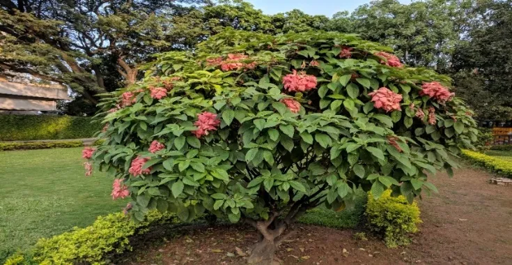
Figure 2: Mussaenda philippica
| Feature | Description |
|---|---|
| Family | Rubiaceae |
| Plant | Shrub |
| Part used | Leaves |
| Major constituents | Flavonoids, phenolics, tannins |
1.6 Research Gap and Scientific Rationale
Although antioxidant and other pharmacological properties of Mussaenda philippica have been investigated, limited evidence is available regarding its potential anti-atherogenic activity. Its effects on lipid abnormalities, oxidative stress, inflammatory biomarkers and aortic tissue changes have not been adequately established together. Therefore, the present study will investigate these parameters to obtain preliminary evidence regarding the possible protective effect of the hydroalcoholic leaf extract against experimental atherosclerotic changes.
1.7 Aim and Objectives of the Study
The present study aims to evaluate the potential anti-atherosclerotic activity of the hydroalcoholic leaf extract of Mussaenda philippica using an appropriate experimental model of atherosclerosis. The study will involve the preparation of the hydroalcoholic leaf extract and preliminary phytochemical screening to identify its major phytochemical constituents. The effects of the extract on serum lipid profile, selected oxidative stress markers, and antioxidant parameters will be investigated to assess its potential protective effects. Furthermore, changes in inflammatory biomarkers and histopathological alterations in aortic tissue will be examined to gain further insight into the possible mechanisms underlying its anti-atherosclerotic activity.
MATERIALS AND METHODS
2.1 Plant Material and Extraction
Fresh leaves of Mussaenda philippica were collected, authenticated by a qualified botanist, washed, shade-dried, and powdered. The powdered material (100 g) was extracted using a hydroalcoholic solvent in a Soxhlet apparatus. The resulting extract was filtered, concentrated by solvent evaporation, and dried. The final extract weighed 13 g, corresponding to a percentage yield of 13% w/w. Preliminary phytochemical screening was performed using standard qualitative tests to detect flavonoids, phenolic compounds, tannins, saponins, and terpenoids.
2.2 Experimental Animals and Study Design
Thirty male Wistar rats were allocated randomly into five groups of six animals each. The animals were maintained under standard laboratory conditions with free access to pellet diet and water and allowed to acclimatize for seven days. Experimental procedures were subject to prior Institutional Animal Ethics Committee (IAEC) approval and applicable CPCSEA requirements. Atherosclerosis was induced in the disease-control and treatment groups using a high-fat/high-cholesterol diet combined with vitamin D₃, following an approved, validated protocol. The groups comprised normal control, disease control, atorvastatin-treated (10 mg/kg), and extract-treated groups receiving 200 or 400 mg/kg orally.
2.3 Biochemical and Inflammatory Assessment
At the end of the experimental period, blood samples were collected according to the approved protocol and centrifuged to separate serum. Total cholesterol, triglycerides, HDL-cholesterol, LDL-cholesterol, and VLDL-cholesterol were measured using standard biochemical methods or commercial diagnostic kits. Oxidative stress and antioxidant status were evaluated by measuring malondialdehyde (MDA), superoxide dismutase (SOD), catalase (CAT), and reduced glutathione (GSH). Serum inflammatory biomarkers, including TNF-α, IL-6, and IL-1β, were assessed using suitable enzyme-linked immunosorbent assay (ELISA) kits.
2.4 Histopathology and Statistical Analysis
Aortic tissues were fixed in 10% neutral buffered formalin, processed, stained with haematoxylin and eosin, and examined microscopically. Data were expressed as mean ± SEM and analysed using one-way ANOVA followed by an appropriate post-hoc test. A p-value < 0.05 was considered statistically significant.
RESULTS AND DISCUSSION
3.1 Percentage Yield of Extract
Hydroalcoholic extraction of 100 g of powdered Mussaenda philippica leaves yielded 13 g of dried extract, corresponding to a percentage yield of 13% w/w.
Percentage yield = (Weight of dried extract / Weight of powdered leaves) × 100
Percentage yield = (13/100) × 100 = 13% w/w
3.2 Preliminary Phytochemical Screening
Preliminary phytochemical screening indicated the presence of flavonoids, phenolic compounds, tannins, saponins, and terpenoids in the hydroalcoholic leaf extract.
Table 3.1: Phytochemical Screening
| Phytochemical | Test | Observation | Result |
|---|---|---|---|
| Flavonoids | Shinoda test | Pink/red colour | Present |
| Phenolic compounds | Ferric chloride test | Green/violet colour | Present |
| Tannins | Ferric chloride test | Blue-black colour | Present |
| Saponins | Foam test | Persistent foam | Present |
| Terpenoids | Salkowski test | Reddish-brown colour | Present |
3.3 Effect on Body Weight
The disease control group showed the highest body-weight gain (39.8 g), whereas the normal control group showed a gain of 16.8 g. Treatment with the extract at 200 and 400 mg/kg reduced weight gain to 30.7 g and 24.9 g, respectively.
Table 3.2: Effect on Body Weight
| Group | Initial weight (g) | Final weight (g) | Weight gain (g) |
|---|---|---|---|
| Normal control | 176.4 ± 2.8 | 193.2 ± 2.9 | 16.8 |
| Disease control | 175.8 ± 2.6 | 215.6 ± 3.7 | 39.8 |
| Atorvastatin 10 mg/kg | 177.1 ± 2.9 | 199.5 ± 3.1 | 22.4 |
| Extract 200 mg/kg | 176.7 ± 2.7 | 207.4 ± 3.4 | 30.7 |
| Extract 400 mg/kg | 176.9 ± 2.8 | 201.8 ± 3.0 | 24.9 |
Values are expressed as mean ± SEM, n = 6.
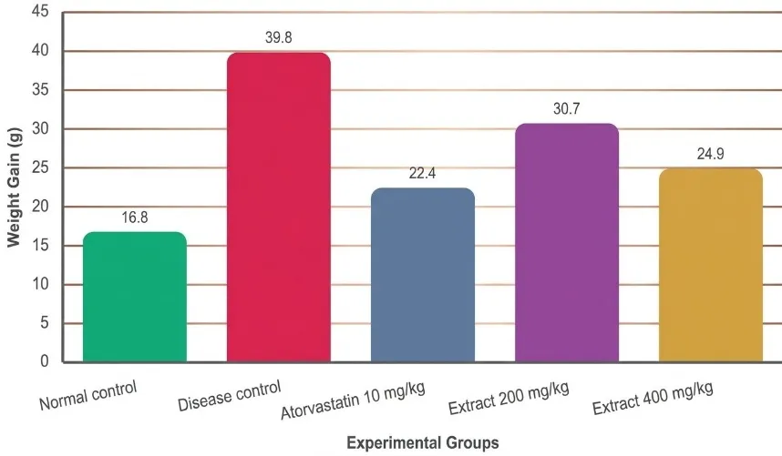
Figure 3: Effect of Plant Extract and Atorvastatin on Total Body Weight Gain (G)
3.4 Effect on Lipid Profile
The disease control group showed elevated total cholesterol, triglycerides, LDL, and VLDL, together with reduced HDL cholesterol. Treatment with the extract improved the lipid profile, with the 400 mg/kg dose producing greater changes than the 200 mg/kg dose.
Table 3.3: Effect on Lipid Profile
| Parameter | Normal control | Disease control | Atorvastatin 10 mg/kg | Extract 200 mg/kg | Extract 400 mg/kg |
|---|---|---|---|---|---|
| Total cholesterol | 82.4 ± 2.1 | 184.6 ± 4.8 | 103.2 ± 3.1 | 142.8 ± 3.9 | 118.6 ± 3.4 |
| Triglycerides | 70.2 ± 2.0 | 142.5 ± 3.9 | 82.6 ± 2.5 | 111.4 ± 3.1 | 91.8 ± 2.7 |
| HDL | 45.6 ± 1.5 | 24.8 ± 1.2 | 41.9 ± 1.4 | 33.7 ± 1.3 | 38.8 ± 1.4 |
| LDL | 22.8 ± 1.4 | 131.3 ± 4.1 | 44.8 ± 2.6 | 86.8 ± 3.5 | 61.4 ± 2.9 |
| VLDL | 14.0 ± 0.4 | 28.5 ± 0.8 | 16.5 ± 0.5 | 22.3 ± 0.6 | 18.4 ± 0.5 |
Values are mean ± SEM, n = 6. Units should be confirmed from the original laboratory records.
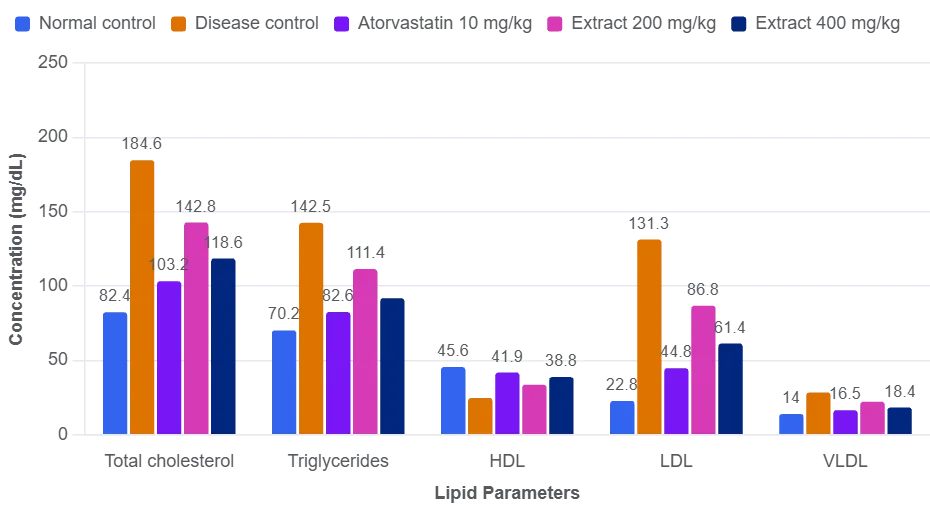
Figure 4: Effect of Plant Extract and Atorvastatin on Lipid Profile
The extract at 400 mg/kg reduced total cholesterol from 184.6 to 118.6, representing a 35.75% reduction. Triglycerides decreased by 35.58%, LDL by 53.24%, and VLDL by 35.44%. HDL increased from 24.8 to 38.8, corresponding to a 56.45% increase compared with the disease control group.
These findings suggest that the extract may improve lipid metabolism in the experimental model.
3.5 Effect on Oxidative Stress Markers
The disease control group showed an increase in MDA from 1.84 to 4.96 and reductions in SOD, CAT, and GSH. Treatment with the extract improved these parameters, particularly at 400 mg/kg.
Table 3.4: Effect on Oxidative Stress Markers
| Parameter | Normal control | Disease control | Atorvastatin 10 mg/kg | Extract 200 mg/kg | Extract 400 mg/kg |
|---|---|---|---|---|---|
| MDA | 1.84 ± 0.12 | 4.96 ± 0.24 | 2.21 ± 0.13 | 3.42 ± 0.18 | 2.71 ± 0.15 |
| SOD | 12.8 ± 0.6 | 5.4 ± 0.3 | 11.6 ± 0.5 | 8.1 ± 0.4 | 10.3 ± 0.5 |
| CAT | 58.4 ± 2.1 | 24.6 ± 1.4 | 52.1 ± 1.9 | 38.5 ± 1.6 | 47.8 ± 1.8 |
| GSH | 8.6 ± 0.4 | 3.2 ± 0.2 | 7.8 ± 0.3 | 5.7 ± 0.3 | 6.9 ± 0.3 |
Values are mean ± SEM, n = 6. Units and assay methods should be specified according to the experimental protocol.
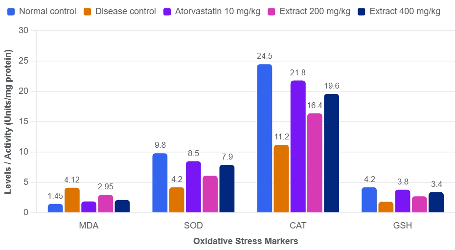
Figure 5: Effect of Plant Extract and Atorvastatin on Oxidative Stress Markers
At 400 mg/kg, the extract reduced MDA by 45.36% compared with the disease control group. SOD increased by 90.74%, CAT by 94.31%, and GSH by 115.63%. These findings suggest a possible improvement in antioxidant defence and reduction in lipid peroxidation.
3.6 Effect on Inflammatory Biomarkers
The disease control group showed increased TNF-α, IL-6, and IL-1β levels. Treatment with the extract reduced all three inflammatory biomarkers, with the higher dose producing greater reductions.
Table 3.5: Effect on Inflammatory Biomarkers
| Parameter (pg/mL) | Normal control | Disease control | Atorvastatin 10 mg/kg | Extract 200 mg/kg | Extract 400 mg/kg |
|---|---|---|---|---|---|
| TNF-α | 32.4 ± 1.8 | 89.6 ± 3.6 | 39.8 ± 2.0 | 61.7 ± 2.8 | 48.3 ± 2.3 |
| IL-6 | 18.2 ± 1.1 | 56.4 ± 2.7 | 24.7 ± 1.4 | 39.5 ± 2.1 | 30.8 ± 1.7 |
| IL-1β | 14.7 ± 0.9 | 43.8 ± 2.2 | 19.9 ± 1.1 | 30.6 ± 1.6 | 24.1 ± 1.3 |
Values are mean ± SEM, n = 6.
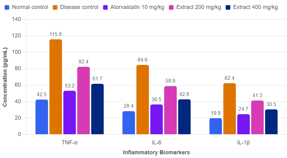
Figure 6: Effect of Plant Extract and Atorvastatin on Inflammatory Biomarkers
At 400 mg/kg, TNF-α decreased by 46.09%, IL-6 by 45.39%, and IL-1β by 44.98% compared with the disease control group. These findings suggest that the extract may help reduce inflammatory responses associated with the experimental model.
3.7 Histopathological Findings
The normal control group showed preserved vascular architecture and an intact intimal layer. The disease control group exhibited intimal thickening, lipid deposition, and inflammatory cell infiltration.
The atorvastatin group showed comparatively better vascular preservation. Moderate improvement was observed with the extract at 200 mg/kg, while the 400 mg/kg dose showed comparatively less lipid deposition and inflammatory infiltration.
These observations were consistent with the improvements in lipid profile, oxidative stress markers, and inflammatory biomarkers. Quantitative lesion scoring and statistical analysis would strengthen the histopathological interpretation.
3.9 Discussion
The findings indicate that the hydroalcoholic leaf extract of Mussaenda philippica improved several parameters associated with experimental atherosclerosis. The improvements included reductions in total cholesterol, triglycerides, LDL, VLDL, MDA, and inflammatory biomarkers, together with increases in HDL, SOD, CAT, and GSH.
The 400 mg/kg dose generally showed greater effects than the 200 mg/kg dose. The preliminary presence of flavonoids and phenolic compounds may be relevant to the observed antioxidant and anti-inflammatory effects, although the active constituents and mechanisms have not been established.
Overall, the biochemical and histopathological findings support further investigation of the extract's potential anti-atherogenic activity.
SUMMARY AND CONCLUSION
The present study evaluated the anti-atherogenic potential of the hydroalcoholic leaf extract of Mussaenda philippica. Extraction of 100 g of powdered leaves yielded 13 g of dried extract (13% w/w). Phytochemical screening revealed flavonoids, phenolics, tannins, saponins, and terpenoids. In Wistar rats, the extract at 400 mg/kg reduced total cholesterol by 35.75%, triglycerides by 35.58%, LDL by 53.24%, MDA by 45.36%, and inflammatory biomarkers by approximately 45%. HDL and antioxidant parameters also improved, with histopathological findings indicating better preservation of vascular architecture.
The hydroalcoholic leaf extract of Mussaenda philippica showed preliminary anti-atherogenic potential through improvements in lipid profile, oxidative stress, inflammatory markers, and vascular histology. The 400 mg/kg dose generally produced greater effects than 200 mg/kg. Further studies are needed to identify the active constituents, confirm statistical significance, investigate underlying mechanisms, and establish safety and therapeutic efficacy.
REFERENCES

Sanskriti Shukla, Ankur Joshi, Dr. Sapna Malviya, Assessment of the Anti-Atherogenic Potential of Hydroalcoholic Leaf Extract of Mussaenda Philippica in an Experimental Model of Atherosclerosis, Int. J. of Pharm. Sci., 2026, Vol 4, Issue 11, 2122-2130. https://doi.org/10.5281/zenodo.24216208
 10.5281/zenodo.24216208