

We use cookies to ensure our website works properly and to personalise your experience. Cookies policy
1Professor & HOD, Department of Pharmaceutical Chemistry and Analysis, Malla Reddy Institute of Pharmaceutical Sciences, Malla Reddy Vishwavidyapeeth (Deemed to be University), Maisammaguda, Secunderabad - 500100, Telangana, India.
2Professor & Dean, Department of Pharmaceutical Analysis, Malla Reddy Institute of Pharmaceutical Sciences, Malla Reddy Vishwavidyapeeth (Deemed to be University), Maisammaguda, Secunderabad - 500100, Telangana, India.
3Department of Pharmaceutical Analysis, Malla Reddy Institute of Pharmaceutical Sciences, Malla Reddy Vishwavidyapeeth (Deemed to be University), Maisammaguda, Secunderabad - 500100, Telangana, India.
A rapid and precise reverse phase high performance liquid chromatographic method has been developed for the validated of Nirmatrelvir and Ritonavir, in its pure form as well as in marketed pharmaceutical dosage forms. This Chromatographic Separation was carried out on Develosil C18 (4.6mm×250mm) 5µm particle size Column using a mixture of Acetonitrile and Acetate buffer (pH-4.3) was taken in the ratio of 35:65% v/v as the mobile phase at a flow rate of 1.0 mL/min, the wavelength detection was carried out at 238nm. The retention time of the Nirmatrelvir and Ritonavir was found to be 2.179, 3.610 ± 0.02 min respectively. The method produce linear responses in the concentration range of 20-60µg/ml of Nirmatrelvir and 10-30µg/ml of Ritonavir respectively. The developed method precision for the determination of assay was below 2.0%RSD. The developed method is useful in the quality control of bulk and pharmaceutical formulations.
Nirmatrelvir (PF-07321332) is an orally bioavailable 3C-like protease (3CLPRO) inhibitor that is the subject of clinical trial NCT04756531. 3CLPRO is responsible for cleaving polyproteins 1a and 1ab of SARS-CoV-2. Without the activity of the SARS-CoV-2 3CLPRO, nonstructural proteins (including proteases) cannot be released to perform their functions, inhibiting viral replication [1]. In the US, Europe, and Canada, Nirmatrelvir, in combination with ritonavir, is indicated for the treatment of mild-to-moderate coronavirus disease 2019 (COVID-19) in adults who are at high risk for progression to severe COVID-19, including hospitalization or death. In Europe, this therapeutic indication is approved under conditional marketing authorization [2]. Nirmatrelvir is a SARS-CoV-2 main protease inhibitor. It works by preventing the growth of the virus that causes COVID-19. Ritonavir increases ("boosts") the levels of Nirmatrelvir. This helps Nirmatrelvir work better [3]. The IUPAC name of Nirmatrelvir is 5 (1R, 2S, 5S)-N-[(1S)-1-cyano-2-[(3S)-2-oxo pyrrolidin-3-yl] ethyl]-3-[(2S)-3, 3-dimethyl-2-[(2, 2, 2-trifluoro acetyl) amino] butanoyl]-6, 6-dimethyl-3-azabi cyclo [3.1.0] hexane-2-carboxamide [3]. The Chemical Structure of Nirmatrelvir is shown in follows
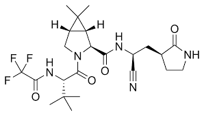
Figure None: 1: Chemical Structure of Nirmatrelvir
Ritonavir is an antiretroviral protease inhibitor that is widely used in combination with other protease inhibitors in the therapy and prevention of human immunodeficiency virus (HIV) infection and the acquired immunodeficiency syndrome (AIDS) [4]. Ritonavir can cause transient and usually asymptomatic elevations in serum aminotransferase levels and, rarely, can lead to clinically apparent acute liver injury. In HBV or HCV coinfected patients, highly active antiretroviral therapy with ritonavir may result of an exacerbation of the underlying chronic hepatitis B or C. Ritonavir is indicated in combination with other antiretroviral agents for the treatment of HIV-1 infection [5]. Ritonavir is used along with other medications to treat human immunodeficiency virus (HIV) infection. Ritonavir is in a class of medications called protease inhibitors. It works by decreasing the amount of HIV in the blood. The IUPAC name of Ritonavir is 1, 3-thiazol-5-ylmethyl N-[(2S, 3S, 5S)-3-hydroxy-5-[[(2S)-3-methyl-2-[[methyl-[(2-propan-2-yl-1, 3-thiazol-4-yl) methyl] carbamoyl] amino] butanoyl] amino]-1, 6-diphenyl hexan-2-yl] carbamate [6]. The Chemical Structure of Ritonavir is shown in following Figure-2.
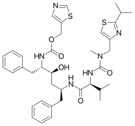
Figure None: 2: Chemical Structure of Ritonavir
MATERIALS AND METHODS
Instruments Used:
Table None: 1: Instruments Used
| S.No. | Instruments and Glasswares | Model |
|---|---|---|
| 1 | HPLC | WATERS Alliance 2695 separation module, Software: Empower 2, 996 PDA detector. |
| 2 | pH meter | Lab India |
| 3 | Weighing machine | Sartorius |
| 4 | Volumetric flasks | Borosil |
| 5 | Pipettes and Burettes | Borosil |
| 6 | Beakers | Borosil |
| 7 | Digital Ultra Sonicator | Labman |
Chemicals Used:
Table None: 2: Chemicals Used
| S.No. | Chemical | Brand Names |
|---|---|---|
| 1 | Nirmatrelvir (Pure) | Synpharma Research Lab, Hyderabad |
| 2 | Ritonavir (Pure) | Synpharma Research Lab, Hyderabad |
| 3 | Water and Methanol for HPLC | LICHROSOLV (MERCK) |
| 4 | Acetonitrile for HPLC | Merck |
| 5 | Acetic Acid | Merck |
HPLC Method Development:
Preparation of Standard Solution:
Accurately weigh and transfer 10 mg of Nirmatrelvir and Ritonavir working standard into a 10ml of clean dry volumetric flasks add about 7ml of Methanol and sonicate to dissolve and removal of air completely and make volume up to the mark with the same Methanol. Further pipette 0.4ml of Nirmatrelvir and 0.2ml of Ritonavir from the above stock solutions into a 10ml volumetric flask and dilute up to the mark with Methanol.
Procedure:
Inject the samples by changing the chromatographic conditions and record the chromatograms, note the conditions of proper peak elution for performing validation parameters as per ICH guidelines [27].
Mobile Phase Optimization:
Initially the mobile phase tried was Methanol: Water and ACN: Water with varying proportions. Finally, the mobile phase was optimized to Acetonitrile and Acetate buffer (pH-4.3) in proportion 35:65% v/v respectively.
Optimization of Column:
The method was performed with various C18 columns like Symmetry, X terra and ODS column. Develosil C18 (4.6mm×250mm) 5µm particle size Column was found to be ideal as it gave good peak shape and resolution at 1ml/min flow [7].
Preparation of Mobile Phase:
Accurately measured 350ml of Acetonitrile (35%) of and 650ml of Acetate buffer (65%) were mixed and degassed in a digital ultra sonicater for 20 minutes and then filtered through 0.45 µ filter under vacuum filtration.
Diluent Preparation:
The Mobile phase was used as the diluent.
Method Validation Parameters
System Suitability
Accurately weigh and transfer 10 mg of Nirmatrelvir and Ritonavir working standard into a 10ml of clean dry volumetric flasks add about 7mL of Diluents and sonicate to dissolve it completely and make volume up to the mark with the same solvent [8]. (Stock solution). Further pipette out 0.4ml of Nirmatrelvir and 0.2ml of Ritonavir from the above stock solutions into a 10ml volumetric flask and dilute up to the mark with Diluent.
Procedure:
The standard solution was injected for five times and measured the area for all five injections in HPLC. The %RSD for the area of five replicate injections was found to be within the specified limits.
Specificity Study of Drug:
Preparation of Standard Solution:
Accurately weigh and transfer 10 mg of Nirmatrelvir and Ritonavir working standard into a 10ml of clean dry volumetric flasks add about 7ml of Diluents and sonicate to dissolve it completely and make volume up to the mark with the same solvent. (Stock solution). Further pipette out 0.4ml of Nirmatrelvir and 0.2ml of Ritonavir from the above stock solutions into a 10ml volumetric flask and dilute up to the mark with Diluent [9].
Preparation of Sample Solution:
Take average weight of Tablet and crush in a mortar by using pestle and weight 10 mg equivalent weight of Nirmatrelvir and Ritonavir sample into a 10mL clean dry volumetric flask and add about 7mL of Diluent and sonicate to dissolve it completely and make volume up to the mark with the same solvent. Filter the sample solution by using injection filter which contains 0.45µ pore size. Further pipette out 0.4ml of Nirmatrelvir and 0.2ml of Ritonavir Sample solution from the above stock solutions into a 10ml volumetric flask and dilute up to the mark with Diluent [10].
Procedure:
Inject the three replicate injections of standard and sample solutions and calculate the assay by using formula [11-13]:
%ASSAY =
Sample area Weight of standard Dilution of sample Purity Weight of tablet
___________ × ________________ × _______________×_______×______________×100
Standard area Dilution of standard Weight of sample 100 Label claim
Preparation of Drug Solutions for Linearity and Range:
Accurately weigh and transfer 10 mg of Nirmatrelvir and Ritonavir working standard into a 10ml of clean dry volumetric flasks add about 7ml of Diluents and sonicate to dissolve it completely and make volume up to the mark with the same solvent [14]. (Stock solution)
Preparation of Level – I (20ppm of Nirmatrelvir and 10ppm of Ritonavir):
Pipette out 0.2ml of Nirmatrelvir and 0.1ml of Ritonavir in to a 10ml volumetric flask and make the volume upto mark by using diluent and sonicate for air entrapment.
Preparation of Level – II (30ppm of Nirmatrelvir and 15ppm of Ritonavir):
Pipette out 0.3ml of Nirmatrelvir and 0.15ml of Ritonavir in to a 10ml volumetric flask and make the volume upto mark by using diluent and sonicate for air entrapment.
Preparation of Level – III (40ppm of Nirmatrelvir and 20ppm of Ritonavir):
Pipette out 0.4ml of Nirmatrelvir and 0.2ml of Ritonavir in to a 10ml volumetric flask and make the volume upto mark by using diluent and sonicate for air entrapment [15].
Preparation of Level – IV (50ppm of Nirmatrelvir and 25ppm of Ritonavir):
Pipette out 0.5ml of Nirmatrelvir and 0.25ml of Ritonavir in to a 10ml volumetric flask and make the volume upto mark by using diluent and sonicate for air entrapment.
Preparation of Level – V (60ppm of Nirmatrelvir and 30ppm of Ritonavir):
Pipette out 0.6ml of Nirmatrelvir and 0.3ml of Ritonavir in to a 10ml volumetric flask and make the volume up to mark by using diluent and sonicate for air entrapment.
Procedure:
Inject each level into the chromatographic system and measure the peak area.
Plot a graph of peak area versus concentration (on X-axis concentration and on Y-axis Peak area) and calculate the correlation coefficient [16-18].
Precision
Repeatability
Preparation of Nirmatrelvir and Ritonavir Product Solution for Precision:
Accurately weigh and transfer 10 mg of Nirmatrelvir and Ritonavir working standard into a 10ml of clean dry volumetric flasks add about 7ml of Diluents and sonicate to dissolve it completely and make volume up to the mark with the same solvent. (Stock solution). Further pipette out 0.4 ml of Nirmatrelvir and 0.2ml of Ritonavir from the above stock solutions into a 10ml volumetric flask and dilute up to the mark with Diluent.
The standard solution was injected for five times and measured the area for all five injections in HPLC. The %RSD for the area of five replicate injections was found to be within the specified limits [19].
Intermediate Precision:
To evaluate the intermediate precision (also known as Ruggedness) of the method, Precision was performed on different days by maintaining same conditions.
Procedure:
Day 1:
The standard solution was injected for Six times and measured the area for all Six injections in HPLC. The %RSD for the area of Six replicate injections was found to be within the specified limits [20].
Day 2:
The standard solution was injected for Six times and measured the area for all Six injections in HPLC. The %RSD for the area of Six replicate injections was found to be within the specified limits.
Accuracy:
For preparation of 50% Standard stock solution:
Accurately weigh and transfer 10mg of Nirmatrelvir and Ritonavir working standard into a 10ml of clean dry volumetric flasks add about 7mL of Diluents and sonicate to dissolve it completely and make volume up to the mark with the same solvent. (Stock solution). Further pipette out 0.2ml of Nirmatrelvir and 0.1ml of Ritonavir from the above stock solutions into a 10ml volumetric flask and dilute up to the mark with Diluent [21].
For preparation of 100% Standard stock solution:
Accurately weigh and transfer 10 mg of Nirmatrelvir and Ritonavir working standard into a 10ml of clean dry volumetric flasks add about 7mL of Diluents and sonicate to dissolve it completely and make volume up to the mark with the same solvent. (Stock solution). Further pipette out 0.4ml of Nirmatrelvir and 0.2ml of Ritonavir from the above stock solutions into a 10ml volumetric flask and dilute up to the mark with Diluent.
For preparation of 150% Standard stock solution:
Accurately weigh and transfer 10 mg of Nirmatrelvir and Ritonavir working standard into a 10ml of clean dry volumetric flasks add about 7mL of Diluents and sonicate to dissolve it completely and make volume up to the mark with the same solvent. (Stock solution). Further pipette out 0.6ml of Nirmatrelvir and 0.3ml of Ritonavir from the above stock solutions into a 10ml volumetric flask and dilute up to the mark with Diluent.
Procedure:
Inject the Three replicate injections of individual concentrations (50%, 100%, 150%) were made under the optimized conditions. Recorded the chromatograms and measured the peak responses. Calculate the Amount found and Amount added for Nirmatrelvir and Ritonavir and calculate the individual recovery and mean recovery values [22].
Robustness:
The analysis was performed in different conditions to find the variability of test results. The following conditions are checked for variation of results.
For preparation of Standard solution:
Accurately weigh and transfer 10 mg of Nirmatrelvir and Ritonavir working standard into a 10ml of clean dry volumetric flasks add about 7mL of Diluents and sonicate to dissolve it completely and make volume up to the mark with the same solvent. (Stock solution). Further pipette out 0.4ml of Nirmatrelvir and 0.2ml of Ritonavir from the above stock solutions into a 10ml volumetric flask and dilute up to the mark with Diluent.
Effect of Variation of Flow Conditions:
The sample was analyzed at 0.9 ml/min and 1.1 ml/min instead of 1ml/min, remaining conditions are same. 20µl of the above sample was injected and chromatograms were recorded [23].
Effect of Variation of Mobile Phase Organic Composition:
The sample was analyzed by variation of mobile phase i.e. Acetonitrile: Acetate buffer was taken in the ratio 40:60 and 30:70 instead of 35:65 remaining conditions are same. 20µl of the above sample was injected and chromatograms were recorded.
RESULTS AND DISCUSSION
Optimization of an Analytical Method:
Optimized Chromatographic Conditions:
Mobile phase ratio : Acetonitrile and Acetate buffer (pH-4.3) (35:65% v/v)
Column : Develosil C18 (4.6mm×250mm) 5µm particle size Column
Column temperature : Ambient
Wavelength : 238nm
Flow rate : 1ml/min
Injection volume : 20µl
Run time : 6minutes
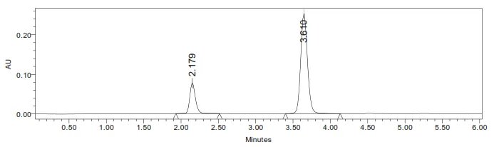
Figure None: 3: Optimized Chromatogram of Nirmatrelvir and Ritonavir
Method Validation
The validation parameters, such as linearity, specificity, precision, and accuracy, limit of detection (LOD), limit of quantification (LOQ), robustness and ruggedness repeatability were assessed in accordance with the ICH Q2 (R1) guidelines [27].
System Suitability:
Five distinct injections of Nirmatrelvir and Ritonavir solutions were used to evaluate the technique's applicability. The retention time, peak area, column efficiency, peak symmetry, and theoretical plates were calculated for standard solutions [24].
Table None: 3: Results of System Suitability for Nirmatrelvir
| S.No. | Peak Name | RT | Area (µV*sec) | Height (µV) | USP Plate Count | USP Tailing |
|---|---|---|---|---|---|---|
| 1 | Nirmatrelvir | 2.152 | 526358 | 86598 | 5695 | 1.56 |
| 2 | Nirmatrelvir | 2.157 | 526548 | 86254 | 5652 | 1.57 |
| 3 | Nirmatrelvir | 2.141 | 526854 | 86598 | 5627 | 1.56 |
| 4 | Nirmatrelvir | 2.133 | 526598 | 86245 | 5692 | 1.57 |
| 5 | Nirmatrelvir | 2.166 | 524874 | 86521 | 5641 | 1.56 |
| Mean | 526246.4 | |||||
| Std. Dev | 787.353 | |||||
| % RSD | 0.149617 |
Table None: 4: Results of System Suitability for Ritonavir
| S.No. | Peak Name | RT | Area (µV*sec) | Height (µV) | USP Plate Count | USP Tailing | Resolution |
|---|---|---|---|---|---|---|---|
| 1 | Ritonavir | 3.674 | 1682821 | 1686958 | 8659 | 1.56 | 9.8 |
| 2 | Ritonavir | 3.631 | 1682726 | 1685745 | 8675 | 1.57 | 9.9 |
| 3 | Ritonavir | 3.625 | 1687361 | 1685421 | 8692 | 1.56 | 9.8 |
| 4 | Ritonavir | 3.692 | 1682811 | 1685242 | 8642 | 1.57 | 9.8 |
| 5 | Ritonavir | 3.629 | 1683816 | 1685364 | 8635 | 1.58 | 9.8 |
| Mean | 1683907 | ||||||
| Std. Dev | 1982.03 | ||||||
| % RSD | 0.117704 |
Specificity (Assay)
In accordance with ICH guidelines, the assay method must be capable of quantifying the active pharmaceutical ingredient in the presence of excipients, impurities, and degradation products without interference. The assay was performed on tablet formulations. The percentage of assay for the drug content was determined. The results demonstrate that the method is both accurate and specific, enabling precise measurement of the active ingredient in the presence of excipients in the tablet matrix [25].
%ASSAY =
Sample area Weight of standard Dilution of sample Purity Weight of tablet
___________ × ________________ × _______________×_______×______________×100
Standard area Dilution of standard Weight of sample 100 Label claim
CONCLUSION:
The % purity of Nirmatrelvir and Ritonavir in pharmaceutical dosage form was found to be 99.89%
Linearity
A precisely weighed amount of 10 mg of the drug was transferred into a 10 mL volumetric flask and diluted with solvent to the calibration mark, yielding a concentration of 1000 μg/mL. To assess the drug’s linear response, concentrations ranging from 20 to 60μg/mL and 10 to 30μg/mL for Nirmatrelvir and Ritonavir respectively were prepared from this stock solution. A calibration curves was constructed by plotting the peak area against the corresponding concentrations. The linearity of the response was evaluated, and the correlation coefficients (r2) and y-intercepts were determined from the resulting curves [26].
Table None: 5: Chromatographic Data for Linearity Study of Nirmatrelvir
| Concentration μg/ml |
Average Peak Area |
|---|---|
| 20 | 272897 |
| 30 | 402986 |
| 40 | 526389 |
| 50 | 649785 |
| 60 | 769287 |
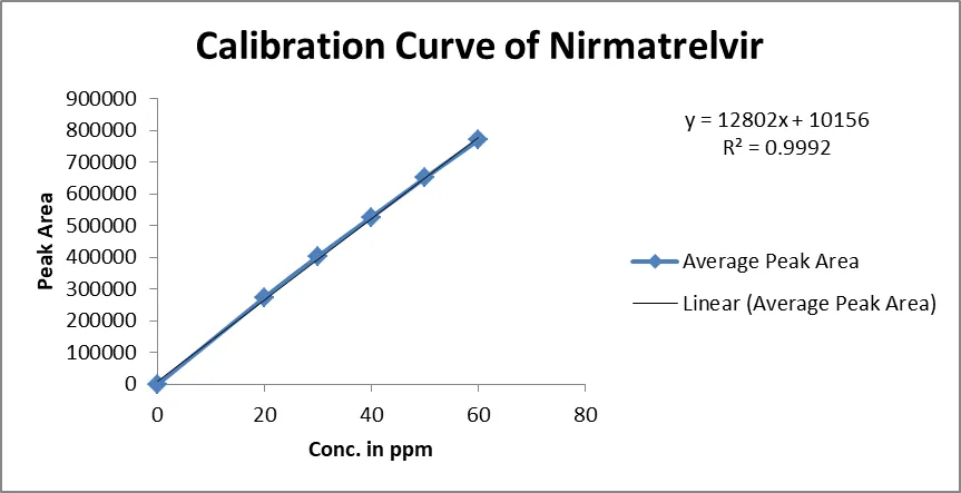
Figure None: 4: Calibration Curve of Nirmatrelvir
Table None: 6: Chromatographic Data for Linearity Study of Ritonavir
| Concentration μg/ml |
Average Peak Area |
|---|---|
| 10 | 1000237 |
| 15 | 1448768 |
| 20 | 1887285 |
| 25 | 2365897 |
| 30 | 2826845 |
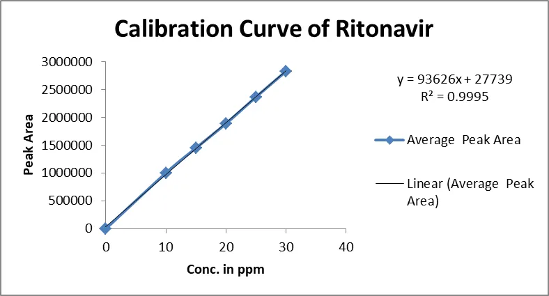
Figure None: 5: Calibration Curve of Ritonavir
Precision
A standard solution of 40μg/ml and 20μg/ml was prepared and injected 5 times into the system. A chromatogram was recorded and the %RSD of the Peak Area was calculated [28].
Table None: 7: Results of Repeatability for Nirmatrelvir
| S. No. | Peak Name | Retention time | Area (µV*sec) |
Height (µV) |
USP Plate Count | USP Tailing |
|---|---|---|---|---|---|---|
| 1 | Nirmatrelvir | 2.157 | 526358 | 86598 | 5689 | 1.56 |
| 2 | Nirmatrelvir | 2.159 | 524856 | 86542 | 5687 | 1.57 |
| 3 | Nirmatrelvir | 2.186 | 526985 | 86578 | 5684 | 1.56 |
| 4 | Nirmatrelvir | 2.160 | 528654 | 86354 | 5689 | 1.56 |
| 5 | Nirmatrelvir | 2.170 | 528457 | 86958 | 5639 | 1.56 |
| Mean | 527062 | |||||
| Std Dev | 1569.114 | |||||
| %RSD | 0.297709 |
Table None: 8: Results of Repeatability for Ritonavir:
| S. No. | Peak Name | Retention time | Area (µV*sec) |
Height (µV) |
USP Plate Count | USP Tailing |
|---|---|---|---|---|---|---|
| 1 | Ritonavir | 3.603 | 1687589 | 367859 | 8659 | 1.79 |
| 2 | Ritonavir | 3.608 | 1685987 | 368547 | 8679 | 1.80 |
| 3 | Ritonavir | 3.600 | 1685987 | 367985 | 8645 | 1.80 |
| 4 | Ritonavir | 3.696 | 1685754 | 365874 | 8695 | 1.79 |
| 5 | Ritonavir | 3.629 | 1685985 | 364589 | 8625 | 1.79 |
| Mean | 1686260 | |||||
| Std Dev | 749.493 | |||||
| %RSD | 0.044447 |
Intermediate Precision
The intermediate precision was calculated by measuring the responses of a standard peak on another day of the same solution concentration.
Day-I:
Table None: 9: Results of Intermediate Precision for Nirmatrelvir (Day-1)
| S.No. | Peak Name | RT | Area (µV*sec) | Height (µV) | USPPlate count | USPTailing | %Assay |
|---|---|---|---|---|---|---|---|
| 1 | Nirmatrelvir | 2.198 | 546585 | 87589 | 5898 | 1.58 | 100% |
| 2 | Nirmatrelvir | 2.196 | 548758 | 87985 | 5879 | 1.59 | 100% |
| 3 | Nirmatrelvir | 2.160 | 549854 | 87452 | 5868 | 1.58 | 100% |
| 4 | Nirmatrelvir | 2.160 | 548798 | 87421 | 5847 | 1.59 | 100% |
| 5 | Nirmatrelvir | 2.160 | 542659 | 87963 | 5896 | 1.58 | 100% |
| 6 | Nirmatrelvir | 2.186 | 548754 | 87254 | 5874 | 1.59 | 100% |
| Mean | 547568 | ||||||
| Std. Dev | 2631.576 | ||||||
| % RSD | 0.480593 |
Table None: 10: Results of Intermediate Precision for Ritonavir
| S.No. | Peak Name | Rt | Area (µV*sec) | Height (µV) | USPPlate Count | USPTailing | Resolution |
|---|---|---|---|---|---|---|---|
| 1 | Ritonavir | 3.623 | 1698587 | 385482 | 8789 | 1.81 | 9.8 |
| 2 | Ritonavir | 3.611 | 1698574 | 385698 | 8759 | 1.80 | 9.8 |
| 3 | Ritonavir | 3.696 | 1698532 | 385748 | 8754 | 1.81 | 9.9 |
| 4 | Ritonavir | 3.696 | 1698574 | 386958 | 8754 | 1.81 | 10.01 |
| 5 | Ritonavir | 3.696 | 1698532 | 385755 | 5798 | 1.80 | 9.98 |
| 6 | Ritonavir | 3.642 | 1698547 | 386558 | 8762 | 1.80 | 10.02 |
| Avg | 1698558 | ||||||
| Std. Dev | 23.77113 | ||||||
| % RSD | 0.001399 |
Day-II:
Table None: 11: Results of Intermediate Precision Day 2 for Nirmatrelvir
| S.No. | Peak Name | RT | Area (µV*sec) | Height (µV) | USPPlate Count | USPTailing |
|---|---|---|---|---|---|---|
| 1 | Nirmatrelvir | 2.198 | 536854 | 8758 | 5789 | 1.58 |
| 2 | Nirmatrelvir | 2.196 | 536985 | 8795 | 5726 | 1.59 |
| 3 | Nirmatrelvir | 2.178 | 536587 | 8746 | 5742 | 1.58 |
| 4 | Nirmatrelvir | 2.142 | 532546 | 8754 | 5746 | 1.59 |
| 5 | Nirmatrelvir | 2.177 | 534587 | 8725 | 5798 | 1.58 |
| 6 | Nirmatrelvir | 2.177 | 538598 | 8726 | 5785 | 1.59 |
| Mean | 536026.2 | |||||
| Std. Dev | 2131.492 | |||||
| % RSD | 0.397647 |
Table None: 12: Results of Intermediate Precision Day 2 for Ritonavir
| S.No. | Peak Name | RT | Area (µV*sec) | Height (µV) | USPPlate Count | USPTailing | Resolution |
|---|---|---|---|---|---|---|---|
| 1 | Ritonavir | 3.611 | 1678598 | 356875 | 8875 | 1.82 | 9.9 |
| 2 | Ritonavir | 3.623 | 1678985 | 358985 | 8856 | 1.83 | 10.01 |
| 3 | Ritonavir | 3.684 | 1678984 | 358754 | 8862 | 1.82 | 9.9 |
| 4 | Ritonavir | 3.697 | 1678985 | 352412 | 8849 | 1.83 | 10.01 |
| 5 | Ritonavir | 3.684 | 1678549 | 358987 | 8873 | 1.82 | 9.9 |
| 6 | Ritonavir | 3.684 | 1678984 | 358986 | 8842 | 1.83 | 10.01 |
| Mean | 1678848 | ||||||
| Std. Dev | 212.8048 | ||||||
| % RSD | 0.012676 |
Accuracy
The accuracy of the method was determined by spiking with a known amount of drug to result in sample solutions with the following levels of drug 0%, 100% and 150% relative to the working concentration in triplicate according to the method of analysis and analyzed as per the method. The % recovery was calculated [29].
Table None: 13: The Accuracy Results for Nirmatrelvir
| %Concentration (at specification Level) |
Area | Amount Added (ppm) |
Amount Found (ppm) |
% Recovery | Mean Recovery |
|---|---|---|---|---|---|
| 50% | 267011.3 | 20 | 20.063 | 100.315% | 100.28% |
| 100% | 523752.3 | 40 | 40.118 | 100.295% | |
| 150% | 778457.3 | 60 | 60.133 | 100.221% |
Table None: 14: The Accuracy Results for Ritonavir
| %Concentration (at specification Level) |
Area | Amount Added (ppm) |
Amount Found (ppm) |
% Recovery | Mean Recovery |
|---|---|---|---|---|---|
| 50% | 972876.3 | 10 | 10.094 | 100.94% | 100.48% |
| 100% | 1900122 | 20 | 19.998 | 99.99% | |
| 150% | 2851152 | 30 | 30.156 | 100.52% |
Limit of Detection: The detection limit of an individual analytical procedure is the lowest amount of analyte in a sample which can be detected but not necessarily quantitated as an exact value.
LOD = 3.3 × σ / s
Where
σ = Standard deviation of the response and S = Slope of the calibration curve
Nirmatrelvir = 1.04µg/ml
Ritonavir = 3.12µg/ml
Quantitation Limit: The quantitation limit of an individual analytical procedure is the lowest amount of analyte in a sample which can be quantitatively determined.
LOQ = 10×σ/S
Where
σ = Standard deviation of the response and S = Slope of the calibration curve
Nirmatrelvir =2.1µg/ml
Ritonavir = 6.3µg/ml
Robustness: Nirmatrelvir and Ritonavir were tested for resilience at flow rates ranging from 0.9 to 1.1 ml/min and mobile phase ratios varying from more to less organic phase. Only at low flow rates is the technique robust, and it is also resistant to a 10% shift in the mobile phase. The standard and samples of Nirmatrelvir and Ritonavir were injected using a variety of chromatographic settings. The resolution, tailing factor, and plate count properties remained unchanged [30].
Table None: 15: Results for Robustness of Nirmatrelvir
| Parameter Used For Sample Analysis | Peak Area | Retention Time | Theoretical Plates | Tailing Factor |
|---|---|---|---|---|
| Actual Flow rate of 1.0 mL/min | 526389 | 2.133 | 5679 | 1.56 |
| Less Flow rate of 0.9 mL/min | 542685 | 2.210 | 5264 | 1.54 |
| More Flow rate of 1.1 mL/min | 526483 | 2.184 | 5426 | 1.52 |
| Less organic phase | 516854 | 2.200 | 5163 | 1.57 |
| More Organic phase | 506898 | 2.172 | 5098 | 1.51 |
Table None: 16: Results for Robustness of Ritonavir
| Parameter Used For Sample Analysis | Peak Area | Retention Time | Theoretical Plates | Tailing Factor |
|---|---|---|---|---|
| Actual Flow rate of 1.0 mL/min | 1687285 | 3.692 | 8685 | 1.79 |
| Less Flow rate of 0.9 mL/min | 1725468 | 4.498 | 8265 | 1.68 |
| More Flow rate of 1.1 mL/min | 1652847 | 3.505 | 8415 | 1.59 |
| Less organic phase | 1687485 | 4.504 | 8326 | 1.62 |
| More organic phase | 1674524 | 3.512 | 8415 | 1.63 |
Stability Studies
The specificity of the method can be demonstrated by applying stress conditions using acid, alkaline, peroxide, thermal, UV, water degradations. The sample was exposed to these conditions the main peak of the drug was studied for peak purity that indicating the method effectively separated the degradation products from the pure active ingredient.
Table None: 17: Results of Forced Degradation Studies of Nirmatrelvir
| S.No. | Stress Condition | Peak Area | % of Degraded Amount | % of Active Amount | Total % of Amount |
|---|---|---|---|---|---|
| 1 | Standard | 526389 | 0 | 100% | 100% |
| 2 | Acidic | 371683.27 | 29.39 | 70.61 | 100% |
| 3 | Basic | 411794.11 | 21.77 | 78.23 | 100% |
| 4 | Oxidative | 480645.79 | 8.69 | 91.31 | 100% |
| 5 | Thermal | 327045.48 | 37.87 | 62.13 | 100% |
| 6 | Photolytic | 477118.99 | 9.36 | 90.64 | 100% |
Table None: 18: Results of Forced Degradation Studies of Ritonavir
| S.No. | Stress Condition | Peak Area | % of Degraded Amount | % of Active Amount | Total % of Amount |
|---|---|---|---|---|---|
| 1 | Standard | 1687285 | 0 | 100% | 100% |
| 2 | Acidic | 1359614.25 | 19.42 | 80.58 | 100% |
| 3 | Basic | 1445497.05 | 14.33 | 85.67 | 100% |
| 4 | Oxidative | 1644427.96 | 2.54 | 97.46 | 100% |
| 5 | Thermal | 1297353.43 | 23.11 | 76.89 | 100% |
| 6 | Photolytic | 1632954.42 | 3.22 | 96.78 | 100% |
SUMMARY AND CONCLUSION
In the present investigation, a simple, sensitive, precise and accurate RP-HPLC method was developed for the quantitative estimation of Nirmatrelvir and Ritonavir in bulk drug and pharmaceutical dosage forms. This method was simple, since diluted samples are directly used without any preliminary chemical derivatization or purification steps. Acetonitrile: Acetate Buffer (pH-4.3) (35:65 v/v) was chosen as the mobile phase. The solvent system used in this method was economical. The %RSD values were within 2 and the method was found to be precise. The results expressed in Tables for RP-HPLC method was promising. The RP-HPLC method is more sensitive, accurate and precise compared to the Spectrophotometric methods. This method can be used for the routine determination of Nirmatrelvir and Ritonavir in bulk drug and in Pharmaceutical dosage forms.
REFERENCES

P. Laxmi Madhuri, H. Ramana, Singam Vinisha, A Validated Stability-Indicating RP-HPLC Method for Simultaneous Determination of Nirmatrelvir and Ritonavir in Bulk Drug and Marketed Pharmaceutical Dosage Forms, Int. J. of Pharm. Sci., 2026, Vol 4, Issue 11, 2131-2143. https://doi.org/10.5281/zenodo.24216352
 10.5281/zenodo.24216352