

We use cookies to ensure our website works properly and to personalise your experience. Cookies policy
Dr. Gurachar Nargund college of Pharmacy, Muradi, under RGUHS University.
Transdermal drug delivery offers a painless alternative to hypodermic injections and avoids the first-pass metabolism associated with oral medications. However, the stratum corneum presents a formidable barrier to macromolecular therapeutics. Microneedle (MN) patches have emerged as a disruptive technology capable of breaching this barrier safely and painlessly. This review provides a comprehensive overview of recent advancements in MN technology. We examine the evolution of MN design, mechanical classification, and cutting-edge fabrication methodologies. Furthermore, we explore their application in precision medicine—including responsive drug delivery, diagnostics, and vaccine formulation—and analyze the translational hurdles, regulatory frameworks, and clinical prospects shaping the future of transdermal therapies.
The largest organ in the human body, the skin controls the interaction between the body's internal and external environments and serves as a protective covering. Its primary function in the body is to provide robust defense against biological, chemical, and physical dangers as well as to maintain proper hydration and temperature regulation. The skin's intricate structure and functions support its function as a selective barrier, but they also provide significant challenges or barriers to the delivery of medications through the skin. The outside epidermis, the middle dermis, and the interior hypodermis also referred to as subcutaneous tissue—are the three separate layers that make up the structure of the skin. 1-3
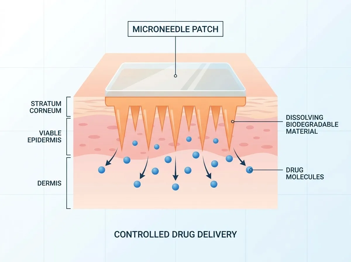
Figure 1: Drug Release and Penetration Mechanism of a Microneedle Patch
The epidermis is made up of several sublayers, including stratum corneum that serves as the main barrier against molecular penetration. This layer is made up of dead, flattened keratinocytes embedded into lipid matrix, to form robust and highly resistant barrier that prevents most molecules, particularly those with significant molecular weight or hydrophilic properties, from diffusing passively into deeper layers. Below this is the viable epidermis, where living keratinocytes and melanocytes carry out immunological and pigment-related functions. The dermis beneath is well-supplied with blood vessels and nerves, that contain fibroblasts, collagen, elastin fibers, and immune cells, which provides structural support that serves as the target for many transdermally delivered therapies and treatments. Lastly, the hypodermis, primarily made up of adipose and connective tissue, offers insulation and cushioning, acting as a reservoir for lipophilic drugs. 4-6
Transdermal drug delivery aims to transport therapeutic agents across the above mentioned layers to achieve systemic or localized effects while avoiding the disadvantages of oral or injectable routes such as first-pass metabolism or invasiveness respectively. But, the considerable barrier nature of the stratum corneum has limited the range of deliverable drugs to small, lipophilic molecules. To overcome all these limitations, various technologies like microneedle patches have introduced a minimally invasive and effective strategy to bypass the stratum corneum by painlessly creating microchannels through the skin’s outer layer (Fig 1). Now microneedles enables enhanced permeability of broad spectrum of pharmacologically active molecules, even the macromolecules and hydrophilic compounds, expanding the therapeutic potential of transdermal delivery. These devices provide controllable drug administration with better patient compliance and lesser side effects by modifications in their geometry, materials, and insertion parameters based on detailed understanding of the skin anatomy and physiology as par need. 7
The engineering of such microneedle systems, from fabrication techniques to their promising clinical applications, emphasizing how the interplay between device design and skin biology can optimize therapeutic outcomes.
2. MICROFABRICATION
Microfabrication technologies, particularly those developed for microelectromechanical systems (MEMS), have played a role in advancing the design and production of miniaturized medical devices. These tools enable the better fabrication of microstructures with dimensions suitable for sharp, robust microneedles capable of penetrating the skin barrier with minimal invasiveness. MEMS-based microfabrication methods have facilitated the manufacture of microneedle patches, enabling efficient transdermal drug delivery, biosensing, and diagnostic sampling by accessing interstitial fluid (ISF) (Fig 2) in a patient-friendly manner, i.e. with no or minimal pain.
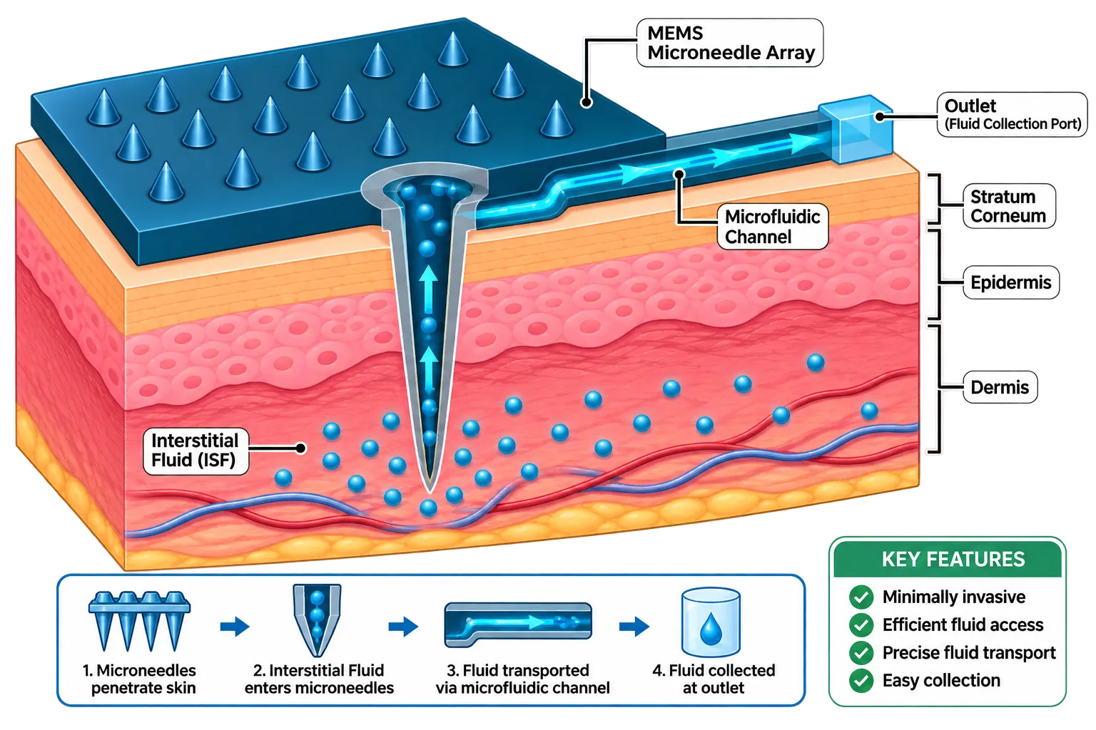
Figure 2: Schematic Representation of MEMS Microneedle Array
Microneedles are a micron-sized devices designed to painlessly penetrate the stratum corneum—the skin’s primary barrier—without damaging underlying tissues. They are fabricated using MEMS techniques such as lithography, etching, micromolding, and additive manufacturing. These techniques enable production of various types of microneedles such as solid, hollow, coated, dissolving, and hydrogel-forming variants, which are mainly made for controlled drug release or biosensing. For example, silicon-based microneedles are mainly shaped using standard MEMS processes to include internal channels for fluid injection and sampling 8.
Recent advances in micro- and nanofabrication have combined 3D printing with lithography to improve microneedle devices, allowing the integration of conductive inks and hybrid materials. This has resulted in improved sensing performance and lower manufacturing costs.9 Also, the use of additive manufacturing techniques provides fast modelling possibilities for optimisation of microneedle designs in terms of mechanical strength and skin penetration efficacy10.
Besides drug delivery, MEMS sensors with microneedles find other applications. They can be used for personalised health monitoring, extracting biomarkers from ISF for real-time physiological monitoring, including cancer biomarker detection and metabolic profiling. MEMS-fabricated microneedle devices are progressing towards industrial-scale production frameworks that allow for patient-centric, personalised microneedle designs specific to individual anatomical and therapeutic needs, thereby advancing clinical translation 11.
So,MEMS-enabled microfabrication provides the foundational platform for developing sophisticated, minimally invasive microneedle systems with applications spanning transdermal drug delivery, diagnostics, and wearable health monitoring. The synergy of microfabrication precision and biomedical engineering continues to propel these devices toward enhanced patient outcomes and widespread clinical use.12
3. Classification and Structural Design of Microneedles
Microneedles are generally classified into five distinct categories (fig 3) based on their materials, structural design, and mechanism of drug release.
3.1 Solid Microneedles
2.2 Coated Microneedles
3.3 Dissolving Microneedles
Mechanism: Encapsulates the therapeutic agent directly within a biodegradable, water-soluble polymer matrix ("poke-and-release"). The entire needle structure degrades within the tissue, leaving no sharp biohazardous waste behind.
MATERIALS
: Hyaluronic acid, carboxymethyl cellulose (CMC), polyvinylpyrrolidone (PVP), silk fibroin, and sugars (maltose, sucrose).
Limitation: Storage stability can be compromised by humidity, and manufacturing temperatures must accommodate fragile biomolecules. 15
3.4 Hollow Microneedles
Mechanism: Functions like a miniaturized array of traditional hypodermic needles ("poke-and-flow"). Liquid formulations are actively pumped or passively infused through an internal lumen directly into the viable skin layers.
MATERIALS
: Glass, silicon, metals, and ceramics.
Limitation: Prone to clogging by tissue fragments during insertion; requires complex microfluidic integration.16
3.5 Hydrogel-Forming Microneedles
Mechanism: Made of crosslinked hydrophilic polymers that contain no drug in their initial dry state, or act as a rate-controlling membrane. Upon skin insertion, they uptake ISF and swell. This swelling creates a continuous aqueous pathway through which drugs diffuse from an attached reservoir into the systemic circulation.
MATERIALS
: Poly (vinyl alcohol) (PVA), poly (methyl vinyl ether-co-maleic anhydride) (PMVE/MA).
Advantage: Intact removal after therapy ensures no polymer residue remains in the skin; excellent for high-dose drug delivery and diagnostic fluid sampling.17
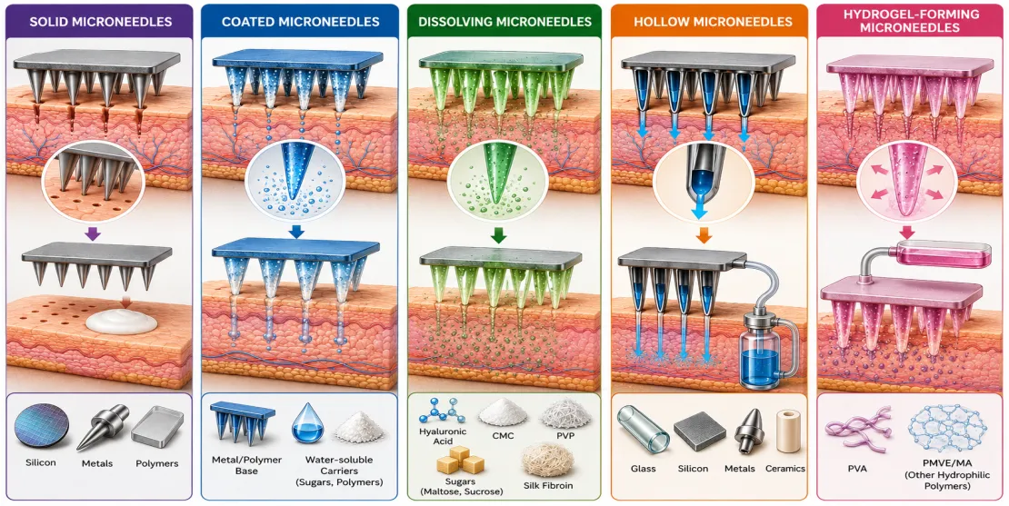
Figure 3: Classification and Structural Features of Different Types of Microneedle Patches
4. Advanced Fabrication Technologies
The transition of MNs from laboratory scale to industrial manufacturing has driven the adoption of highly precise microfabrication methods (Fig 4).18
| Fabrication Method | Primary Materials | Key Advantages | Major Limitations |
|---|---|---|---|
| Photolithography & Etching | Silicon, Glass | Sub-micron precision; highly reproducible shapes. | Expensive; requires cleanroom access; multi-step chemical waste. |
| Micro-Molding (Cast Molding) | Polymers, Hydrogels | Low cost; scalable; mild conditions preserve protein stability. | Relies on a master mold; high risk of air bubbles or incomplete filling. |
| 3D Printing (SLA, TPP) | Photopolymers, Ceramics | Rapid prototyping; custom geometry for personalized medicine. | Limited selection of biocompatible resins; slow throughput for mass production. |
| Laser Ablation | Metals, Polymers | Rapid production of master molds without chemicals. | Thermal damage to edges; surface roughness requires secondary polishing. |
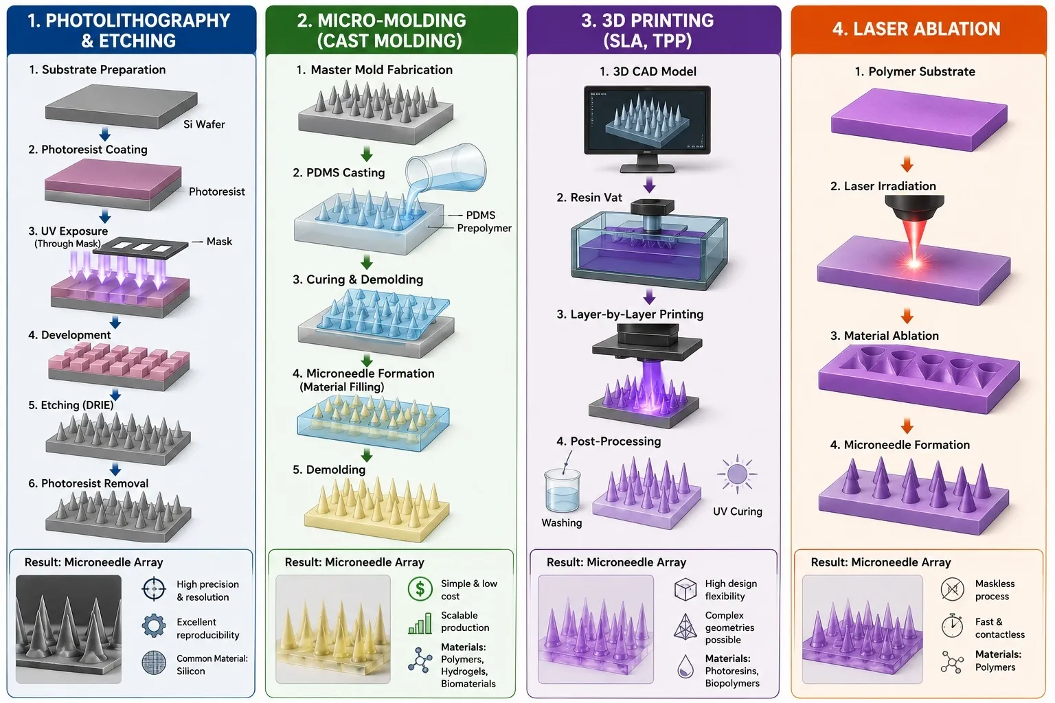
Figure 4: Different Fabrication Methods Used for MNs
5. Precision Transdermal Delivery Applications
5.1 Stimuli-Responsive "Smart" Drug Delivery
Modern microneedle systems are engineered to release therapeutics in response to specific physiological or external triggers, minimizing off-target side effects.
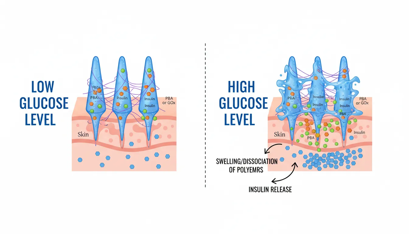
Figure 5: Stimuli-responsive Behavior of Glucose-Sensitive Microneedles Under Elevated Glucose Levels.
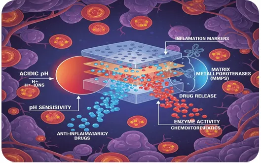
Figure 6: Microenvironment-targeted Drug Delivery via pH- and Enzyme-Sensitive Patches.
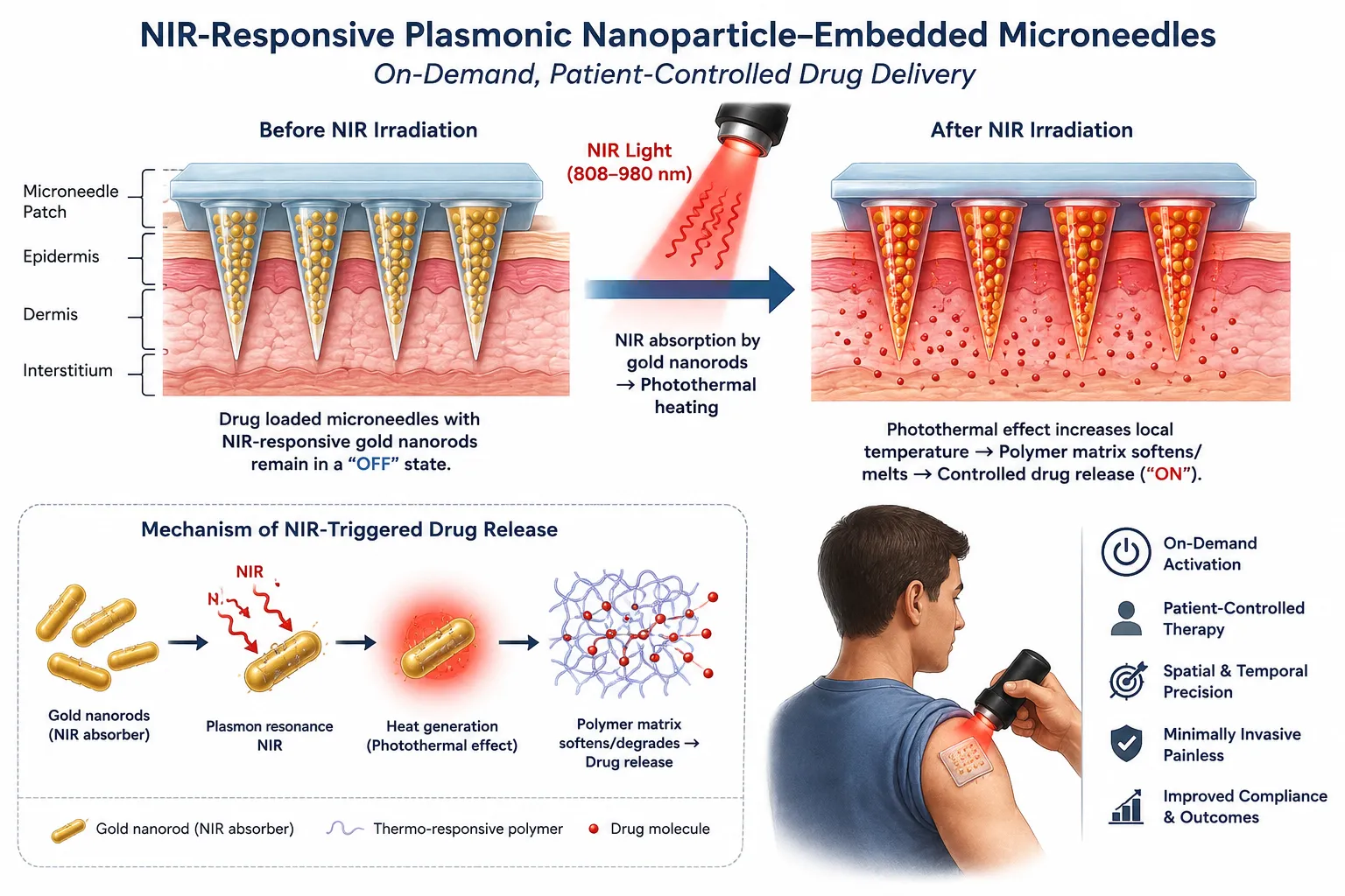
Figure 7: Photothermally Triggered Drug Delivery via NIR-responsive Plasmonic Nanoparticles (Patient Controlled)
5.2 Vaccine Delivery and Immunomodulation
The skin is highly immunogenic, crowded with Langerhans cells and dermal dendritic cells.
MN patches target these antigen-presenting cells directly, generating robust mucosal and systemic immune responses (Fig 8) at a fraction of the dosage required by intramuscular injections (dose sparing).
Solid MN formulations eliminate the need for a cold chain, offering dry-state stability for vaccines targeted at developing regions.
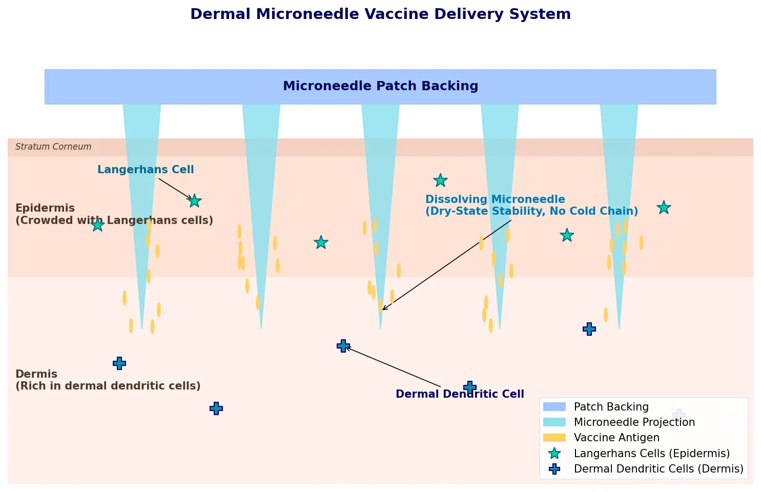
Figure 8: Mechanism of Direct Antigen-Presenting Cell (APC) Targeting by Microneedle (MN) Patches.
5.3 Non-Dermal and Emerging Applications of Microneedle Technology
Microneedle technology, initially developed for transdermal drug delivery, has expanded significantly beyond skin applications to various other tissues and organs, including the eye and several less explored sites. This broadening of applications leverages the minimally invasive, targeted, and efficient delivery capabilities of microneedles to overcome biological barriers that traditionally limit therapeutic efficacy.
5.4 Ocular Applications of Microneedles
Ocular drug delivery presents unique challenges due to multiple physiological barriers, limited permeability, rapid clearance, and poor bioavailability with traditional topical and systemic formulations. Frequent administration of eye drops or invasive intraocular injections are often required, both presenting drawbacks like discomfort, low patient compliance, inflammation, and risk of infection.
Microneedles offer a promising alternative for ocular drug delivery by enabling minimally invasive, localized, and enhanced penetration through ocular barriers such as the cornea and sclera. Various studies and reviews have highlighted the advantages of microneedle arrays that are capable of delivering a wide range of therapeutics—including monoclonal antibodies, small molecules, and sustained-release formulations—directly into ocular tissues with improved bioavailability and reduced side effects compared to traditional methods [30] [31] [32] [33] [34] .
In particular, responsive microneedles—those that release drugs upon physiological or external stimuli such as pH, temperature, enzymes, or light—enhance precision in ocular therapeutics, enabling personalized and on-demand treatment for various eye diseases [31]. These technologies improve patient compliance and treatment efficacy for conditions affecting both the anterior and posterior segments of the eye, including age-related macular degeneration and glaucoma [33].
The exploration of microneedles for ocular use has also advanced toward clinical translation, with ongoing efforts to address manufacturing, regulatory, and biocompatibility challenges. The minimally invasive application and capability to sustain drug release highlight microneedles as a revolutionary approach in eye care [30] [35] .
5.5 Other Non-Dermal Applications of Microneedles
Beyond ocular sites, microneedles have been adapted for drug delivery in a variety of other unconventional and poorly accessible sites, where conventional administration routes are limited or ineffective. A comprehensive review of non-dermal microneedle applications includes the oral mucosa, vaginal mucosa, gastric mucosa, nail, scalp, vascular tissue, and even cardiac tissue:
These applications exploit the microneedles’ ability to pierce biological barriers that are typically challenging to traverse non-invasively, thereby maximizing drug efficacy while reducing required doses and systemic side effects.
6. Translational Hurdles and Regulatory Frameworks
Despite promising preclinical successes, translating microneedle patches into commercial clinical products presents several unique hurdles:
7. Clinical Trials: Current Status
The translation of microneedle (MN) patches from preclinical research to human use is meticulously tracked across various global clinical trials. Regulatory bodies like the FDA classify these innovative systems as combination products, requiring extensive evaluation of both device mechanical safety and therapeutic pharmacokinetics. [46, 47, 48, 49, 50]
The table below compiles specific, well-documented human clinical trials tracking different microneedle patch systems across various evaluation phases:
| Clinical Trial Identifier | Trial Phase | Therapeutic Agent / Indication | Microneedle Type & Material | Key Findings / Clinical Status |
|---|---|---|---|---|
| NCT02438423 | Phase 1 | Inactivated Influenza Vaccine (Flu Immunization) | Dissolving (Polymers via Micromolding) | Found to be safe, well-tolerated, and generated robust immune responses similar to standard IM injections. Strongly preferred by >70% of participants. |
| NCT03607903 | Phase 1/II | Adalimumab / Auto-immune diseases | Hollow (Silicone, 3 pyramid-shaped needles) | Successfully completed; evaluated continuous micro-infusion delivery kinetics for macromolecular biologics. |
| NCT02192021 | Phase 1 | Doxorubicin / Cutaneous T-cell Lymphoma (CTCL) | Dissolving (Polymers via Micromolding) | Completed localized oncology assessment evaluating targeted intradermal delivery to skin lesions while minimizing systemic toxicities. |
| NCT05078463 | Phase 2 | Topical Anesthesia Enhancement / Paediatric Thalassemia | Dissolving (400 μm length) | Evaluated for rapid, localized skin numbing prior to clinical needle procedures to reduce procedural pain and anxiety in pediatric patients. |
| NCT02955576 | Clinical Evaluation | Hyaluronic Acid Patch / Topical Psoriatic Plaques | Dissolving (Hyaluronic acid matrix) | Completed randomized controlled trial utilizing a split-body design to assess enhanced transdermal delivery of topical psoriasis treatments. |
| NCT03207763 | Phase 1 | Placebo Microneedle / Safety & Reactogenicity in Infants | Solid, water-soluble conical matrix | Completed pediatric safety trial evaluating skin tolerance, wearability, and acceptability profiles in infants and young children. |
| NCT05108714 | Clinical Evaluation | Lidocaine Hydrochloride / Local Anesthesia | Hollow (Silicone, 0.6 mm length) | Completed micro-infusion study evaluating immediate local numbing on human skin. |
8. Clinical Prospects and Conclusion
The horizon for microneedle patches is rapidly expanding. Dozens of clinical trials are underway globally, investigating patches for influenza vaccination, continuous glucose monitoring, osteoporosis management (parathyroid hormone delivery), and localized dermatological cosmetics.
As material science converges with digital healthcare, future microneedles will likely incorporate wireless microfluidic components, allowing patches to communicate directly with smartphone apps to log dosing data and update physician networks. By getting high patient compliance with absolute dosing accuracy, engineered microneedle patches are positioned to redefine the landscape of precision medicine and self-administered therapeutics.
The clinical trial data for microneedle patches demonstrates a successful transition from laboratory concepts to validated transdermal medical platforms, highlighting proven patient safety, broad therapeutic versatility, and advancements in targeted micro-infusion systems. These early-phase human trials establish the necessary clinical foundation for upcoming Phase 3 efficacy studies and decentralized healthcare applications.
REFERENCES


Manjunath Halkeri, Priyaganga, Dr. Shravan L. Nargund, Engineering Microneedle Patches for Precision Transdermal Delivery: From Fabrication to Clinical Prospects, Int. J. of Pharm. Sci., 2026, Vol 4, Issue 10, 3703-3714. https://doi.org/10.5281/zenodo.23640336
 10.5281/zenodo.23640336