

We use cookies to ensure our website works properly and to personalise your experience. Cookies policy
Department of Pharmaceutics, Bharathi College of Pharmacy, Bharathinagara, Mandya, Karnataka, India-571422.
Background: Invasomes are flexible vesicular drug delivery systems containing phospholipids and terpenes that can provide controlled drug release and enhance topical drug delivery. However, the performance of invasomal systems is strongly influenced by the concentration of formulation components. Therefore, systematic optimization of formulation variables is essential for obtaining a formulation with desirable drug-loading and release characteristics. Objective: The present study was undertaken to optimize Clotrimazole-loaded invasomes using a Face-Centred Composite Design (FCCD) and to investigate the individual and interactive effects of soya lecithin, menthol and thymol on drug content and in-vitro drug release. Methods: Clotrimazole-loaded invasomes were prepared by the mechanical dispersion technique. A three-factor FCCD was employed with soya lecithin, menthol and thymol as independent formulation variables. Twenty experimental formulations were prepared according to the design matrix. Drug content and cumulative drug release were selected as the principal responses for optimization. Design-Expert software was used for sequential model selection, analysis of variance, factor profiling, interaction analysis, contour-plot analysis, diagnostic evaluation and numerical optimization. The optimized formulation was subsequently incorporated into Carbopol 934 gel and evaluated for physicochemical characteristics and drug-release behaviour. Results: The linear model was selected for drug content, whereas a quadratic model was selected for drug release. The drug-content model was significant (F = 5.17, p = 0.0109), with menthol and thymol significantly influencing the response. The quadratic drug-release model was highly significant (F = 26.67, p < 0.0001), with an r² of 0.9600, adjusted r² of 0.9240 and predicted r² of 0.7703. Soya lecithin showed a negative effect on drug release, whereas menthol and thymol showed positive effects. Significant AC and BC interactions were observed. Numerical optimization generated a formulation containing 150 mg soya lecithin, 299.47 mg
Topical drug delivery offers several advantages for the treatment of localized skin disorders. However, the effectiveness of topical therapy can be influenced by the physicochemical properties of the drug and the barrier characteristics of the skin.1
Clotrimazole is an antifungal drug widely used in the treatment of superficial fungal infections. Its poor aqueous solubility presents a challenge in the development of effective topical delivery systems.2
Invasomes are deformable vesicular drug delivery systems containing phospholipids and terpenes. The incorporation of terpenes into the vesicular system provides flexibility to the vesicular membrane and makes invasomes a potential carrier for topical drug delivery.3
The performance of an invasomal system depends considerably on the concentration of its formulation components. Soya lecithin contributes to the formation of the vesicular structure, while terpenes such as menthol and thymol influence the characteristics of the vesicular system and drug-release behaviour. Consequently, changes in the concentration of these components can produce considerable variation in formulation responses.4
Traditional formulation development based on changing one variable at a time is time-consuming and does not adequately demonstrate interactions between formulation variables. Design of Experiments (DoE) provides a systematic approach for investigating several formulation variables simultaneously. Response Surface Methodology (RSM) further permits mathematical modelling of responses and identification of formulation conditions within the investigated design space.5
In the present study, a Face-Centred Composite Design (FCCD) was employed to optimize Clotrimazole-loaded invasomes. Soya lecithin, menthol and thymol were selected as independent variables, while drug content and cumulative drug release were selected as formulation responses.
The objective of the present investigation was to systematically evaluate the influence of soya lecithin, menthol and thymol on the critical responses of Clotrimazole-loaded invasomes and to identify an optimized formulation using numerical optimization in Design-Expert software.6
Materials Used
Clotrimazole was purchased from Balaji drug store Mumbai, Menthol and Thymol collected from SD Fine Chem., Mumbai, Soya lecithin collected from Sonic Biochem Extraction Ltd., Indore and Ethanol collected from Thermo Fisher Scientific India Pvt. Ltd. Mumbai.
Preformulation studies7-14
Determination of melting point of the drug
Melting point of Clotrimazole was determined by using capillary tube method using melting point apparatus and DSC.
Fourier transforms infrared spectroscopy (FTIR) studies
Drug-excipients compatibility studies were carried out using FTIR. Infrared spectrum of pure drug (Clotrimazole) and physical mixture of Clotrimazole and Menthol, Thymol, Soya Lecithin was recorded from the ranges to 400 – 4000 cm⁻¹. FTIR analysis was carried out using a Shimadzu FTIR spectrophotometer.
Determination of absorption maximum (λ max) of the drug
Clotrimazole 100 mg/ml concentration was prepared in methanol and buffer pH 6.8 (1:1) the solution was scanned from 200-400 nm by UV Spectrophotometer and a spectrum was observed for absorption maxima.
Method of Preparation
Preparation of invasomes by mechanical dispersion technique
Invasomes were prepared by the mechanical dispersion technique. Accurately weighed phospholipid was dissolved in ethanol to form the organic phase. To this mixture, terpene and the drug sample were added and mixed thoroughly. The resulting mixture was stirred using a magnetic stirrer until all the components were completely dissolved. The organic phase was then added slowly in a dropwise manner to the aqueous phase under continuous stirring to obtain a uniform dispersion. Finally, the dispersion was sonicated using a probe sonicator for 5–10 minutes to obtain uniform Invasomes. 15
Preparation of clotrimazole loaded invasomal gel
Carbopol 934 was hydrated in purified water, followed by slow addition of clotrimazole-loaded invasomal suspension containing 100 mg drug with gentle stirring. Triethanolamine was added dropwise for neutralization, and the mixture was gently stirred to obtain a homogeneous, lump-free gel.
Table 1: Formulation Design by Using Face Central Composite Design (FCCD)
| Formulation code | Run | Factor 1 | Factor 2 | Factor 3 |
|---|---|---|---|---|
| A (Soya lecithin) | B (Menthol) | C (Thymol) | ||
| F1 | 1 | -1 | -1 | -1 |
| F2 | 2 | +1 | -1 | -1 |
| F3 | 3 | -1 | +1 | -1 |
| F4 | 4 | +1 | +1 | -1 |
| F5 | 5 | -1 | -1 | +1 |
| F6 | 6 | +1 | -1 | +1 |
| F7 | 7 | -1 | +1 | +1 |
| F8 | 8 | +1 | +1 | +1 |
| F9 | 9 | -1 | 0 | 0 |
| F10 | 10 | +1 | 0 | 0 |
| F11 | 11 | 0 | -1 | 0 |
| F12 | 12 | 0 | +1 | 0 |
| F13 | 13 | 0 | 0 | -1 |
| F14 | 14 | 0 | 0 | +1 |
| F15–20 | 15–20 | 0 | 0 | 0 |
Evaluation of Invasomes16-20
Surface morphology by scanning electron microscopy (SEM), Vesicle size analysis, Vesicle size analysis by zeta analyser, Zeta potential determination, Drug content determination, Entrapment Efficiency determination, In-vitro diffusion study.
Surface morphology by scanning electron microscopy (SEM)
The surface morphology of Clotrimazole Invasomes was determined by SEM. Invasomes were coated with Gold-palladium alloy coating done by sputter coater (Polaron SEM coating system). Then the samples were observed under the SEM (JSMT330A, JEOL) at a beam voltage of 15 KV.
Vesicle size analysis by optical microscopy
A drop of Invasomes with distilled water formulation is added on glass slide without cover slip to observe the formation of Invasomes. Vesicle size analysis was carried out using an optical microscope with a calibrated eye piece micrometer. About 200 Invasomes were measured individually, average was taken and their size distribution range and mean diameter were calculated.
Vesicle size analysis by zeta analyser
The vesicle size of the vesicle after hydration of Invasomes was also determined by Microtac Zeta potential Analyzer optimized formulation. Particle size measurements were carried at 25 °C by photon correlation spectroscopy on Malvern Zetasizer Nano ZR instrument (Malvern, Model ZEN3600) armed with a 4 mW He-Ne laser (633 nm). Samples were put in transparent disposable cuvette and the dispersant viscosity and refractive index was set to 0.8872 cP and 1.330 at 25 °C. vesicle size was analyzed by the Dispersion Technology Software provided by Malvern Instruments. All samples were kept in refrigerator at 4 °C prior to characterization.
Zeta potential determination
The surface charge of Clotrimazole loaded Invasomes prepared with soya lecithin was determined to evaluate their stability. Zeta potential of the Invasomal vesicles was measured using a Microtrac instrument. The obtained values reflected the electrostatic repulsion between vesicles, which plays a critical role in preventing aggregation and ensuring colloidal stability of the Invasomal dispersion.
Drug content determination
An invasomal formulation equivalent to 10 mg of Clotrimazole was accurately transferred into a volumetric flask and dissolved in 10 mL of methanol. The resulting dispersion was centrifuged at an appropriate speed for 10–15 minutes to separate the vesicles and obtain a clear supernatant containing the drug. The supernatant was collected and suitably diluted with phosphate buffer (pH 6.8). The absorbance of the diluted solution was measured using a UV–Visible spectrophotometer at 260 nm.
Entrapment efficiency determination
The entrapment efficiency of Clotrimazole Invasomes was determined after hydration with distilled water. 2 ml of distilled water was added to the Invasomes equivalent to 2 mg of drug then the mixture was shaken manually for 2min. For the separation of unentrapped Clotrimazole, the Invasomal suspension was subjected to centrifugation on a cooling centrifuge at 25000 rpm for The For the separation of un entrapped drug. The clear supernatant (0.1 ml) was taken and diluted to 10ml with methanol and water solution and absorbance was recorded at 260 nm using UV visible spectrophotometer then calculates the percentage drug in each formulation.
Vesicle size analysis
A drop of Invasomes with distilled water formulation is added on glass slide without cover slip to observe the formation of Invasomes. Vesicle size analysis was carried out using an optical microscope with a calibrated eye piece micrometer. About 200 Invasomes were measured individually, average was taken and their size distribution range and mean diameter were calculated.
In-vitro drug release
The drug release was evaluated using a treated cellophane membrane attached to one end of a tube containing the formulation. The tube was suspended in 200 mL of pH 6.8 buffer and maintained at 37 ± 0.5°C with continuous stirring at 100 rpm. Samples (1 mL) were withdrawn at predetermined intervals (1, 2, 4, 6, and 8 hours) and replaced with an equal volume of fresh buffer to maintain sink conditions. The withdrawn samples were diluted to 10 mL with pH 6.8 buffer and analyzed using a UV spectrophotometer at 260 nm. The cumulative percentage of drug released was calculated and plotted against time. Release kinetics and the mechanism of Clotrimazole release were determined by fitting the data to various kinetic models.
Release kinetic analysis
The drug release kinetic studies were done by various mathematical models. The model that gives high ‘r’ value is considered as he best fit of the release data. The release constant was calculated from the slope of the appropriate plots, and the regression coefficient (r2) was determined.
Stability studies as per ICH guidelines
Intermediate stability studies were performed for 2 months as per ICH guidelines. The optimized formulation was kept at 30°C±2°C and 65 ± 5% RH in stability chamber. At the end of 0, 30, 60 days intervals regularly tested for % drug content and drug release were fixed as physical parameters for stability testing.
Evaluation of Invasomal Gel
Appearance
The prepared gels were examined for clarity, Color, homogeneity, presence of foreign particles and fiber.
pH
1gm of gel was dispersed in 100 ml distilled water and the pH was examined using digital pH meter.
Spreadability study
The spreadability of the Invasomal gel was studied by measuring the change in diameter when 500 mg of gel was placed between 2 horizontal plates of 6cm with a standardized weight of 20g placed over it. Calculate the time required for 6cm replacement while applying 20gm of weight (El-Tokhy et al., 2021). Calculate the spreadability using formula:
Spreadability=(weight tied to upper slide (g) X length of glass slide (cm))/(Time taken in sec)
Extrudability study
The prepared Invasomal gel was filled in collapsible tubes and its extrudability was estimated in terms of weight in grams required to produce a 0.5 cm ribbon of gel in 10 seconds.
Viscosity
For determining the viscosity of the invasomal gel Brookfield viscometer (DV-E Brookfield Engineering Laboratories, MA, USA) at 37°C with spindle No.64 was used. An appropriate amount of gel was placed onto the centre of the viscometer plate directly below the spindle using the spatula and viscosities were measured.
Drug content determination (%)
Clotrimazole loaded Invasomal gel was assayed by an UV spectrophotometric method. Invasomal gel containing equivalent to 10 mg of drug were dissolved in a 10 ml of methanol. After suitable dilution absorbance was measured by UV spectrophotometer against blank at λ max 260 nm and drug content was calculated.
%Drug content=(Actual Drug)/(Theoretical Drug)×100
In-vitro diffusion study
The diffusion medium used was phosphate buffer pH 6.8. Assembly of diffusion cell for in-vitro diffusion studies the diffusion cell was designed as per the dimension given. Diffusion cell with an effective diffusion area of 3.14 cm² was used for in-vitro permeation studies. One gm of Invasomal gel containing Clotrimazole was placed in the donor compartment and the receptor compartment was filled with receptor fluid. The egg membrane was mounted carefully and ensured intimate contact with the receptor fluid. The speed of stirring was kept constant throughout the experiment. At 0.5, 1, 2, 4, 6 and 8 h, 1 ml sample was withdrawn and replaced with an equal volume of receptor fluid to maintain sink conditions. The samples were suitably diluted and absorbance was measured at 260 nm using a UV spectrophotometer.
Stability studies as per ICH guidelines
Intermediate stability studies were performed for 2 months as per ICH guidelines for optimized formulation. Drug content and % CDR at 8hrs were fixed as evaluation parameter for stability study. Stability study was carried out by storing the prepared Invasomal gel at intermediate stability study condition 30°C±2°C and 65 ± 5% RH for a period of 2 months duration. The parameters were checked at 30 days intervals as per ICH guidelines.
RESULT
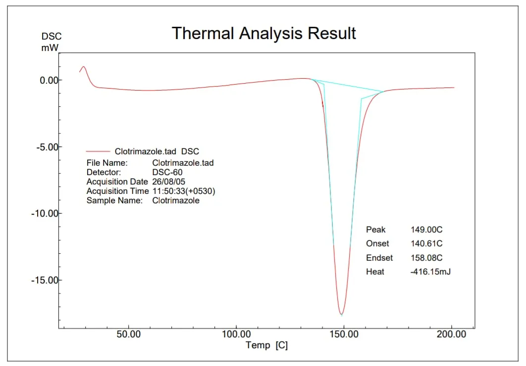
Figure 1: DSC Graph of Pure Clotrimazole
Table 2: Melting Point Report of Clotrimazole
| Reported | Method | Results observed | |||
|---|---|---|---|---|---|
| 140-150℃ | Thiele’s tube method | Trail 1 | Trail 2 | Trail 3 | Average |
| 148℃ | 147℃ | 148℃ | 147.6℃ | ||
| DSC | 149℃ | ||||
Table 3: Solubility Analysis Report
| Media | Solubility (mg/ml) | Parts of solvent required for 1 part of solute | Solubility Criteria |
|---|---|---|---|
| Water | 0.096 | 10416 | Practically insoluble |
| pH 1.2 | 0.062 | 15976 | Practically insoluble |
| pH 4.6 | 0.076 | 13044 | Practically insoluble |
| pH 6.8 | 0.127 | 7874 | Very slightly soluble |
| pH 7.4 | 0.119 | 8403 | Very slightly soluble |
| Methanol | 125.30 | 7.98 | Freely soluble |
Determination of Absorption Maxima (λmax)
Absorption Maxima (λ max) of the Clotrimazole was found to be 260nm in Methanol and buffer pH 6.8 (1:1) by using UV spectrophotometry.
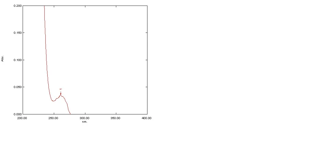
Figure 2: UV Spectrum of Clotrimazole at 10 Μg/ml Concentration.
Table 4: Standard Calibration Curve of Clotrimazole
| Absorbance at λmax 260 nm | ||||
|---|---|---|---|---|
| Conc (µg/ml) | T-1 | T-2 | T-3 | AVG ± SD |
| 0 | 0 | 0 | 0 | 0 |
| 50µg/ml | 0.151 | 0.153 | 0.152 | 0.152±0.001 |
| 100µg/ml | 0.283 | 0.285 | 0.281 | 0.283±0.002 |
| 150µg/ml | 0.42 | 0.423 | 0.422 | 0.422±0.002 |
| 200µg/ml | 0.542 | 0.543 | 0.543 | 0.543±0.001 |
| 250µg/ml | 0.675 | 0.677 | 0.675 | 0.676±0.001 |
| 300µg/ml | 0.816 | 0.817 | 0.816 | 0.816±0.001 |
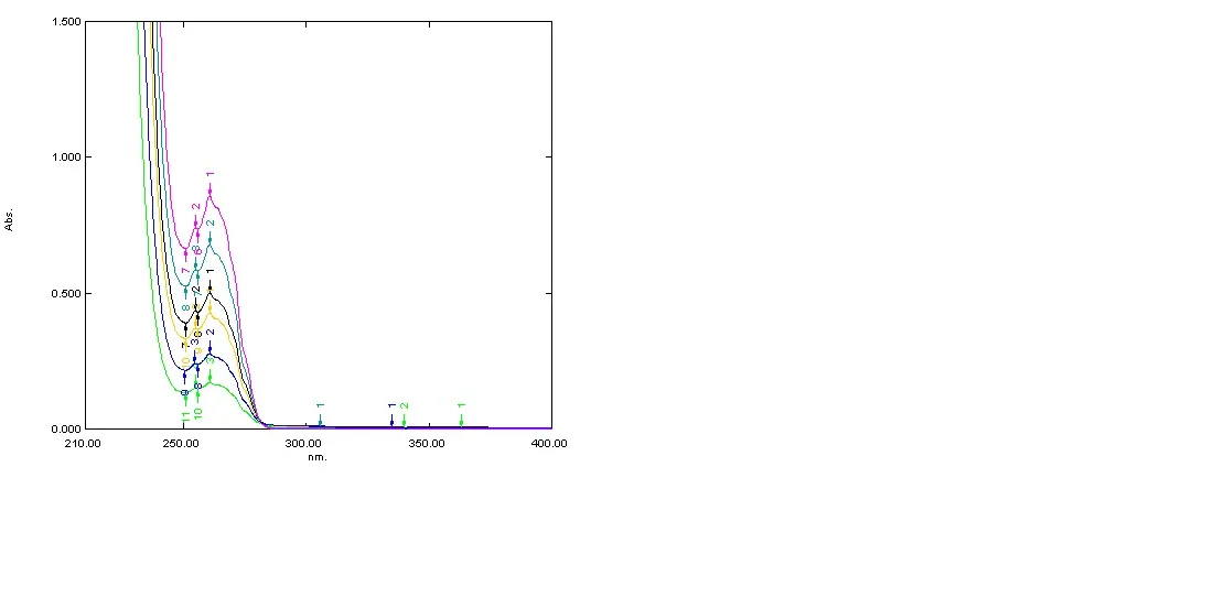
Figure 3: UV Spectrum of Clotrimazole at Different Concentration of 0-300 Μg/ml
[Figure - see the formatted PDF]
Figure 4: Plot of Standard Calibration Curve of Clotrimazole
Compatibility studies by FTIR spectrophotometer

Figure 5: FT-IR Spectrum of Pure Clotrimazole Drug
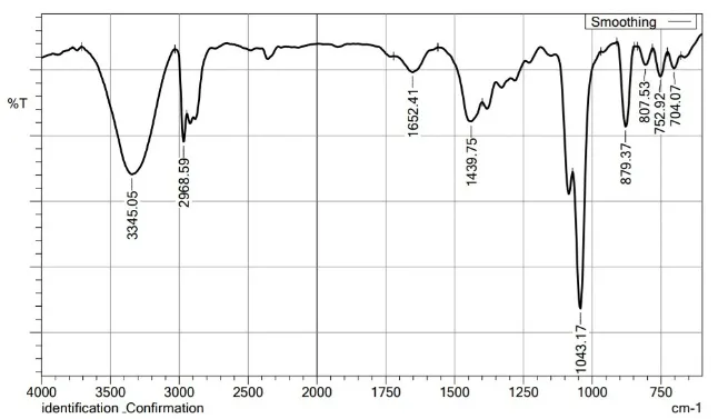
Figure 6: FTIR Spectrum of Drug and Invasome Formulation
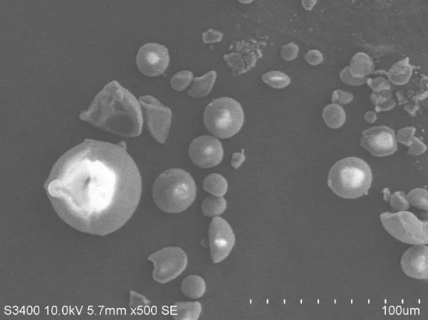
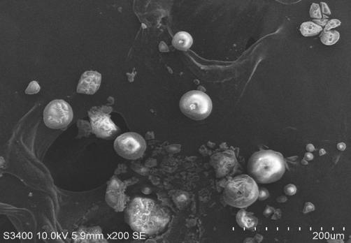
Figure 7: SEM Image of Optimized Invasomes
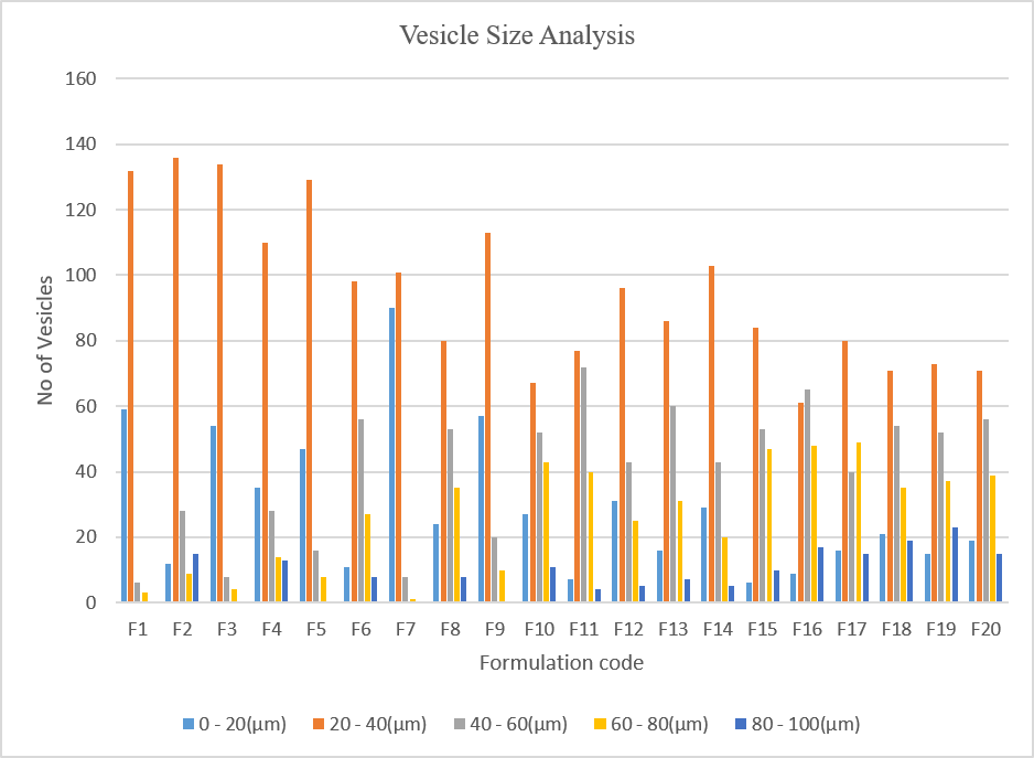
Figure 8: Vesicle Size Analysis for Formulation F1-F20
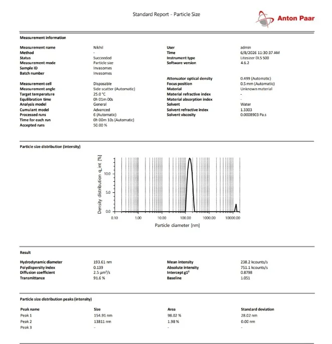
Figure 9: Vesicle Size Analysis of Optimized Invasomes by Zeta Analyser

Figure 10: Zeta Potential of Optimized Invasomes
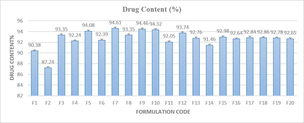
Figure 11: Drug Content for Formulated Invasomes F1-F20
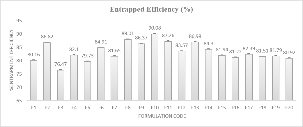
Figure 12: Entrapment Efficiency for Formulated Invasomes F1-F20
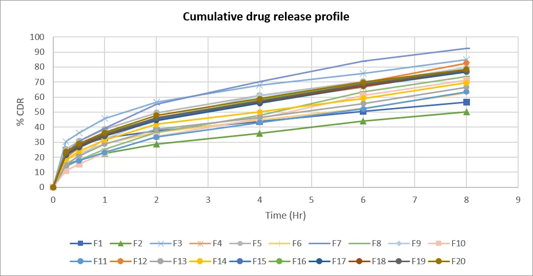
Figure 13: In-vitro Diffusion Study of Formulation F1-F20
Table 5: Release Kinetic Analysis
| Formulation code | Zero order | First order | Higuchi | Peppas model | |
|---|---|---|---|---|---|
| R2 | n value | ||||
| F1 | 0.7273 | 0.9458 | 0.9140 | 0.9850 | 0.2467 |
| F2 | 0.8585 | 0.9338 | 0.9181 | 0.9830 | 0.2632 |
| F3 | 0.7629 | 0.8435 | 0.9195 | 0.9255 | 0.2909 |
| F4 | 0.8519 | 0.8364 | 0.8956 | 0.8975 | 0.2614 |
| F5 | 0.7966 | 0.8936 | 0.9219 | 0.9835 | 0.3358 |
| F6 | 0.8452 | 0.9306 | 0.9314 | 0.9277 | 0.2810 |
| F7 | 0.8601 | 0.9225 | 0.9650 | 0.9781 | 0.3695 |
| F8 | 0.9336 | 0.8907 | 0.9114 | 0.9662 | 0.2584 |
| F9 | 0.8778 | 0.7633 | 0.9068 | 0.9639 | 0.2392 |
| F10 | 0.9460 | 0.9023 | 0.8880 | 0.9378 | 0.2140 |
| F11 | 0.9106 | 0.8679 | 0.9228 | 0.9424 | 0.2893 |
| F12 | 0.8864 | 0.8845 | 0.9218 | 0.9567 | 0.2839 |
| F13 | 0.8664 | 0.8459 | 0.9428 | 0.9629 | 0.3271 |
| F14 | 0.8443 | 0.8378 | 0.8703 | 0.8997 | 0.2323 |
| F15 | 0.8531 | 0.8699 | 0.9170 | 0.9695 | 0.2718 |
| F16 | 0.8455 | 0.9639 | 0.9782 | 0.9985 | 0.3575 |
| F17 | 0.8598 | 0.9628 | 0.9656 | 0.9534 | 0.3598 |
| F18 | 0.8543 | 0.9928 | 0.9756 | 0.9463 | 0.3434 |
| F19 | 0.8479 | 0.9246 | 0.9559 | 0.9484 | 0.3512 |
| F20 | 0.8696 | 0.9284 | 0.9666 | 0.9669 | 0.3612 |
Table 6: Results of Intermediate Stability Studies of Optimized Invasome Formulation at 30°C±2°C and 65 ± 5% RH.
| Parameters | Duration in days | ||
|---|---|---|---|
| 0 | 30 | 60 | |
| % DC | 94.81 | 93.86 | 92.71 |
| % CDR | 92.47 | 91.84 | 90.89 |
Optimization by DoE Software – Design Expert
a) Optimization analysis of response % drug content
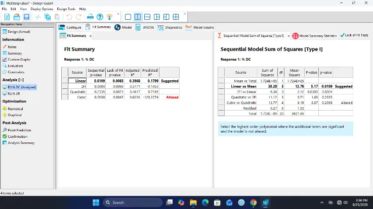
Figure 14: Model Selection Summary on Response % DC
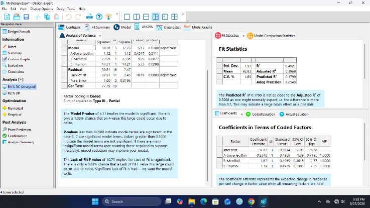
Figure 15: ANOVA Results on Response % DC
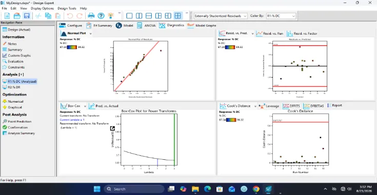
Figure 16: The Diagnostic Plot on Response % DC

Figure 17: Factor Profiler of Different Factors on Response % DC
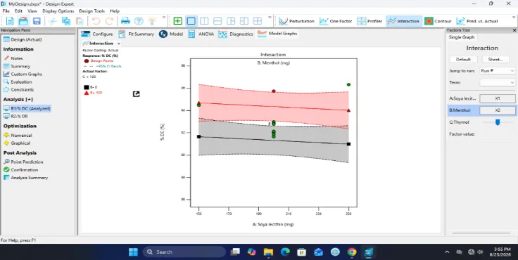
Figure 18: The Interaction Plot Illustrates the Combined Effect of Soya Lecithin (a) and Menthol (B) on Response % DC
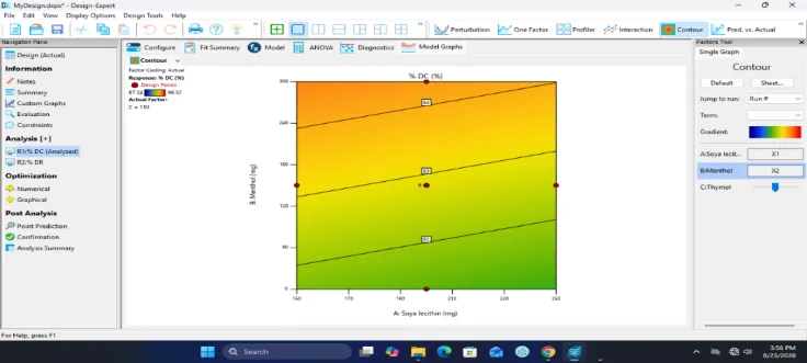
Figure 19: The Contour Plot Represents the Combined Effect of Soya Lecithin (a) and Menthol (B) on Response % DC
b) Optimization analysis of response % drug release

Figure 20: Model Selection Summary on Response % DR
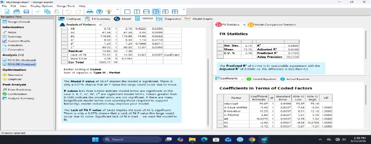
Figure 21: ANOVA Results on Response % DR
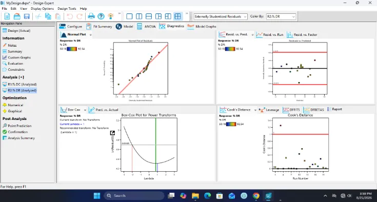
Figure 22: The Diagnostic Plot on Response % DR
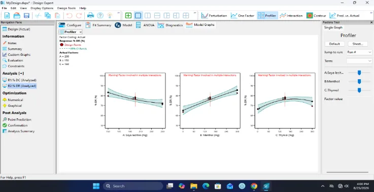
Figure 23: Factor Profiler of Different Factors on Response % DR
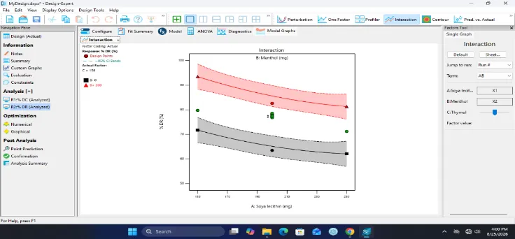
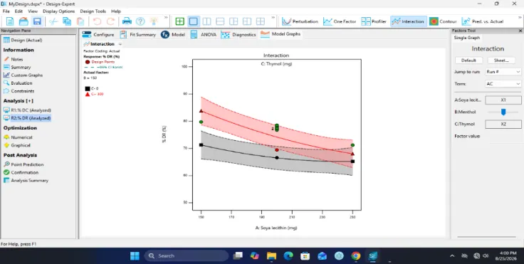
Figure 24: The Interaction Plot Demonstrates the Combined Effect of Soya Lecithin (a) and Menthol (B) on Response % DR
Figure 25: The Interaction Plot Between Soya Lecithin (a) and Thymol (C) on Response % DR
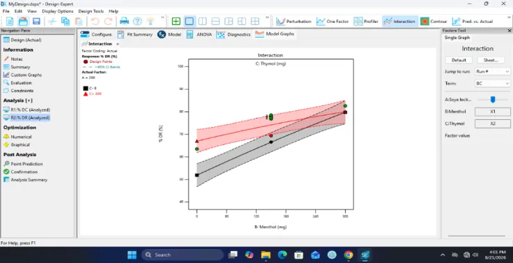
Figure 26: The Interaction Plot Between Menthol (B) and Thymol (C) on Response % DR
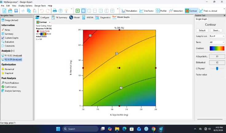
Figure 27: The Soya Lecithin and Menthol Contour Plot on Response % DR
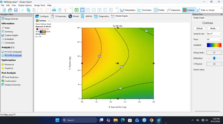
Figure 28: The Soya Lecithin and Thymol Contour Plot on Response % DR
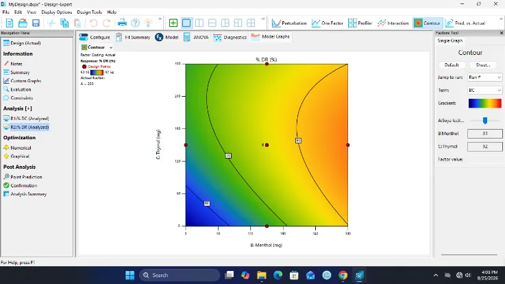
Figure 29: The Menthol and Thymol Contour Plot on Response % DR

Figure 30: The Predicted vs. Actual Plot on Response % DR
c) Predicted optimized invasome formulation obtained by design expert
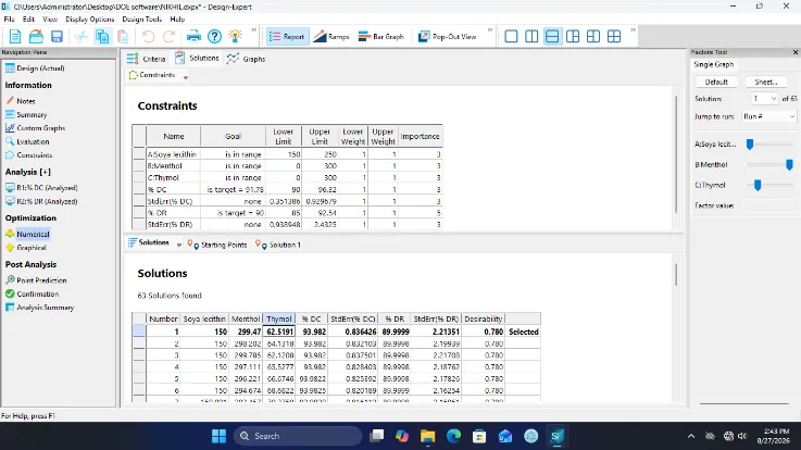
Figure 31: Design Space Solution for Optimized Invasome Formulation
Table 7: Optimized Invasome Formulation Result
| Formulation Code | Entrapment efficiency (%) | % CDR at 8 hours | Drug content (%) |
|---|---|---|---|
| IF | 94.71±0.10 | 92.47 | 94.75±0.14 |
Table 8: Results of Intermediate Stability Studies of Optimized Invasome Formulation at 30°C±2°C and 65 ± 5% RH
| Parameters | Duration in days | ||
|---|---|---|---|
| 0 | 30 | 60 | |
| % DC | 94.81 | 93.86 | 92.71 |
| % CDR | 92.47 | 91.84 | 90.89 |
Evalaution of Clotrimazole Invasomal Gel
The optimized formulation was incorporated into the gel by the use of Carbopol 934 polymer and was found to be suitable for topical delivery because of the desirable consistency.
Physical appearance
Physical appearance of formulation was checked and the results are given below table
Table 9: Physical Appearance of Invasomal Gel
| Formulation code | Appearance |
|---|---|
| IG | Transparent Color, odorless with smooth and uniform appearance |
Table 10: pH, Viscosity (cps), Spreadability, Extrudability and Drug Content of Invasomal Gel
| Formulation code | pH | Viscosity(cps) | Spreadability (g.cm/sec) |
Extrudability (g) |
Drug Content (%) |
|---|---|---|---|---|---|
| IG | 6.67±0.02 | 3563.33±2.08 | 11.63±0.63 | 123.33±5.77 | 92.88± 0.09 |
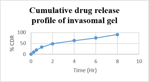
Figure 32: In-vitro Diffusion Study of Invasomal Gel
Table 11: Release Kinetic Analysis
| Formulation code | Zero order (r2) | First order (r2) | Higuchi (r2) | Peppas model | |
|---|---|---|---|---|---|
| r2 | n value | ||||
| F7IG | 0.9141 | 0.9654 | 0.9949 | 0.9853 | 0.529 |
DISCUSSION
Preformulation Studies
The melting point of Clotrimazole was found to be 147.6°C by Thiele’s tube method and 149°C by DSC, showing a sharp endothermic peak and confirming the crystalline nature and purity of the drug. The values were within the reported pharmacopoeial range of 140–150°C.
Clotrimazole exhibited poor aqueous solubility, with solubility of 0.119, 0.127, 0.076, 0.062 and 0.096 mg/mL in pH 7.4, pH 6.8, pH 4.6, pH 1.2 buffers and water, respectively. In contrast, its solubility in methanol was 125.30 mg/mL. According to IP solubility criteria, the drug was very slightly soluble in pH 6.8 and 7.4 buffers and practically insoluble in water, pH 1.2 and pH 4.6 buffers, indicating the need for a suitable drug-delivery approach to improve solubilization and topical permeation.
The UV spectrum showed a maximum absorption at 260 nm. The calibration curve demonstrated linearity over the concentration range of 0–300 µg/mL, with an R² value of 0.999 and a slope of 0.0027, confirming its suitability for quantitative analysis.
FTIR analysis showed that the characteristic absorption bands of Clotrimazole were retained in the physical mixture without disappearance of major drug peaks or appearance of new prominent bands. This indicated the absence of significant chemical interaction between Clotrimazole and the formulation components and supported their compatibility for invasome development.
Formulation Development and Evaluation of Invasomes
Clotrimazole-loaded invasomes were prepared by the mechanical dispersion technique using a Face-Centred Composite Design (FCCD). Soya lecithin, menthol and thymol were selected as formulation variables to evaluate their influence on the formulation responses.
SEM analysis confirmed the formation of invasomal vesicles with a porous surface morphology. Microscopic evaluation showed variation in vesicle size among the formulations, indicating that the concentrations of soya lecithin, menthol and thymol influenced vesicle formation and membrane organization.
The optimized formulation exhibited a mean hydrodynamic vesicle size of 193.61 nm and a zeta potential of −31.9 ± 0.5 mV, indicating nanosized vesicles with good electrostatic stability. Drug content among the experimental formulations ranged from 87.24% to 94.61%, demonstrating efficient drug incorporation. Entrapment efficiency ranged from 76.47% to 90.08%, suggesting effective retention of Clotrimazole within the vesicular system.
All formulations exhibited controlled drug release over 8 h. The differences in release profiles demonstrated the influence of soya lecithin, menthol and thymol concentrations on Clotrimazole release. The sustained release behaviour may be attributed to gradual diffusion of the drug through the vesicular membrane and the influence of phospholipid and terpene components on membrane characteristics.
Release kinetic analysis indicated that the formulations showed good correlation with the Higuchi and Korsmeyer–Peppas models. The Korsmeyer–Peppas n values ranged from 0.2140 to 0.3695, indicating predominantly Fickian diffusion as the major release mechanism.
Optimization by Design of Experiments
A Face-Centred Composite Design was employed using soya lecithin, menthol and thymol as independent variables, while drug content and drug release were selected as responses.
For drug content, the linear model was selected as the most suitable model, with a significant p-value of 0.0109. Menthol and thymol significantly influenced drug content, whereas soya lecithin showed no significant effect. The model exhibited an R² of 0.4921, adjusted R² of 0.3968 and predicted R² of 0.1799. Increasing menthol and thymol concentrations generally increased drug content, while increasing soya lecithin produced a slight decreasing tendency.
For drug release, the quadratic model was selected based on sequential model analysis. The model was highly significant (p < 0.0001), with R², adjusted R² and predicted R² values of 0.9600, 0.9240 and 0.7703, respectively. Soya lecithin showed a negative effect on drug release, whereas menthol and thymol exhibited positive effects. Significant interactions were observed between soya lecithin–thymol and menthol–thymol, while the soya lecithin–menthol interaction was not significant. The quadratic effect of thymol was also significant, indicating a nonlinear influence on drug release.
The optimized Clotrimazole-loaded invasome formulation obtained by numerical optimization consisted of 100 mg Clotrimazole, 150 mg soya lecithin, 299.47 mg menthol and 62.51 mg thymol. The optimized formulation showed 94.71 ± 0.10% entrapment efficiency, 94.75 ± 0.14% drug content and 92.47% cumulative drug release at 8 h, indicating efficient drug incorporation and sustained release characteristics.
Evaluation of Clotrimazole Invasomal Gel
The optimized invasomes were incorporated into Carbopol 934 gel to improve consistency and suitability for topical application. The prepared gel was transparent, smooth, uniform and odourless, with no visible lumps or phase separation. The pH was 6.67 ± 0.02, indicating suitability for topical application. The viscosity was 3563.33 ± 2.08 cps, providing adequate consistency while maintaining satisfactory spreadability.
The spreadability and extrudability were found to be 11.63 ± 0.63 g·cm/sec and 123.33 ± 5.77 g, respectively, indicating convenient application and handling. The drug content was 92.88 ± 0.09%, demonstrating satisfactory incorporation and uniform distribution of Clotrimazole within the gel.
The invasomal gel exhibited progressive and sustained drug release, increasing from 10.41% at 0.25 h to 90.73% at 8 h. The controlled release may be attributed to the combined effect of drug diffusion from the invasomal vesicles and subsequent diffusion through the Carbopol gel matrix.
Release kinetic analysis showed that the Higuchi model provided the highest correlation (r² = 0.9949), followed by the Korsmeyer–Peppas model (r² = 0.9853). The Korsmeyer–Peppas n value of 0.529 indicated non-Fickian transport, suggesting that drug diffusion occurred along with other contributing release mechanisms.
Stability studies demonstrated only minor changes in drug content and drug release during the investigated storage period, indicating that the optimized invasomal gel maintained satisfactory drug-retention and release characteristics and exhibited acceptable stability under the studied conditions.
CONCLUSION
The present study successfully developed and optimized Clotrimazole-loaded invasomes using the mechanical dispersion technique and Face-Centred Composite Design. The preformulation studies confirmed the crystalline nature, poor aqueous solubility, and compatibility of Clotrimazole with the selected formulation components. The optimized invasomal formulation containing Clotrimazole, soya lecithin, menthol and thymol exhibited satisfactory vesicle characteristics, with a mean particle size of 193.61 nm, zeta potential of −31.9 ± 0.5 mV, drug content of 94.75 ± 0.14%, and entrapment efficiency of 94.71 ± 0.10%. It achieved 92.47% drug release over 8 h, indicating sustained drug release.
The optimized invasomes were successfully incorporated into Carbopol 934 gel, producing a smooth and uniform formulation with suitable pH, viscosity, spreadability, extrudability and drug content. The invasomal gel showed 90.73% drug release at 8 h and followed Higuchi kinetics with non-Fickian transport, indicating diffusion along with other release mechanisms. Stability studies showed only minor changes in the evaluated parameters during storage. Overall, the developed Clotrimazole invasomal gel demonstrated promising physicochemical characteristics, efficient drug entrapment and sustained drug release, suggesting its potential as a suitable topical drug-delivery system for improved Clotrimazole therapy.
REFERENCES


Parthiban S., Nikhilgowda Y. V., Formulation and Optimization of Clotrimazole Invasomal Gel Using Response Surface Methodology, Int. J. of Pharm. Sci., 2026, Vol 4, Issue 10, 3526-3544. https://doi.org/10.5281/zenodo.23633590
 10.5281/zenodo.23633590