

We use cookies to ensure our website works properly and to personalise your experience. Cookies policy
Department of Pharmaceutics, Bharathi College of Pharmacy, Bharathinagara, Mandya, Karnataka, India-571422.
Aceclofenac is a widely used non-steroidal anti-inflammatory drug; however, its poor aqueous solubility may limit effective drug delivery. The present study aimed to develop and optimize Aceclofenac-loaded Pharmacosomes using a Quality by Design (QbD) approach and incorporate the optimized formulation into a topical gel. Pharmacosomes were prepared by the solvent evaporation method using soya lecithin and chloroform. A Central Composite Design (CCD) was employed to investigate the effects of formulation variables. Preformulation studies, including melting point, UV spectroscopy, FTIR and DSC, were performed. The prepared Pharmacosomes were evaluated for particle size, zeta potential, entrapment efficiency, drug content and in-vitro drug release. Release kinetics and numerical optimization were carried out using Design-Expert® software. The optimized formulation was incorporated into Carbopol 934 gel and evaluated for pH, viscosity, spreadability, extrudability and drug content. Aceclofenac exhibited a melting point of 150°C and a DSC endothermic peak at 147.65°C. The calibration curve demonstrated good linearity (R² = 0.9996), while FTIR studies indicated drug–excipient compatibility. The optimized Pharmacosomes exhibited a particle size of 187.52 nm and a zeta potential of ?24.9 mV. Entrapment efficiency and drug content ranged from 78.46–91.49% and 83.58–94.55%, respectively. Controlled drug release was observed for up to 8 hours. Numerical optimization identified 434 mg soya lecithin, 15 mL chloroform and 90 minutes hydration time, with a desirability of 0.880. The optimized gel exhibited a pH of 6.73, viscosity of 5463 ± 3.42 cps, spreadability of 13.47 ± 0.53 g·cm/sec and drug content of 93.48 ± 0.06%. Intermediate stability studies indicated satisfactory stability of the optimized formulation.
Pain is an unpleasant sensory and emotional experience associated with actual or potential tissue damage. It acts as a warning signal and significantly affects the quality of life. Chronic inflammatory conditions and musculoskeletal disorders are commonly managed using non-steroidal anti-inflammatory drugs (NSAIDs), which exert their action by inhibiting cyclooxygenase (COX) enzymes and reducing prostaglandin synthesis.1 Aceclofenac is a widely used NSAID possessing analgesic and anti-inflammatory properties; however, its poor aqueous solubility, gastrointestinal side effects, and limited bioavailability restrict its therapeutic efficacy.2-4
Vesicular drug delivery systems have emerged as promising carriers to enhance drug delivery and reduce adverse effects. Among them, Pharmacosomes are colloidal dispersions in which the drug is covalently linked to phospholipids, forming amphiphilic complexes that self-assemble into vesicular structures in aqueous media. Pharmacosomes improve drug solubility, membrane permeability, bioavailability, and provide controlled release while minimizing drug leakage and toxicity.5-6
Pharmacosomes offer several advantages over conventional vesicular systems such as liposomes and niosomes due to the formation of a stable drug–phospholipid complex through covalent bonding. This unique structure enhances the physicochemical stability of the formulation and eliminates the problem of drug leakage commonly observed in other vesicular carriers. In addition, Pharmacosomes improve the dissolution rate of poorly water-soluble drugs, facilitate better absorption across biological membranes, and increase therapeutic efficacy. Their biocompatible and biodegradable nature, combined with the ability to provide sustained drug release and targeted delivery, makes Pharmacosomes an attractive approach for improving the performance of poorly soluble drugs such as Aceclofenac.7-9
Topical gel formulations are suitable for the local delivery of drugs to the skin because of their ease of application, good spreadability, and acceptable consistency. Incorporation of Pharmacosomes into a gel base provides a convenient topical dosage form and combines the advantages of the vesicular drug delivery system with the desirable properties of a gel. A suitable gelling agent such as Carbopol 934 can provide the required viscosity, consistency, and spreadability for topical application. Therefore, incorporation of Aceclofenac Pharmacosomes into a topical gel may provide controlled drug release and improve the topical delivery of Aceclofenac.10-12
The present study was undertaken to develop and evaluate Aceclofenac-loaded Pharmacosomes using the solvent evaporation method with soya lecithin. The prepared Pharmacosomal formulations were evaluated for their physicochemical characteristics, entrapment efficiency, drug content, and in-vitro drug release. The F10 Pharmacosomal formulation was subsequently incorporated into a Carbopol 934-based topical gel and evaluated for physical appearance, pH, viscosity, spreadability, extrudability, drug content, and in-vitro drug release.
MATERIALS AND METHODS
MATERIALS
Aceclofenac was obtained from Embiotic Laboratories Pvt. Ltd. Soya lecithin was procured from Sonic Biochem Extraction Ltd., Indore. Chloroform and potassium dihydrogen orthophosphate were obtained from S D Fine Chem Limited, Mumbai. Methanol and sodium hydroxide were procured from Thermo Fisher Scientific India Pvt. Ltd., Mumbai. Hydrochloric acid was obtained from S D Fine Chem Limited, Mumbai. All chemicals and reagents used in the study were of pharmaceutical or laboratory reagent (LR) grade and were used as received without further purification.
Preformulation studies12-15
Determination of melting point of the drug
The melting point of Aceclofenac was determined by the capillary tube method using a digital melting point apparatus. The observed melting point was compared with the reported value to confirm the identity and purity of the drug.
Fourier Transform Infrared Spectroscopy (FTIR) Studies
Drug–excipient compatibility studies were carried out using FTIR spectroscopy. The infrared spectra of pure Aceclofenac and its physical mixture with Soya lecithin were recorded using the Potassium bromide (KBr) pellet technique. The spectra were recorded over the range of 4000–500 cm⁻¹ using an FTIR spectrophotometer. The characteristic peaks of the drug and physical mixture were compared to evaluate any possible drug–excipient interactions.
Determination of absorption maximum (λmax) of the drug
A solution of Aceclofenac (10 µg/mL) was prepared using phosphate buffer pH 6.8 and methanol (80:20). The solution was scanned in the wavelength range of 200–400 nm using a UV–Visible spectrophotometer to determine the absorption maximum (λmax). The maximum absorbance of Aceclofenac was observed at 273 nm.
Preparation of Pharmacosomes
Pharmacosomes were formulated and optimized using a Central Composite Design (CCD). The optimized formulations were prepared by the solvent evaporation method. Accurately weighed quantities of Aceclofenac (100 mg) and soya lecithin, as specified in the experimental design, were dissolved in the required volume of chloroform to obtain a clear solution. The resulting solution was transferred to a round-bottom flask and subjected to solvent evaporation under reduced pressure using a rotary evaporator to form a thin, uniform lipid film on the inner wall of the flask. The dried film was further kept under vacuum to ensure complete removal of residual solvent. Subsequently, the film was hydrated with distilled water for the predetermined hydration time according to the experimental design, resulting in the spontaneous formation of Pharmacosomes.16
Table 1: Pharmacosomes Were Formulated Using Central Composite Design (CCD) and Prepared by the Solvent Evaporation Method for Formulation
| Formulation code | Run | Factor 1 | Factor 2 | Factor 3 |
|---|---|---|---|---|
| A (Soya lecithin) | B (Chloroform) | C (Time) | ||
| F1 | 1 | -1 | -1 | -1 |
| F2 | 2 | +1 | -1 | -1 |
| F3 | 3 | -1 | +1 | -1 |
| F4 | 4 | +1 | +1 | -1 |
| F5 | 5 | -1 | -1 | +1 |
| F6 | 6 | +1 | -1 | +1 |
| F7 | 7 | -1 | +1 | +1 |
| F8 | 8 | +1 | +1 | +1 |
| F9 | 9 | - α | 0 | 0 |
| F10 | 10 | + α | 0 | 0 |
| F11 | 11 | 0 | - α | 0 |
| F12 | 12 | 0 | + α | 0 |
| F13 | 13 | 0 | 0 | - α |
| F14 | 14 | 0 | 0 | + α |
| F15-20 | 15-20 | 0 | 0 | 0 |
A = Drug : Lecithin ratio [−α = 1:0.5 (108 mg), −1 = 1:1 (217 mg), 0 = 1:2 (434 mg), +1 = 1:3 (651 mg), +α = 1:3.5 (759 mg)]
B = Chloroform volume (−α = 3 mL, −1 = 5 mL, 0 = 10 mL, +1 = 15 mL, +α = 17 mL)
C = Hydration time (−α = 20 min, −1 = 30 min, 0 = 60 min, +1 = 90 min, +α = 100 min)
Evaluation of Pharmacosomes17-21
Drug content
Pharmacosomal formulation equivalent to 10 mg of Aceclofenac was accurately transferred into a volumetric flask and dissolved in 10 mL of methanol. The resulting dispersion was centrifuged at an appropriate speed for 10–15 minutes to separate the vesicles and obtain a clear supernatant containing the drug. The supernatant was collected and suitably diluted with phosphate buffer (pH 6.8). The absorbance of the diluted solution was measured using a UV–Visible spectrophotometer at 273 nm.
Entrapment Efficiency determination
The entrapment efficiency of Aceclofeanc Pharmacosomes was determined after hydration with distilled water. 2 ml of distilled water was added to the Pharmacosome equivalent to 2 mg of drug then the mixture was shaken manually for 2 min. For the separation of unentrapped Aceclofeanc, the Pharmacosomal suspension was subjected to centrifugation on a cooling centrifuge at 25000 rpm for the separation of unentrapped drug. The clear supernatant (0.1 ml) was taken and diluted to 10ml with methanol and water solution and absorbance was recorded at 273 nm using UV visible spectrophotometer then calculates the percentage drug in each formulation.
Vesicle size analysis
A drop of Pharmacosomes with distilled water formulation is added on glass slide without cover slip to observe the formation of liposome. Vesicle size analysis was carried out using an optical microscope with a calibrated eye piece micrometer. About 200 Pharmacosomes were measured individually, average was taken and their size distribution range and mean diameter were calculated.
In-vitro drug release
The release of drug was determined by using the treated cellophane membrane mounted on the one end of open tube, containing formulations. The dialysis tube was suspended in 500 ml beaker, containing 150 ml pH 6.8 buffer. The solution was stirred at 100 rpm with the help of magnetic stirrer at 37 ± 0.5 °C. Perfect sink conditions were maintained during the drug release testing. The samples were withdrawn at suitable time interval (at 1, 2, 4, 6, 8 hrs). The diffusion medium was replaced with same amount of fresh pH 6.8 buffer solutions to maintain the volume 200 ml throughout the experiment. The drug content in the withdrawn samples (1ml) were analyzed by UV spectrophotometer at λmax 273 nm after making the volume up to 10 ml with pH 6.8 buffer and cumulative % of drug released was calculated and plotted against time (t). The rate and release mechanism of Aceclofenac from the formulations were analyzed by fitting the release data in to various kinetic model.
Release Kinetic Analysis
The drug release kinetics studies were done by various mathematical models. The model that gives high ‘r’ value is considered as the best fit of the release data. The release constant was calculated from the slope of the appropriate plots and the regression coefficient (r2) was determined.
RESULT
Table 2: Melting Point Report of Aceclofenac by Theile’s Tube Method:
| Trials | Melting Point | Average |
|---|---|---|
| 1 | 152°C | 150°C |
| 2 | 150°C | |
| 3 | 149°C |
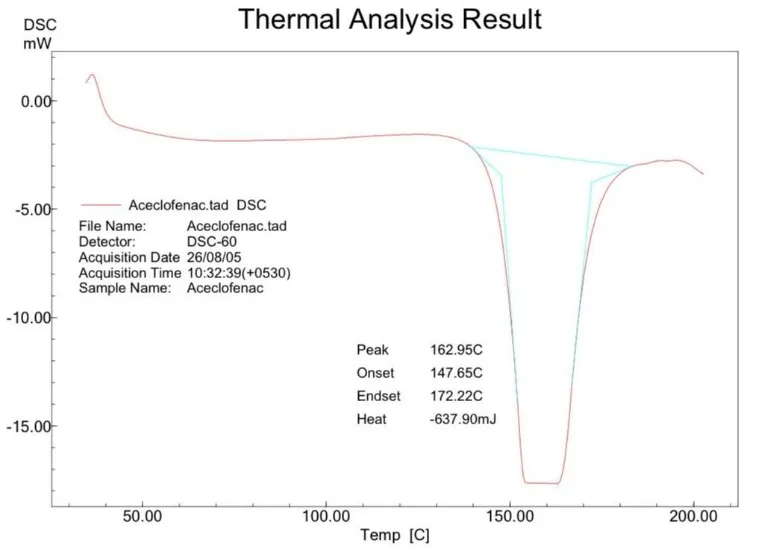
Figure 1: DSC of Pure Aceclofenac
Table 3: Solubility Analysis Report
| Solvents | Solubility (mg/ml) | Volume of solvent in ml/gm (ml) |
Solubility Criteria |
|---|---|---|---|
| Water | 0.07472 | 13383 | Practically insoluble |
| Buffer pH 1.2 | 0.06413 | 15593 | Practically insoluble |
| Buffer pH 4.6 | 0.07805 | 12812 | Practically insoluble |
| Buffer pH 6.8 | 0.3502 | 2855 | Very slightly soluble |
| Buffer pH 7.4 | 0.40084 | 2494 | Very slightly soluble |
Determination of Absorption Maxima (λmax)
Absorption Maxima (λ max) of the Aceclofenac was found to be 273nm in Methanol by using UV spectrophotometry.

Figure 2: UV Spectrum of Aceclofenac at 10 Μg/ml Concentration.
Table 4: Standard Calibration Curve of Aceclofenac
| Absorbance at λmax 273 nm | ||||
|---|---|---|---|---|
| Conc. | T-1 | T-2 | T-3 | AVG ± SD n = 3 |
| 0 | 0 | 0 | 0 | 0 |
| 8 | 0.189 | 0.194 | 0.196 | 0.194 ± 0.0025 |
| 16 | 0.393 | 0.365 | 0.385 | 0.381 ± 0.0143 |
| 24 | 0.572 | 0.551 | 0.564 | 0.564 ± 0.0106 |
| 32 | 0.756 | 0.747 | 0.746 | 0.746 ± 0.0055 |
| 40 | 0.938 | 0.915 | 0.952 | 0.956 ± 0.0186 |
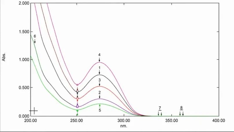
Figure 3: UV Spectrum of Aceclofenac at Different Concentration of 0-40 Μg/ml
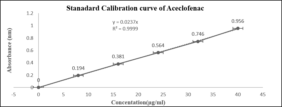
Figure 4: Plot of Standard Calibration Curve of Aceclofenac
Compatibility studies by FTIR spectrophotometer
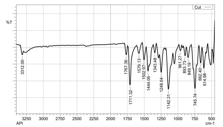
Figure 5: FTIR Spectra of Aceclofenac
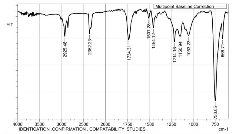
Figure 6: FTIR Spectra of Pharmacosomes (F10 Formulation)
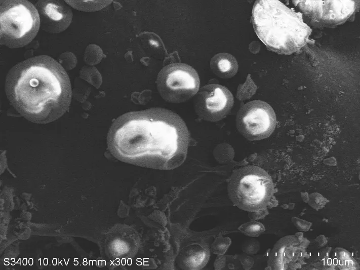
Figure 7: SEM Image of Optimized Formulation
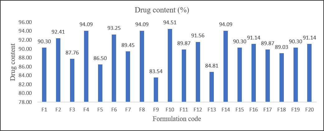
Figure 8: Drug Content of Pharmacosomal Formulation F1-F20

Figure 9: Entrapment Efficiency Pharmacosomal Formulation F1-F20
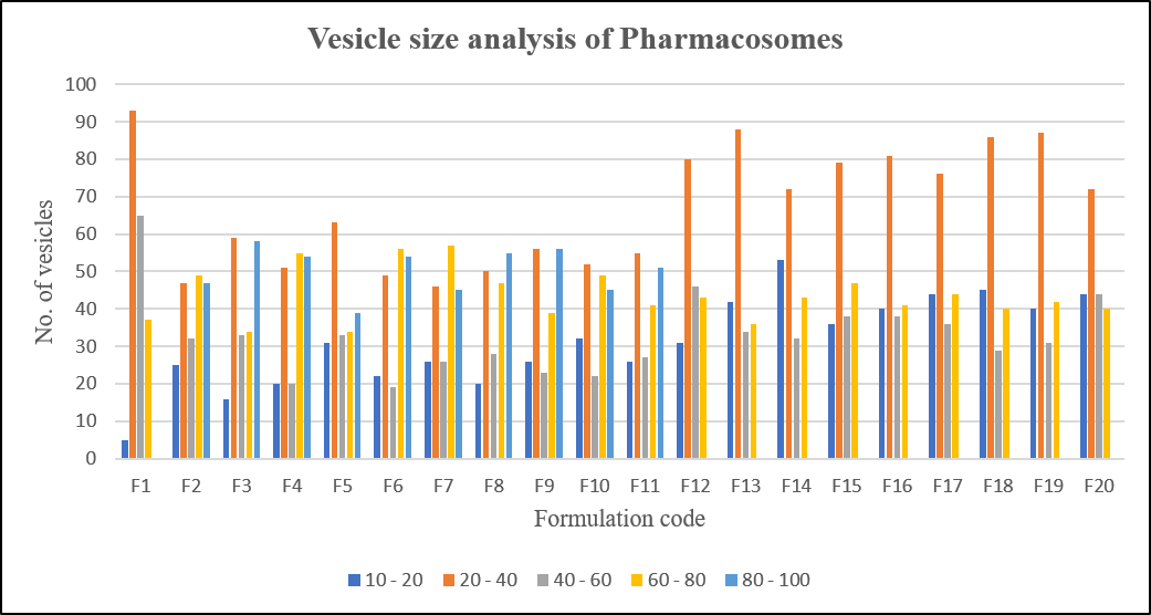
Figure 10: Vesicle Size Analysis of Pharmacosomes Formulation F1-F20
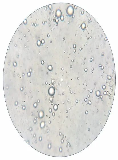
Figure 11: Microphotograph of Pharmacosomes (F10 Formulation)
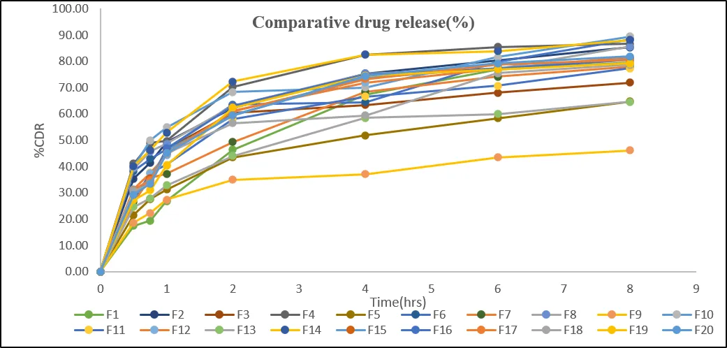
Figure 12: In Vitro Diffusion Study of Formulation F1-F20
Table 5: Release Kinetics of Pharmacosomes Formulation
| Formulation code | Zero order | First order | Higuchi | Peppas model | |
|---|---|---|---|---|---|
| r2 | n value | ||||
| F1 | 0.8513 | 0.9526 | 0.9774 | 0.9567 | 0.5274 |
| F2 | 0.6932 | 0.9038 | 0.9181 | 0.9830 | 0.2632 |
| F3 | 0.5956 | 0.7435 | 0.8495 | 0.9255 | 0.1909 |
| F4 | 0.6429 | 0.8364 | 0.8856 | 0.8975 | 0.2614 |
| F5 | 0.7788 | 0.8930 | 0.9619 | 0.9835 | 0.3358 |
| F6 | 0.7402 | 0.9306 | 0.9314 | 0.9277 | 0.2810 |
| F7 | 0.7866 | 0.9225 | 0.965 | 0.9781 | 0.3695 |
| F8 | 0.6855 | 0.8907 | 0.9114 | 0.9662 | 0.2584 |
| F9 | 0.6848 | 0.7633 | 0.9068 | 0.9639 | 0.2392 |
| F10 | 0.6628 | 0.9023 | 0.8880 | 0.9378 | 0.2140 |
| F11 | 0.7056 | 0.8679 | 0.9228 | 0.9424 | 0.2893 |
| F12 | 0.7003 | 0.8845 | 0.9218 | 0.9567 | 0.2839 |
| F13 | 0.7402 | 0.8459 | 0.9428 | 0.9629 | 0.3271 |
| F14 | 0.6208 | 0.8378 | 0.8703 | 0.8997 | 0.2323 |
| F15 | 0.6906 | 0.8699 | 0.9170 | 0.9695 | 0.2718 |
| F16 | 0.6550 | 0.8309 | 0.8946 | 0.9362 | 0.2543 |
| F17 | 0.6886 | 0.8628 | 0.9156 | 0.9534 | 0.2798 |
| F18 | 0.7254 | 0.8928 | 0.9256 | 0.9430 | 0.2634 |
| F19 | 0.6798 | 0.8246 | 0.9059 | 0.8840 | 0.3112 |
| F20 | 0.7071 | 0.8840 | 0.9266 | 0.9669 | 0.2912 |
Optimization by DoE software – design expert
a) Optimization analysis of response % drug content
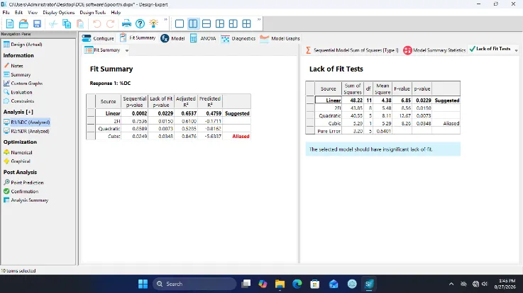
Figure 13: Model Selection Summary on Response % Drug Content
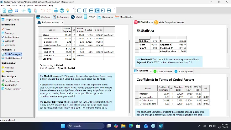
Figure 14: ANOVA Results on Response % Drug Content
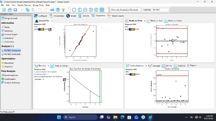
Figure 15: The Diagnostic Plot on Response % Drug Content
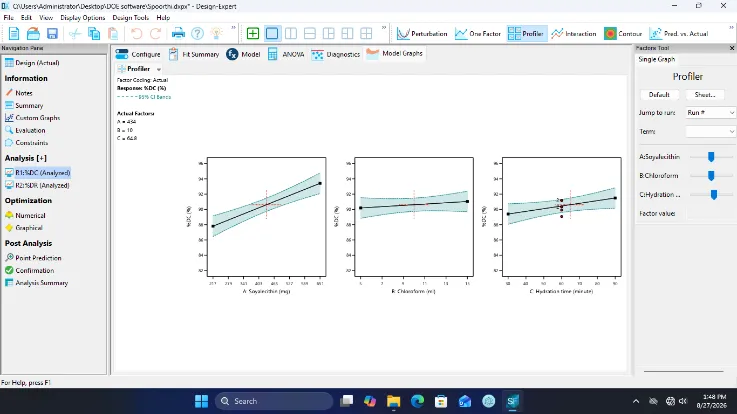
Figure 16: Factor Profiler of Different Factors on Response % Drug Content
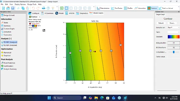
Figure 17: Contour Plot Represent the Combine Effect of Soya Lecithin (a) and Chloroform (B) on Response % Drug Content
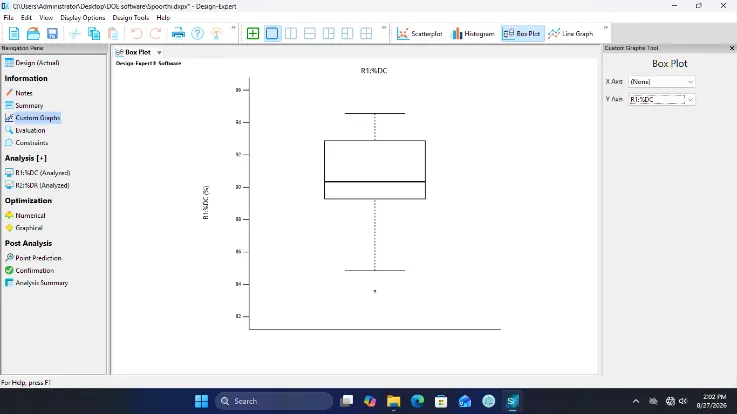
Figure 18: Box-plot on Response % Drug Content
b) Optimization analysis of response % drug release
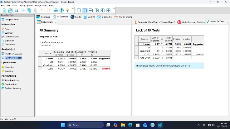
Figure 19: Model Selection Summary on Response % Drug Release
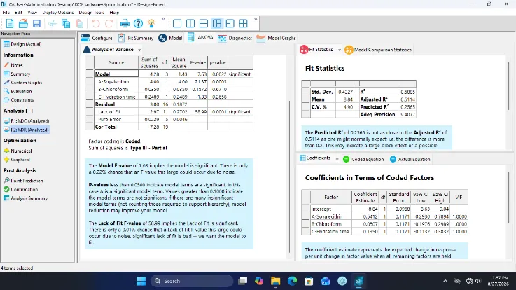
Figure 20: ANOVA Results on Response % Drug Release

Figure 21: The Diagnostic Plot on Response % Drug Release

Figure 22: Factor Profiler of Different Factors on Response % Drug Release

Figure 23: The Soya Lecithin and Choloroform Contour Plot on Response % Drug Release
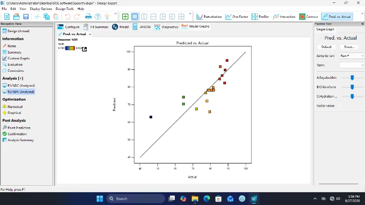
Figure 24: The Predicted V/S Actual Plot on Response % Drug Release

Figure 25: Box Plot of Main Effect on % Drug Release
c) Predicted Optimized Pharmacosome formulation obtained by design expert
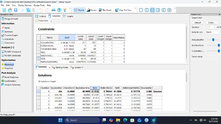
Figure 26: Design Space Solution for Optimized Pharmacosome Formulation
Table 6: Optimized Formulation Result
| Entrapment efficiency(%) | % CDR at 8 hours | Drug content(%) |
|---|---|---|
| 82.95 | 83.82 | 92.40 |
Evaluation of Pharmacosomal Gel
The optimized formulation was incorporated into the gel by the use of Carbopol 934 polymer and was found to be suitable for topical delivery because of the desirable consistency.
Physical appearance
Physical appearance of formulation was checked and the results are given below table
Table 7: Physical Appearance of Pharmacosomal Gel
| Formulation | Appearance |
|---|---|
| Pharmacosomal gel | Transparent Color, odorless with smooth and uniform appearance |
Table 8: pH, Viscosity, Spreadability, Extrudability of Gel
| Code | pH | Viscosity(cps) | Spreadability gm*cm /sec | Extrudability | Drug content(%) |
|---|---|---|---|---|---|
| Gel | 6.73 | 5463 ±3.42 | 13.47±0.53 | 118±4.56 | 93.48±0.06 |
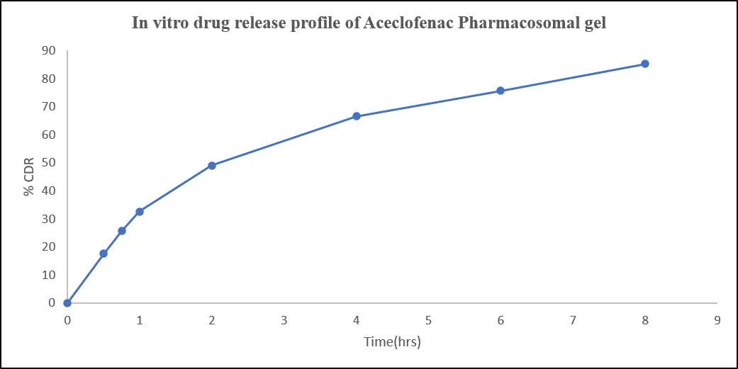
Figure 27: Invitro Drug Release Profile of Aceclofenac Pharmacosomal Gel
Release kinetics
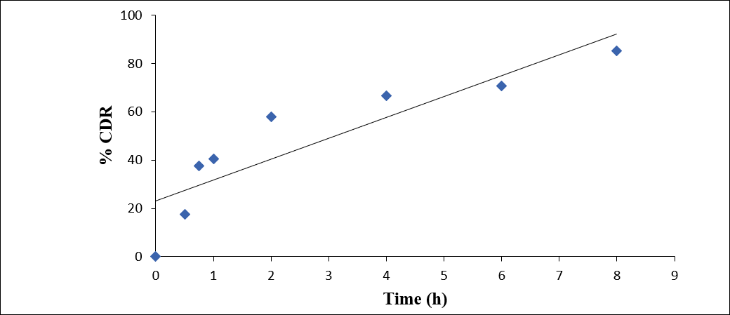
Figure 28: Zero Order Release Kinetic Profile of Gel Formulation
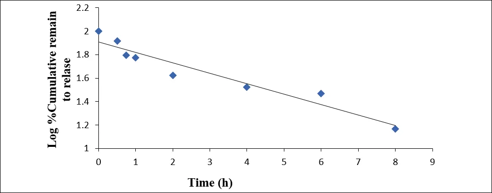
Figure 29: First Order Release Kinetic Profile of Gel Formulation
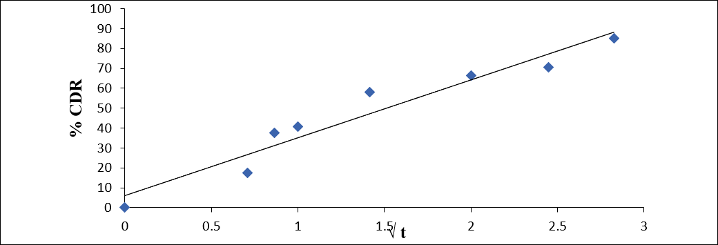
Figure 30: Higuchi Release Kinetic Profile of Gel Formulation
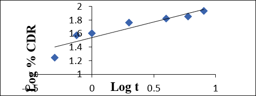
Figure 31: Peppas Release Kinetic Profile of Gel Formulation
Table 9: Data for Different Kinetic Model for Gel
| Formulation | Zero order (r2) |
First order (r2) |
Higuchi (r2) |
Peppas model | |
|---|---|---|---|---|---|
| r2 | n value | ||||
| Gel | 0.7855 | 0.9275 | 0.9363 | 0.8508 | 0.5374 |
Stability studies of Pharmacosomal gel
Table 10: Results of Intermediate Stability Studies of Pharmacosomal Gel at 30°C+2°C and 65 ± 5% RH
| Parameters | Duration in days | ||
|---|---|---|---|
| 0 | 30 | 60 | |
| % DC | 90.82 | 89.72 | 88.43 |
| % CDR | 87.65 | 85.56 | 84.76 |
DISCUSSION
The melting point of Aceclofenac was determined by Thiele’s tube method and found to be 150°C (149–152°C). DSC showed a characteristic endothermic peak, confirming the thermal characteristics of the drug.
Aceclofenac was practically insoluble in water and acidic phosphate buffers, while slightly better solubility was observed in alkaline medium.
UV spectroscopy showed a λmax of 273 nm, and the calibration curve exhibited excellent linearity (r² = 0.9996). FTIR studies confirmed the compatibility of Aceclofenac with the selected excipients.
Vesicle size analysis showed that the majority of vesicles were distributed within the 20–40 µm range.
Entrapment efficiency and drug content ranged from 78.39–91.41% and 83.54–94.51%, respectively.
F10 showed the highest drug content (94.51%) and cumulative drug release of 89.36% after 8 hours and was selected for further formulation development.
The in-vitro release data indicated diffusion-controlled drug release based on kinetic analysis.
A Quality by Design approach using Central Composite Design was applied to investigate the influence of soya lecithin, chloroform and hydration time on drug content and drug release.
Numerical optimization using the desirability function identified the optimized formulation comprising 434 mg soya lecithin, approximately 15 ml chloroform and 90 minutes hydration time, with a desirability of 0.880.
The optimized Pharmacosomes were incorporated into Carbopol 934 gel, which exhibited satisfactory pH (6.58), viscosity (5365 ± 3.42 cps), spreadability (13.17 ± 0.53 g·cm/sec), extrudability (108 ± 4.56) and drug content (93.18 ± 0.06%).
The optimized Pharmacosomal gel demonstrated controlled drug release and satisfactory physicochemical properties, indicating its potential as a topical delivery system for Aceclofenac.
CONCLUSION
The study successfully developed and optimized Aceclofenac-loaded Pharmacosomes using the solvent evaporation method and Quality by Design approach, followed by evaluation of their physicochemical properties and controlled drug release. Numerical optimization identified the optimized Pharmacosomal formulation using soya lecithin, chloroform, and hydration time as critical formulation variables. The optimized Pharmacosomes were incorporated into a Carbopol 934-based topical gel. The prepared gel showed satisfactory appearance, pH, viscosity, spreadability, extrudability, and drug content. Overall, the optimized Aceclofenac Pharmacosomal gel demonstrated suitable physicochemical characteristics and controlled drug release, indicating its potential as a topical drug delivery system.
Acknowledgement
I express my sincere gratitude towards Dr. Parthiban S, Head of the department, Department of Pharmaceutics and Bharathi College of Pharmacy who has been abundantly helpful in numerous ways for this research work. The authors also thank Bharathi College of Pharmacy, Mandya, Karnataka, India, for providing the required facilities to carry out this research work.
Funding
No funding was provided for this study
REFERENCES


Parthiban S., Spoorthi G. R., Pharmacosomal Gel Formulation of Aceclofenac : Design, Optimization and Evaluation, Int. J. of Pharm. Sci., 2026, Vol 4, Issue 10, 3511-3526. https://doi.org/
 10.5281/zenodo.23633455