

We use cookies to ensure our website works properly and to personalise your experience. Cookies policy
Sri Ramakrishna Hospital, Coimbatore.
Introduction: Aspirin-exacerbated respiratory disease (AERD, also called aspirin-induced asthma or Samter's triad) is an asthma phenotype in which aspirin and other COX-1 inhibitors can trigger bronchospasm, usually alongside chronic rhinosinusitis with nasal polyps. It creates a real prescribing problem when the patient also has coronary artery disease, because the drug that protects the heart may be the one that provokes the airway. Choosing a safe antiplatelet drug, while keeping the asthma regimen safe too, calls for close cooperation between the treating team and the clinical pharmacist. Main symptoms and clinical findings: A 71-year-old woman with long-standing asthma, a previous episode of pulmonary infiltrates with eosinophilia (PIE), recurrent rhinitis and coronary artery disease came in with two to three days of worsening wheeze, chest tightness and increased sputum. She had bilateral wheeze but stable vital signs, with an oxygen saturation of 98% on room air. High-sensitivity troponin I was normal (1.9 pg/mL), and her renal and liver function were normal. Diagnoses, interventions and outcomes: The working diagnosis was an acute exacerbation of asthma with possible AERD. She was treated with nebulised levosalbutamol–ipratropium, formoterol–budesonide, tiotropium, intravenous hydrocortisone, etofylline/theophylline, montelukast–levocetirizine and antibiotics. Sinus CT showed no polyps and no aspirin reaction was documented, so the AERD label stayed presumptive. After cardiology review, aspirin was stopped on Day 4, while clopidogrel and rosuvastatin were continued. Her wheeze, breathlessness and sputum improved, and she went home on Day 6. Conclusion: When an older patient with asthma and coronary disease may have AERD, the aspirin question is a medicines problem as much as a diagnostic one. Here, a presumptive label made without nasal polyps or a documented aspirin reaction led to aspirin being stopped and clopidogrel continued.
Aspirin-exacerbated respiratory disease (AERD) is also known as aspirin-induced asthma or NSAID-exacerbated respiratory disease. It is usually described as asthma with chronic rhinosinusitis and nasal polyps, together with respiratory reactions to aspirin and other COX-1 inhibitors [1–3]. The reaction happens in an airway that is already primed for it. The AERD airway makes a lot of cysteinyl leukotrienes and very little protective prostaglandin E2, and its eosinophils and mast cells are highly active. A COX-1 inhibitor strips away what protection is left [2,3,5]. Reactions usually start within minutes to hours of a dose, and the disease is often harder to control than aspirin-tolerant asthma [1,3].
Aspirin is a cornerstone of secondary prevention in coronary artery disease, so a possible AERD diagnosis puts prescribers in a difficult position. They can stop aspirin, switch to another antiplatelet drug, or consider supervised desensitisation, and each option has consequences for both the heart and the lungs [4,6,7
We report this case because it brings several medicine-related questions together in one admission: an acute asthma exacerbation treated with a long list of drugs, a possible AERD label without nasal polyps, and coronary artery disease that needed continued antiplatelet cover. The drug decisions mattered as much as the diagnosis. This report has three aims: to show how a presumptive AERD label, made without nasal polyps or a documented aspirin reaction, led to aspirin being withdrawn in a patient with proven coronary atherosclerosis; to explain, with two mechanism diagrams (Figures 1 and 2), why that choice makes pharmacological sense; and to argue for a structured aspirin and NSAID history early in admission, which is work a clinical pharmacist is well placed to do.
Patient Information
Clinical Findings
She was alert, oriented and afebrile on arrival, with no pallor, jaundice or lymphadenopathy. Her blood pressure was 120/70 mmHg, pulse 88/min, respiratory rate 20/min and SpO2 98% on room air. Heart sounds were normal with no murmur, and both lungs had good air entry with bilateral wheeze. The abdomen was soft and non-tender, and the neurological examination was unremarkable.
Timeline
| Time | Event |
|---|---|
| Before admission (≈3.5 weeks) | Transthoracic echocardiogram: sclerotic aortic valve, adequate LV systolic function, LV diastolic dysfunction, normal pulmonary artery pressures. |
| Day −3 to −2 | Onset and progression of wheeze, chest tightness and sputum. |
| Day 1 | Admitted. Chest radiograph, ECG, blood tests and cultures sent. Treatment for the acute exacerbation started.Treatment for acute exacerbation started. |
| Day 2 | Urinalysis and abdominal ultrasound. |
| Day 3 | CT of the paranasal sinuses and thorax (non-contrast). |
| Day 4 | Cardiology opinion sought; aspirin stopped, clopidogrel and rosuvastatin continued. Pulmonary function testing showed small airway disease. |
| Day 6 | Discharged clinically improved. |
| Day 16 | Follow-up chest radiograph (PA view). |
Diagnostic Assessment
Investigations
| Test | Result | Reference (as reported by laboratory) |
|---|---|---|
| Haemoglobin | 10.3 g/dL (low) | 12.0–15.0 |
| RBC / haematocrit | 3.49 ×10⁶/µL (low) / 29.6% (low) | 3.8–4.8 / 36–46 |
| MCV | 84.8 fL | 83–101 |
| WBC | 5.54 ×10³/µL | 4.5–10.0 |
| Platelets | 184 ×10³/µL | 150–410 |
| Eosinophils (differential) | 7.0% (high) | 1–6 |
| Absolute eosinophil count | 390 cells/µL | 40–440 |
| Total IgE | 126.97 IU/mL | <158 |
| Sodium / potassium | 140 / 4.3 mmol/L | normal |
| Chloride / bicarbonate | 108 (high) / 22 (low) mmol/L | 98–107 / 23–30 |
| Creatinine / eGFR | 0.7 mg/dL / >60 mL/min/1.73 m² | 0.5–0.9 |
| Random glucose / HbA1c | 101 mg/dL / 5.4% | normal |
| Liver function | Bilirubin, transaminases, ALP, GGT, albumin normal; globulin 2.3 g/dL (low) | – |
| TSH | 0.6981 mIU/L | 0.4–4.5 |
| PT / INR | 13.6 s / 0.88 | 12–15 s |
| hs-troponin I | 1.9 pg/mL | female <15.6 |
| NT-proBNP | 329 pg/mL (flagged high) | <125 (<75 y) |
| ANA (IF) | Negative | – |
| ANCA profile (PR3, MPO, lactoferrin, elastase, cathepsin G, BPI) | All negative | – |
| HBsAg, anti-HCV, HIV I/II | Negative / non-reactive / negative | – |
| Blood cultures (right and left brachial) | No growth at 48 h (preliminary result) | – |
| Urinalysis | Specific gravity 1.005; protein, glucose, nitrite negative; RBC 2–4/hpf | – |
| Aspergillus fumigatus-specific IgE | 0.25 kUA/L (negative) | – |
Imaging and other tests in brief
Diagnostic reasoning in brief
The treating team considered an asthma exacerbation first, supported by wheeze, sputum, a history of asthma and a good response to bronchodilators and corticosteroids. AERD stayed possible but unproven. The sinus CT showed no polyps, the absolute eosinophil count was normal (prior steroid use was not recorded), and no aspirin-provoked reaction is documented . No oral aspirin challenge was done. Eosinophilic or vasculitic disease was screened for with IgE, Aspergillus-specific IgE, ANA and ANCA, all unremarkable, and infection was thought unlikely (afebrile, normal white cell count, no growth in blood cultures at 48 hours). A cardiac contribution could not be fully excluded, given the T-wave inversion, an NT-proBNP of 329 pg/mL and diastolic dysfunction on echocardiography, although the single hs-troponin I was normal and no serial values are in the records provided.
Final diagnosis
Acute exacerbation of bronchial asthma with possible AERD, in a patient with coronary artery disease, a previous episode of pulmonary infiltrates with eosinophilia, and mild fibrotic change in the right middle lobe and lingula on CT.
Therapeutic Intervention
Bronchodilators and inhaled corticosteroid: nebulised levosalbutamol–ipratropium (Duolin), nebulised formoterol–budesonide (Formonide) and tiotropium by metered-dose inhaler (Tiova).
Systemic corticosteroid: intravenous hydrocortisone (Efcorlin).
Add-on asthma drugs: oral etofylline/theophylline (Deriphyllin-R) and montelukast–levocetirizine (Telekast-L).
Anti-infective drugs: injectable piperacillin–tazobactam (Tazar) and azithromycin (Azee).
Cardiovascular drugs: clopidogrel and rosuvastatin (continued), and aspirin (stopped on Day 4 on cardiology advice). Torsemide (Dytor) was also given .
Supportive drugs: pantoprazole (Panzeal) and paracetamol (Calpol). No oxygen requirement was recorded (SpO2 98% on room air).
Follow-up and Outcomes
By Day 6 her cough, breathlessness and wheeze had eased. She was producing less sputum, and it was lighter in colour. She was sleeping better and could speak in full sentences. She went home in stable condition, and no in-hospital complications were recorded. A follow-up chest radiograph was taken on Day 16. Clinical status at review, spirometry, symptom control score, adherence, steroid taper, tolerance of the discharge drugs, any further exacerbation or aspirin-related event, and the cardiology follow-up plan .
DISCUSSION
How aspirin triggers respiratory disease in AERD. Aspirin does not create the underlying disease. It sets off a reaction in an airway that is already primed for it, which is why the condition is also called aspirin-induced asthma. The underlying problem is a type 2 inflammatory state in which mast cells, eosinophils, platelet-adherent leukocytes and group 2 innate lymphoid cells (ILC2) already make large amounts of cysteinyl leukotrienes (LTC₄, LTD₄ and LTE₄) through 5-lipoxygenase and leukotriene C₄ synthase [2,3,13]. At the same time the airway is short of its main internal brake. Prostaglandin E₂ (PGE₂) is low, partly because COX-2 expression is low, and signalling through its EP2 receptor is weak [12,13]. PGE₂ normally keeps mast-cell activation, leukotriene synthesis and ILC2 activity in check [12,13]. Aspirin and other strong COX-1 inhibitors remove the small amount of COX-1-derived prostanoid that is left, most likely PGE₂, and the brake gives way [10]. Mast cells activate, leukotriene output surges, PGD₂ is released, and the airway narrows within minutes to hours of the dose [2,3,10,11] (Figure 1).
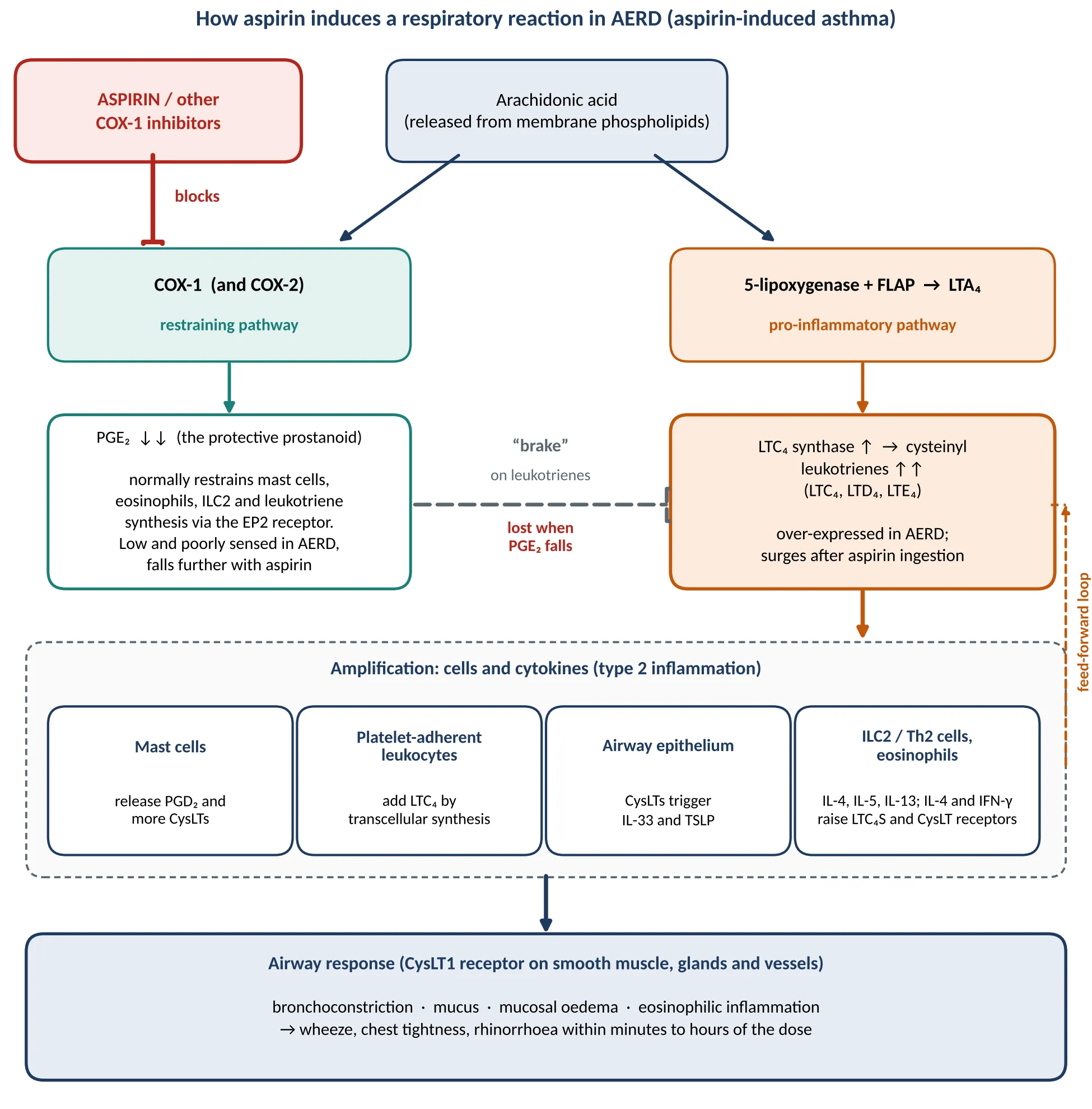
Figure 1: Mechanism by Which Aspirin Induces a Respiratory Reaction in AERD (Aspirin-Induced Asthma). Schematic Synthesised from Published Reviews [2,3,10–13]; It Is Simplified and Is Not a Finding of This Case. COX, Cyclooxygenase; CysLT, Cysteinyl Leukotriene; FLAP, 5-Lipoxygenase-Activating Protein; ILC2, Group 2 Innate Lymphoid Cell; LT, Leukotriene; pg, Prostaglandin; TSLP, Thymic Stromal Lymphopoietin.
The process then feeds itself. Cysteinyl leukotrienes released by mast cells, eosinophils and adherent platelets prompt the epithelium to release IL-33 and thymic stromal lymphopoietin. These activate mast cells and ILC2, which produce IL-4, IL-5 and IL-13 and recruit more eosinophils [11,12]. IL-4 in turn raises leukotriene C₄ synthase and CysLT-receptor expression [13]. This loop is why AERD stays active between reactions, why leukotriene-receptor antagonists are used [1,5], and why avoiding aspirin may not control the disease on its own. In our patient no aspirin reaction is documented, so the record cannot show that this pathway was active in her. Figure 1 describes the published mechanism, not a finding of this case.
.
The aspirin dilemma. Aspirin was stopped on cardiology advice and clopidogrel was continued. This avoids a possible COX-1-driven respiratory reaction and keeps antiplatelet cover in place, but it takes aspirin away from a woman with proven coronary atherosclerosis. For AERD patients who truly need aspirin, supervised desensitisation followed by daily maintenance is an established option, and it may also settle airway inflammation. It needs an experienced centre, and reactions can occur during the procedure [4,6,7]. Registry data in coronary patients with aspirin sensitivity support rapid desensitisation, although only about one in five of that cohort had asthma as the aspirin reaction [7]. Which path was best here (desensitisation, clopidogrel alone, or an oral challenge to settle the diagnosis) depends on how strong the aspirin-reaction history is and how high her coronary risk is, which is why documenting the original reaction matters. Clopidogrel acts on the platelet P2Y12 receptor and does not inhibit COX-1, so it leaves COX-1-derived PGE₂ alone and fits the mechanism in Figure 1. Figure 2 maps each drug in this case to the step it acts on. Clopidogrel is also a prodrug activated by CYP2C19, and pantoprazole interferes with that step less than omeprazole does, which makes it a sensible gastroprotective partner [18,19].
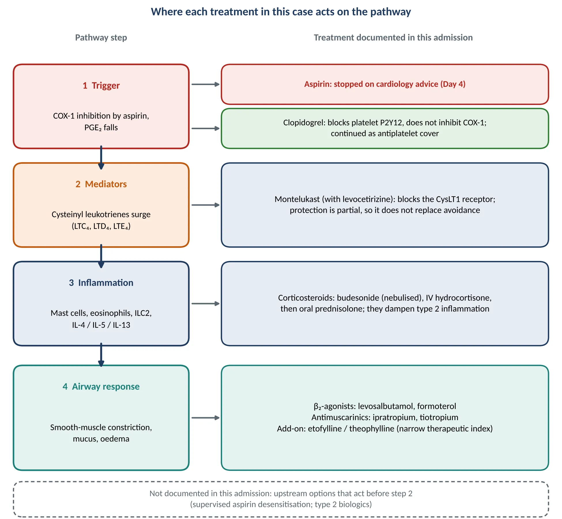
Figure 2: Where Each Treatment Documented in This Admission Acts on the Pathway in Figure 1. Drug Names Are as Recorded in the Discharge Summary; the Mapping to Pathway Steps Is the Authors’ Pharmacological Interpretation.
Role of the clinical pharmacist. Several points in this case are where pharmacist input changes what happens to the patient: reconciling aspirin and other COX-1 inhibitors at admission (including combination analgesics and topical or over-the-counter NSAIDs); counselling on avoidance; checking inhaler and spacer or nebuliser technique; monitoring theophylline levels and interacting drugs; watching for steroid-related effects such as low potassium, raised glucose and bone loss; spotting duplicated drug classes, for example ipratropium with tiotropium; checking antibiotic use against the clinical picture; and reviewing the whole medicine list at discharge. The records provided do not show which of these steps were carried out in this case.
.
Strengths and limitations. The case was assessed from several angles (cardiac, immunological, infective, sinus and chest imaging) by several specialties, and the drug choices can be traced step by step to the mechanism. The limits are plain. This is a single case, the AERD diagnosis is presumptive with no aspirin challenge or documented reaction
CONCLUSION
This case describes a 71-year-old woman with long-standing asthma, a previous episode of pulmonary infiltrates with eosinophilia, recurrent rhinitis and coronary artery disease, who improved after an acute exacerbation and went home on Day 6. AERD was suspected, but the diagnosis stayed presumptive: sinus CT showed no nasal polyps and no aspirin-related respiratory reaction was documented. After cardiology review on Day 4, aspirin was stopped while clopidogrel and rosuvastatin were continued. The case shows how a presumptive label can lead to an antiplatelet change in a patient with proven coronary atherosclerosis, and why a detailed history of previous aspirin and NSAID use should be recorded at admission, before antiplatelet therapy is altered.
Overall, the case supports an individualised, multidisciplinary approach in which the respiratory and cardiology teams and the clinical pharmacist agree the antiplatelet plan together, and in which a pharmacist-led aspirin and NSAID history and medication review help avoid unnecessary changes to cardiovascular treatment while keeping the airway safe. This single case does not establish that AERD was present or that clopidogrel was the better choice, and longer follow-up is needed to see how she does off aspirin.
REFERENCES

Dr. Ramanaprasanth G., Marin Aravindh A. D., Mirdula Mary E., Mourishnawin R. K., The Aspirin Dilemma in an Older Women with Asthma and Coronary Artery Disease : A Case Report of Suspected Aspirin Exacerbated Respiratory Disease, Int. J. of Pharm. Sci., 2026, Vol 4, Issue 11, 2267-2274. https://doi.org/10.5281/zenodo.24270998
 10.5281/zenodo.24270998