

We use cookies to ensure our website works properly and to personalise your experience. Cookies policy
1,2,3,4,5,6,7Research Scholar, Siddhi's Institute of Pharmacy, Nandgaon, Murbad, Thane – 421401, Maharashtra, India.
8Assistant Professor, Siddhi's Institute of Pharmacy, Nandgaon, Murbad, Thane – 421401, Maharashtra, India.
Diabetic foot ulcers (DFUs) are a common complication of diabetes mellitus and can lead to serious problems such as slow wound healing, persistent infection and amputation of the lower limb. Treatment is difficult because recovery can be hindered by a number of conditions such as poor blood circulation, peripheral neuropathy, poor wound healing and antibiotic resistance. Biofilms may also play a role in the ability of bacteria to persist in chronic wounds. This could make the body’s immune system and antimicrobial drugs more tolerant of bacteria that form in biofilms, making it harder to manage infections. However, classical antimicrobial resistance and tolerance associated with biofilms are different mechanisms and should be treated separately. This review highlights the common bacteria responsible for diabetic foot infections, pathophysiology and risk factors of diabetic foot ulcers and the role of biofilms in chronic wounds. It also discusses antibiotic resistance, biofilm detection methods and how these may relate to clinical outcomes including amputation, recurrent infection and delayed healing. The review additionally emphasises the drawbacks of modern diagnostic techniques, and the difficulty of comparing findings from studies using different methodologies. Standardised methods for detection of biofilm need to be established. More prospective clinical studies need to be carried out and whether biofilm results and antibiotic resistance can accurately predict treatment outcomes needs to be established. A better understanding of these variables may facilitate improved diagnosis, appropriate antibiotic selection, and more successful treatment of diabetic foot infections.
Diabetes mellitus is a chronic metabolic disease resulting in damage to blood vessels and nerves. Diabetic foot ulcers (DFUs) are one of the main side effects of diabetes and are open wounds that often appear on the feet of people with the disease. Poor blood circulation can delay wound healing, and peripheral neuropathy can decrease the sense of pain or injury. When these sores become infected there can be serious consequences such as prolonged hospital stays and amputation of the lower limb. Therefore, early diagnosis and proper management of diabetic foot infections are needed to improve patient outcomes [1].
Diabetic foot ulcers are difficult to heal as the process of wound healing is dependent on many factors such as blood flow, glycaemic control, depth of the wound, severity of infection and appropriate wound care. The presence of bacteria in an ulcer does not automatically mean that the patient needs antibiotic treatment. Bacteria may be present in an ulcer without there being an active infection. Identification of the microorganisms causing the disease helps health care providers to choose the most appropriate antimicrobial therapy for clinically infected ulcers. The Infectious Diseases Society of America and the International Working Group on the Diabetic Foot recommend that the infection should be clinically evaluated and appropriate microbiological sampling should be performed to allow for better diagnostic and therapeutic options [1].
Biofilm formation is another factor that can lead to a long-term wound infection. A biofilm is a congregation of microorganisms adherent to a surface and embedded in an extracellular matrix of their own production. The structure shielded the microbes from environmental stress, while also rendering them less vulnerable to host immune responses and antimicrobial therapies. Biofilms have been studied in chronic wounds as they may be involved in chronic inflammation and delayed healing. However, the therapeutic relevance of biofilm detection in diabetic foot ulcers needs to be further studied and it is not always possible to prove that biofilm is the cause of treatment failure in a specific patient [2, 3].
Microbial resistance is another major problem in diabetic foot infections. Some bacteria are resistant to the effects of commonly used antibiotics making finding the right medication hard. Resistant microbes may be linked to persistent infection and poor outcomes, but peripheral artery disease, wound severity, inadequate pressure off-loading, and delayed treatment may also impact these outcomes. Furthermore, biofilm-associated antimicrobial tolerance and genetic antimicrobial resistance are related but distinct processes and should be considered separately when interpreting study results [1,3].
Biofilms, antimicrobial resistance and delayed wound healing have been studied individually but the combined effects on treatment outcomes should be considered carefully. Studies applied different techniques for biofilm detection, microbe identification and wound healing assessment. This makes it difficult to assess outcomes and determine whether detection of biofilms or antimicrobial resistance can be used to accurately predict treatment failure. Therefore, the objective of this review is to explore biofilm detection methods and antimicrobial resistance in diabetic foot ulcers, to examine the association between these and clinical outcomes and to identify evidence gaps that require further research.
2. Pathogenesis and risk factors for diabetic foot ulcers
2.1 Pathophysiology of Diabetic Foot Ulcer .
Diabetic foot ulcers (DFUs) are chronic lesions that may develop in people with diabetes, mainly due to peripheral neuropathy, reduced blood flow, and repeated pressure or injury to the foot. All of these combined can make the skin more susceptible to damage. Subsequent infection and delayed wound healing can make it more difficult to recover from an ulcer [1, 4].
Long term diabetes can damage the nerves that supply the feet, a condition called peripheral neuropathy. Sensory neuropathy reduces sensation of pain, heat, pressure and minor injuries. This means that a person can walk with a cut, a blister or pressure damage and not know it. Motor neuropathy can also cause foot problems by affecting how pressure is distributed when walking. These alterations increase the risk of developing skin degeneration and ulceration [4, 5].
Poor circulation and peripheral arterial disease Diabetes increases the risk for peripheral arterial disease (PAD) or narrowing or blockage of arteries that limits blood flow to the lower limbs. Poor circulation limits the supply of oxygen and nutrients which are required to repair tissues. Thus it may impair the wound healing and reduce the capacity of the injured tissue to heal.PAD is a risk factor for poor healing and amputation in the setting of a foot infection [1, 4].
Poor wound healing: Chronic hyperglycemia may disrupt normal wound healing processes such as inflammatory reactions, immune cell activity, collagen synthesis and neovascularization. Such disorders can delay the formation of healthy tissue and prolong the inflammatory period.This can lead to an ulcer being open for a long period of time and being more prone to infection and re-injury [4, 5].
Microbial infection and biofilm formation Open ulcers can be infected by microorganisms but this does not necessarily imply that there is a clinical infection. As the infection progresses, the microorganisms may invade surrounding tissue and add to the local damage. Some bacteria form biofilms, which are microbial cells protected by an extracellular matrix. This structure can make germs more resistant to antimicrobial treatments and more difficult to kill . However, biofilm formation is only one possible cause of chronic wounds and sufficient human clinical evidence must be used to assess its association with clinical treatment failure [1, 6].
combined effect of all these processes: Poor blood circulation may slow the healing process; neuropathy may prevent the injury from being detected; and repeated pressure may cause the lesion to enlarge. Infection due to impaired host defence and microbial persistence may make healing more difficult. This interaction results in diabetic foot ulcers often needing full care, not just treatment of infection [1, 4].
2.2. Risk Factors of Diabetic Foot Ulcers
The risk factors for DFU can be grouped into three main categories: patient-related variables, foot-related factors, and factors that increase the risk of infection or delayed healing.
Factors related to the patient: Key issues to consider include peripheral neuropathy, peripheral vascular disease, chronic renal disease, long standing diabetes and poor glycaemic control. Previous foot ulcers or lower limb amputation also indicate an increased risk of further ulceration. Smoking and limited movement can make poor circulation or foot care problems worse [1, 5].
Foot-related factors Local tissue stress may be increased by foot deformity, prominent bony prominences, severe callus, inappropriate footwear and repeated mechanical pressure. Fungal infections, minor cuts, blisters, cracked skin and problems with the nails can cause infections or skin breakdown. Routine foot examinations are important because some of these changes may be painless in persons with neuropathy [5].
Factors related to infection and poor outcome: A deep, persistent or recurrent ulcer, poor tissue perfusion, inadequate pressure off-loading, and impaired immunological responses can all hamper healing. The history of antibiotic exposure and presence of antimicrobial resistant organisms can influence the treatment decision and outcome. People with diabetes who have foot infections may also have chronic kidney disease. The severity of the ulcer and the patient's general clinical condition should be considered in conjunction with these factors [5, 7].
2.3. Implications for Antimicrobial Resistance, Biofilm Identification and Treatment Failure
Understanding the pathophysiology and risk factors of DFUs is important to interpret research on biofilms, antibiotic resistance and clinical outcomes . There are many reasons why a wound can take a long time to heal . Those reasons include lack of wound care , neuropathy , poor blood flow , repeated pressure or infection . Thus, delayed healing per se cannot be interpreted as proof of antimicrobial resistance or biofilm formation.
Treatment failure should be evaluated based on the size and duration of the ulcer, the presence of peripheral vascular disease, the glycaemic state, the microbiological findings and the course of treatment. Researchers can compare these parameters to determine if findings related to biofilm or antimicrobial resistance are independently associated with outcomes such as delayed healing, persistent infection, recurrence, or amputation [1, 5, 6].
3. Microbial Profile of Diabetic Foot Infections
3.1. Microorganisms Frequently Recovered from Diabetic Foot Infections
Damage to the skin’s barrier allows germs to enter the underlying tissues, forming diabetic foot ulcers.
“People with diabetes who have peripheral neuropathy, poor blood supply and slow healing wounds are at greater risk of infection. Microorganisms in these wounds may be influenced by the depth and severity of the ulcer, previous antibiotic therapy, length of the wound, and geographic location. Some diseases can be caused by several bacterial species and can be both Gram-positive and Gram-negative [1, 6].
Staphylococcus aureus is one of the most common bacteria associated with the diabetic foot infections. It may cause skin and soft tissue infections and it is present in deeper infected wounds. Methicillin resistant Staphylococcus aureus (MRSA): MRSA is resistant to some of the antibiotics that are often used and can be harder to treat. However, the mere presence of S. aureus in a wound sample does not implicate it as an active pathogen [1, 8].
Streptococcus species, especially beta-haemolytic streptococci are also a frequent cause of diabetic foot infections. These bacteria can cause cellulitis and spread the disease to neighbouring tissues. They are especially relevant in the evaluation of mild infections [1], although the involved organisms may be more diversified in deeper or previously treated wounds.
Enterobacterales are a group of Gram-negative bacteria, including Escherichia coli, Klebsiella species and Proteus species, which can cause deep/complex/chronic wounds in diabetic foot infections. Fewer viable antibiotic alternatives may be left by resistant strains. Findings from a single hospital or region should not be generalised to all patients as the prevalence differs across patient populations and healthcare environments [8].
Another Gram-negative bacteria involved in diabetic foot infections is Pseudomonas aeruginosa. This is important as it becomes resistant to several antimicrobial agents. But it isn’t that common in every field or type of wound. In 2023, IWGDF/IDSA recommended that adequate tissue samples be taken for culture as needed, preferably after washing and debridement, rather than a superficial wound swab [1].
3.2 Polymicrobial Aetiology of Diabetic Foot Infections
Diabetic foot infections may be caused by one species of bacteria or several germs at the same time. In polymicrobial infections bacteria can interact with each other in the wound environment leading to a prolonged infection. As the wound becomes more severe, the infection advances, or the patient is given antibiotics, the microbial composition can change.
In some chronic wounds, bacteria can grow in a biofilm, which is an extracellular matrix that protects the germs. This structure can assist the bacteria to be less susceptible to antimicrobial therapy and to persist in the wound. However, antibiotic resistance acquired genetically differs from tolerance related to biofilm. The two mechanisms will be reviewed separately in this review.
Knowing which microorganisms are involved can help in selecting the right treatment, especially for infections of moderate or severe intensity. The 2023 IWGDF/IDSA guideline recommends obtaining adequate tissue samples for culture when needed, preferably after washing and debridement, rather than relying only on a superficial wound swab [1].
3.3. Difference Between Wound Colonisation and True Infection
In treating diabetic foot, it is important to realise that the presence of bacteria does not always mean a lesion is infected. There may be micro-organisms in open wounds without injuring the surrounding tissue. That is the colonisation we talk about. A true infection occurs when bacteria penetrate into tissue and provoke a detrimental host response [1].
Table 1: Differences Between Wound Colonisation and True Infection
| Feature | Wound colonisation | True wound infection |
|---|---|---|
| Microorganisms | Present on the wound surface | Invade and multiply in tissue |
| Tissue damage | No infection-related tissue damage | Tissue damage may occur |
| Clinical signs | No clear signs of infection | May show redness, swelling, warmth, pain or purulent discharge |
| Diagnosis | Culture may detect bacteria without infection | Primarily based on clinical examination |
| Antibiotic treatment | Not indicated solely because bacteria are present | May be required following clinical assessment |
3.4. Connection to Treatment Failure and Biofilm Detection
Understanding the microbial profile to investigate treatment failure in diabetic foot ulcers. Some wounds may have persistent microbial communities associated with biofilm production, or may harbour resistant bacteria. Both can make therapy more difficult, but neither should be blamed for poor healing.
Treatment outcome is affected by several factors including poor blood supply, inadequate off-loading pressure, uncontrolled blood glucose, wound depth and inadequate wound care. Future research should examine microbial findings in conjunction with clinical parameters [1] to determine which microbial findings are most useful in predicting delayed healing, recurrence and other adverse outcomes.
4. Biofilm Formation and Contribution to Chronicity of Diabetic Foot Ulcers
4.1. Biofilm development
Biofilm is a collection of microorganisms that are attached to a surface and grow together in a self-produced matrix of bacteria. In diabetic foot ulcers (DFUs), bacteria are able to form biofilms on wound tissue, complicating the wound environment and its treatment. These communities may include many different bacterial species that may interact with each other to prolong the infection. It is believed that biofilm formation has a major role in chronic wounds by making germs more difficult to kill by the body’s natural defences and standard antibiotic treatment [2, 9].
Biofilm formation occurs through four distinct phases: initial attachment, production of extracellular matrix, maturation and dispersal. These phases are important for the establishment of bacteria in the wound and maintenance of long-lasting microbial population [2, 9].
4.2. Phases of Biofilm Development
1. The first attachment
In the first stage free living bacteria contact the wound surface and adhere to the injured tissue . Bacterial surface structures and sticky substances may help the organisms to stay on the surface even if the adhesion is poor at first. If the local environment is favourable the connected bacteria begin to grow and form tiny groups. Damaged tissue and recurrent wound injuries in diabetic foot ulcers may provide a favourable niche for bacterial adhesion [2, 9].
2. Formation of the extracellular matrix
After attachment, the bacteria secrete an extracellular polymeric substance (EPS) that encases and supports the developing microbial community. The matrix can consist of polysaccharides, proteins, extracellular DNA, and other materials. It provides a protective environment and promotes tenacious attachment of bacteria to the wound surface. Protection is not only provided by physical blocking but also by bacterial physiology and the wound environment [2, 9]. The matrix can also influence the mobility and activity of antimicrobial compounds inside the biofilm.
3. Development of biofilms
The biofilm becomes more mature and structured as the matrix increases in size and the bacterial cells continue to multiply. Within the biofilm, bacterial activity, oxygen levels and availability of nutrients can vary. Some bacteria grow slowly or enter periods of low activity, while others grow actively. Such differences may lead to a microbial community that is less susceptible to antimicrobial treatment. In addition, interactions between several microbial species may contribute to the persistence of the community in a chronic wound [2, 9].
4. Dispersion
In the final stage, some of the microbes detach from the developed biofilm and migrate to nearby sites. These cells may attach to different parts of the wound and be able to form new microbial communities. Dispersal may thus promote growth or re-establishment of biofilms. How much this happens in a particular diabetic foot ulcer, however, depends on the microbial community and the local conditions of the wound [2, 9].
4.3 Tolerance to antimicrobial agents in biofilms
One of the major problems with biofilms is that the bacteria growing in biofilms may be more resistant to antimicrobial treatment than free-living bacterial cells. The extracellular matrix can affect antimicrobial penetration and some antibiotics may be less effective against slow growing bacteria and metabolically dormant cells. Persister cells are also known to be present in biofilms, which are small populations of bacteria that can survive exposure to antibiotics without necessarily having a resistance mechanism conferred genetically [2, 9].
Such a distinction is important for diabetic foot ulcers because standard testing for antibiotic sensitivity generally involves free living bacteria . The results might not be representative of the behaviour of bacteria within a mature biofilm. However, biofilm tolerance does not mean that all antibiotics are ineffective and the impact of biofilm on the course of treatment of a patient must be considered along with other clinical factors [9, 10].
4.4. Interaction with the body's defence systems
Biofilms may also help microbes evade the immune response. The surrounding matrix and structure of the microbial community may limit effective contact between immune cells and bacteria. Neutrophils and other immune cells may continue to react to the microbes, causing inflammation, but may not completely clear the microbial community. Persistent inflammation can lead to damage of the surrounding tissue and disturb normal phases of wound healing [2, 9].
Diabetics who already have poor wound healing because of neuropathy, poor blood circulation, and decreased immunological function. When these elements exist side by side with a chronic microbial community they may be more difficult to heal. However, biofilms are not the only possible cause of delayed healing, inadequate off-loading, recurrent pressure, poor blood supply and poor metabolic regulation can all be significant factors [2, 9].
4.5. Biofilms and Their Role in Chronic Wounds
Biofilms are implicated in chronic wound persistence and can promote local inflammation and the presence of bacteria. Even when treatment decreases the number of free-living bacteria in a wound, some species may live in protected microbial communities. The injured foot may take longer to heal because of constant pressure, poor blood flow, and repeated tissue damage [2, 10, 11, 12].

Wound isolates from diabetic foot ulcer studies have demonstrated antimicrobial resistance and biofilm forming organisms. For example, in a prospective study of 100 patients with persistent diabetic foot ulcers in India, 38 of 82 culture-positive isolates were biofilm-forming organisms. The results of the present study cannot be generalised for all the diabetic foot ulcer populations, but it does emphasise the need to consider the biofilm formation in addition to the traditional microbiological findings [11].
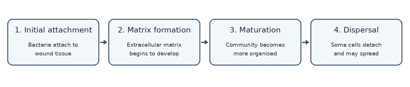
Figure 1: Stages of Biofilm Formation
5. Antimicrobial Resistance in Diabetic Foot Infections
Antimicrobial resistance (AMR) presents a considerable challenge in the management of diabetic foot infections (DFIs). This phenomenon occurs when bacteria develop resistance to or show reduced susceptibility against commonly used antibiotics.. The presence of resistant bacteria in diabetic foot ulcer (DFU) patients restricts treatment options and complicates the control of infection. However, treatment failure may also result from insufficient wound care, delayed wound healing, poor blood circulation and biofilm formation. Thus, antimicrobial resistance may be considered as one of the possible reasons for suboptimal clinical outcomes [1, 13, 14].
5.1. Patterns of Resistance in Infections of the Diabetic Foot
Infections of the diabetic foot are caused by Gram-positive and Gram-negative bacteria. Staphylococcus aureus is a major Gram-positive pathogen, however Gram-negative organisms such as Escherichia coli, Klebsiella species, Pseudomonas aeruginosa and Acinetobacter species have also been reported. The patterns of bacterial distribution and resistance are affected by patient demographics, severity of infection, prior therapy, and geographic location [8, 13, 14].
Methicillin-resistant Staphylococcus aureus (MRSA) is of major concern because it is resistant to the most commonly used beta-lactam antibiotics that are effective against methicillin-sensitive staphylococci. For MRSA infection, different antibiotics may be selected depending on the patient’s clinical condition and susceptibility findings. In an Indian tertiary-care research, Gadepalli et al. studied 56% of the bacterial isolates that were resistant to methicillin. This outcome should not be viewed as a representation of all diabetic foot infections in India as only 80 hospitalised patients with advanced diabetic foot ulcers were included in this study [13].
Another big problem is resistance in gram-negative bacteria. Some strains produce extended-spectrum beta-lactamases (ESBLs) which can decrease the efficacy of many of the cephalosporin and penicillin drugs. Some strains may be resistant to aminoglycosides, fluoroquinolones or other groups of antibiotics. According to Rastogi et al., P. aeruginosa and Acinetobacter species had significant resistance to quinolones and cephalosporins in a prospective Indian study of patients who had recently received antibiotic therapy. For example, only 23% of P. aeruginosa isolates and 5.6% of Acinetobacter isolates were sensitive to the quinolones . These results indicate the presence of bacteria with reduced sensitivity to commonly used antibiotics for previously treated diseases [14].
5.2. Antibiotic Susceptibility Testing
Antibiotic susceptibility testing can help determine which antibiotics are most likely to work against bacteria isolated from an infected ulcer. Two commonly used techniques are disc diffusion method and minimum inhibitory concentration (MIC) determination. The disc diffusion method involves the use of standard laboratory criteria to quantify and interpret bacterial growth around a disc containing an antibiotic. MIC testing is done to find the lowest concentration of the antibiotic that prevents visible growth of the bacteria.
In suspected diabetic foot infection, a culture obtained from a correctly obtained tissue sample is preferred to a superficial swab. The sample should be taken after the wound has been cleaned and debridement where appropriate to minimise contamination. Culture confirms the bacterium [1] and susceptibility testing guides the choice of antibiotics.
However, susceptibility testing has its limitations. It may not truly represent bacterial behaviour in a chronic wound as it examines bacterial response in a laboratory setting. Bacteria when tested in a free-living state might respond differently to exposure to antibiotics than bacteria in a biofilm. False-negative culture results may occur due to prior antibiotic therapy, which may inhibit bacterial growth in culture.Therefore laboratory findings should be interpreted in the light of clinical symptoms, wound severity and other relevant patient characteristics [1, 11].
5.3. Multidrug Resistance and Prior Antibiotic Exposure
Multidrug resistance (MDR) is defined as non-susceptibility to at least one agent in three or more antimicrobial classes. MDR microorganisms are difficult to manage because the therapeutic options that are effective may be limited. The causative bacteria for MDR in diabetic foot infections may be Gram-positive or Gram-negative bacteria which can make the choice of initial antibiotic treatment difficult [15].
Key is looking at previous exposure to antibiotics. Prolonged or repeated exposure may favour the survival and growth of drug-resistant microorganisms. Patients may also be on antibiotics which can skew the culture results and make it difficult to ascertain the full microbial composition.In Indian perspective investigation by Rastogi et al, gram-negative isolates from patients with recently treated diabetic foot infections demonstrated significant resistance which emphasizes the necessity of taking previous treatment into account while interpreting susceptibility results [14].
But not every infection in a patient with a history of antibiotic use will be caused by MDR bacteria. The association may be influenced by the severity of infection, length of hospital stay, prior surgeries and regional patterns of antibiotic use. Therefore, instead of assuming resistance in every patient who has undergone therapy, antibiotics should be selected based on clinical judgement, available culture data, and established treatment guidelines [1].
5.4. Regional Differences in Antimicrobial Resistance
Antimicrobial resistance patterns varied by country, hospital and even within the same area between healthcare facilities. Some studies from India have shown a higher proportion of Gram-positive species while others have shown a significant role of Gram-negative bacteria. The prevalence of P. aeruginosa, MRSA and ESBL-producing bacteria also varies between study populations [8,13,14].
These differences may be due to local antibiotic prescribing patterns, referral practice, exposure to hospital, severity of wound, sampling strategies and laboratory methodology. Although the local environmental conditions and climate may influence the organisms found in wounds, these should not be the only factors used to identify the causative bacteria. According to IWGDF/IDSA recommendations [1], antibiotic selection should be guided by susceptibility data, severity of infection, prior antibiotic exposure, likely or proven pathogens, and local epidemiology.
Therefore, it is not suitable to generalise resistance rates from a particular hospital to all patients or regions. Susceptibility data should be reviewed regularly and local surveillance used to improve empiric antibiotic selection. When culture results are available, therapy can be modified to treat the identified infections and limit the unnecessary use of broad-spectrum antibiotics.
5.5. Importance of Treatment Failure
Antimicrobial resistance occurs when the antibiotic you receive is not effective against the microorganisms that are causing the infection. Biofilm formation can complicate treatment, as bacteria in biofilms may be more resistant to exposure to antimicrobials. However, biofilm-associated tolerance and resistance are different processes. Biofilm tolerance is the reduced susceptibility of bacteria growing in a biofilm condition, while resistance is usually measured against defined susceptibility criteria .
These characteristics should be interpreted with caution to understand their association with clinical outcomes. Proper wound care and good therapy, however, do not always result in treatment failure with a resistant isolate. Similarly, a susceptible isolate will not ensure healing if blood flow or wound care is inadequate. Additional clinical studies are necessary to assess whether the addition of biofilm assessment to traditional susceptibility testing can better predict delayed healing, recurrent infection and other adverse outcomes.
To summarise, management of diabetic foot infections requires good specimen collection, reliable susceptibility testing, local resistance surveillance and judicious use of antibiotics. Such actions should be combined with pressure off-loading, vascular assessment, wound debridement where indicated and glycaemic control to maximise the chances of a successful healing [1].
6. Relationship Between Biofilms, Antimicrobial Resistance, and Treatment Failure in Diabetic Foot Ulcers
6.1. Role of Biofilms in Treatment Failure
Diabetic foot ulcers (DFUs) are chronic wounds that are prone to infection and associated with delayed healing. A factor that could lead to delayed healing is the development of bacterial biofilms. A biofilm is a community of microorganisms that is attached to a surface and embedded within a self-produced matrix of extracellular polymeric substances. The matrix can protect bacteria from external stress and host immunological reactions, and also helps them to remain adhered to wound tissue [2,3,9].
Bacteria growing freely may behave differently from bacteria in a biofilm. Slow-growing bacteria may be less susceptible to antibiotics that work best on actively proliferating cells . The biofilm structure can reduce the penetration or action of some antimicrobial drugs . Thus, bacteria can be resistant to treatment [1, 16] even if the same organisms are sensitive under the standard laboratory testing conditions.
We conducted a prospective study in India on 100 chronic diabetic foot ulcer patients. Out of the 100 samples, 82 were positive for bacterial culture and 38 of the cultured isolates showed signs of biofilm formation. Furthermore, the organisms were found to be multidrug resistant including bacteria producing biofilms. These results indicate the coexistence of resistance and biofilm development in diabetic foot infections. However, the study does not show that treatment failure was only due to biofilm formation or that all isolates that formed biofilms were antibiotic resistant.
This is important for the analysis of results of antibiotic susceptibility. While laboratory studies may not always accurately replicate chronic wound environments, they can be useful in determining resistance and guiding antibiotic use. In this context, laboratory findings and the clinical condition of the patient should be taken into account in the assessment of possible causes of a poor response to treatment [1, 16].
Biofilms however cannot be the only cause for delayed healing in the practical setting. Poor circulation limits the amount of antibiotic that gets to the affected tissue and reduces the delivery of nutrients and oxygen to the wound. Poor glycaemic control can further impair healing, and neuropathy may lead to repeated pressure or unnoticed injury. A wound may remain open due to insufficient pressure off-loading, untreated infection, and presence of dead tissue [1, 3].
The 2023 IWGDF/IDSA guideline recommends a clinical assessment of diabetic foot infections, alongside the management of these infections with antibiotics and appropriate microbiological sampling.It also emphasises the need for metabolic control, vascular assessment, pressure off loading and wound care [1]. This supports the concept that treatment failure is usually a complex process rather than a result of a single microbiological event.
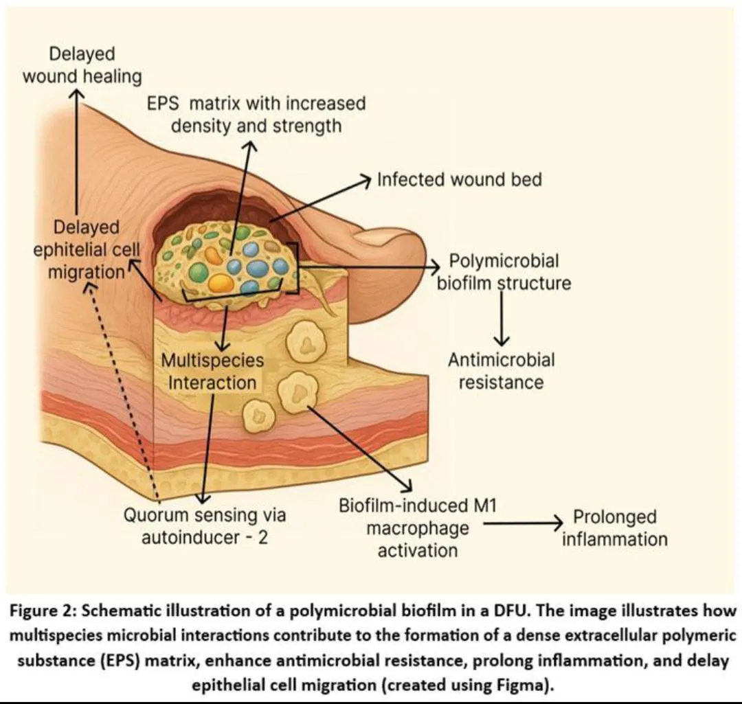
Persistent infection can occur when bacteria persist in deeper tissues or bone, if the antibiotic selected is unable to eradicate the causative organisms, or if necrotic and infected tissue is inadequately treated. Biofilms may contribute to persistence but it is difficult to measure their discrete role in the delayed healing of diabetic foot ulcers in humans [3, 8].
Studies differ in patient selection, sampling techniques, diagnostic criteria, and outcome definitions, which makes it difficult to interpret evidence linking biofilm-producing organisms and antibiotic resistance to amputation, mortality, recurrence, or prolonged treatment. Microbial results should not be considered as independent evidence for causality because these results are also very much influenced by ulcer severity, ischaemia, extent of infection, comorbidities and the care provided [3, 8, 11, 17].
Differences in sampling strategies, diagnostic approaches, study populations and outcome definitions continue to limit the evidence linking biofilm identification with recurrence, length of therapy and hospitalisation [3, 8]. Isolation of a biofilm producing organism in the laboratory does not, in particular, prove that a biofilm is responsible for a poor clinical response in a patient.
7. Future Directions and Research Gaps
7.1. Lack of Standardised Methods for Biofilm Detection
Biofilm formation is known to play an important role in chronic wound infection, however, detection of biofilms in diabetic foot ulcers (DFUs) remains difficult. In different researches, it is used with different methodologies such as molecular techniques, bacterial culture and microscopy. These approaches do not always produce comparable results, making it difficult to compare results across studies. Furthermore, germs in a wound do not always mean that the wound is clinically infected.
The IWGDF/IDSA guidelines mention there is insufficient evidence to recommend routine use of quantitative microbiological analysis to diagnose diabetic foot infections at present [1]. Therefore, future studies should aim to develop reliable and reproducible techniques for identification of clinically significant biofilms and their distinction from normal wound colonisation. [17] In addition, standardised sampling techniques and accurate standards for the evaluation of test results are required.
7.2. Need for Further Research in Humans
Many studies on biofilm and antibiotic resistance have relied on single time-point observations, small cohorts of patients, or laboratory tests. These investigations contribute to the understanding of the possible role of biofilms in chronic wounds but are not able to determine definitively their impact on patient healing and treatment outcomes.
Further prospective clinical trials are needed in which patients are followed from time of initial diagnosis until time of healing, recurrence, or other predetermined endpoint. Biofilm results, antimicrobial susceptibility, wound severity, blood circulation, glycaemic control and medication received should be recorded in such investigations. This would allow researchers to establish whether resistance patterns and detection of biofilm are independently associated with adverse outcomes.
7.3. Difference between test results and clinical outcomes
Another major research gap is the lack of data on whether biofilm detection improves clinical decision making. A test may show microorganisms or biofilm-like structures but that does not mean the results will show delayed healing, treatment failure or amputation.
Future research should establish whether findings related to biofilm are associated with quantifiable outcomes such as hospitalisation, lower limb amputation, recurrence, time to full wound healing and persistent infection. Researchers also should determine whether standard clinical assessment and culture-directed therapy are superior to directing therapy on the basis of these test results. This matters because diagnostic techniques are of no value unless the results can translate into meaningful improvements in patient care.
7.4. Limitations of diagnostic accuracy
There is currently no evidence to support the routine clinical application of biofilm detection methods in real diabetic foot wounds. The results may be affected by laboratory processes, wound depth, previous antibiotic therapy and sampling technique. While traditional culture may not pick up organisms that are hard to grow under routine lab conditions, some methods may be able to pick up bacteria that are present but not actively infecting.
Future research should compare potential diagnostic techniques with appropriate reference techniques and clinical evaluations. Reports of studies should include data on sensitivity, specificity, reproducibility, cost, and turnaround time for results. It is also important to determine if these methods can be successfully implemented in typical clinical laboratories, especially in resource-limited settings.
7.5. The Need for Geographically Representative Data on Antimicrobial Resistance
Geographical location, healthcare environment, previous antibiotic exposure and patient demographics influence patterns of antimicrobial resistance. One hospital's or one country's data may not be a good representation of microorganisms and resistance patterns in other countries. This limits the generalisability of some published research.
More representative data can be generated by multicenter studies covering several hospitals and geographical areas. The bacterial species isolated, the antibiotic susceptibility pattern, the study site, the sample type and relevant patient characteristics should be indicated by the researchers. Regional surveillance may be useful to identify differences between local resistance patterns and those reported globally in India and other countries with different healthcare settings. The data can be used to reduce the overuse of antibiotics and to select the appropriate antibiotic.
7.6. Clinical Validation of Antibiofilm Treatments
Several strategies are being investigated to disrupt biofilms, including enzymes, bacteriophages, antimicrobial peptides and specific wound-dressing technologies. Success in the lab however doesn’t necessarily mean these methods translate to better patient outcomes. More research is needed to establish their safety, efficacy, optimal dose, and compatibility with standard wound management.
Future research should move on from well-designed lab tests to proper clinical trials. These trials should assess complete wound healing, resolution of infection, recurrence, side effects and need for further surgery. Debridement, pressure off-loading, vascular management and glycaemic control should also be considered as these can influence healing without the need for antimicrobial therapy.
7.7. Future Research Directions
Future research should focus on the development of standardised methods for biofilm detection, prospective clinical studies and clear links between diagnostic findings and patient outcomes. Clinical trials to evaluate the efficacy and safety of new antibiofilm treatments are also needed, as is enhanced regional surveillance of antibiotic resistance.
The main question is whether identification of biofilms adds information to improve the management of diabetic foot ulcers. Answering this question could allow researchers to develop better evidence-based diagnostic and therapeutic approaches in people with chronic or difficult to heal wounds.[1, 3, 16]
CONCLUSIONS
Diabetic foot ulcers (DFUs) are complicated to treat due to a number of factors affecting wound healing, including peripheral neuropathy, poor blood flow, slow wound healing, biofilm formation and antimicrobial resistance. Biofilms may help germs thrive in chronic wounds and resist antimicrobial treatment. Resistant bacteria could make selecting effective drugs difficult. However, both factors should be considered, since delayed healing is not necessarily indicative of the formation of biofilms or antimicrobial resistance, among other clinical problems.
This review stresses the importance of adequate biofilm detection methods, bacterial identification and antibiotic susceptibility testing for the understanding of treatment failure in diabetic foot ulcers. Because of variations in detection techniques and study design, it is challenging to compare results and evaluate the predictive value of these traits for clinical outcomes. Further well-designed human studies are needed to assess the association of biofilm identification, antimicrobial resistance, and outcomes such as delayed healing, persistent infection, recurrence, and amputation. In conclusion, a good infection screening, adequate wound care, pressure off-loading, blood glucose management and prompt treatment may result in better outcomes and fewer complications of diabetic foot ulcers.
REFERENCES


Raj Dohale, Yash Mulay, Sumit Kor, Aditya Ghavat, Yash Ghodke, Aditya Jamghare, Avadhoot Partole, Shreyash Pardeshi, Treatment Failure in Diabetic Foot Ulcers: Linking Biofilm Detection, Antimicrobial Resistance, and Clinical Outcomes – Review Article, Int. J. of Pharm. Sci., 2026, Vol 4, Issue 11, 2253-2265. https://doi.org/10.5281/zenodo.24270907
 10.5281/zenodo.24270907