
View Article

Abstract

Wound healing is a complex biological process which involves four distinct phases like hemostasis, inflammation, proliferation and tissue remodeling. The various phytoconstituents present in the medicinal plants made them a potential wound healing agent. This review summarizes the wound healing potential of medicinal plants based on various phytoconstituents like alkaloids, flavonoids, tannins, saponins, phenolics terpenoids and glycosides present in it, which exhibits antioxidant, antimicrobial, anti-inflammatory, angiogenic and collagen promoting effect.

Keywords

Wound healing, Medicinal plants, Antioxidant, Antimicrobial, Tissue regeneration

Introduction

× Popup Image

Globally, wounds particularly chronic wounds are a major source of expense for healthcare systems. According to estimates, at least 2% of people in the United States of America (USA) may suffer from a chronic wound of some kind. According to a study by Queen and Harding, the three nations that spent the most on wound care in 2022—the United States, China, and Japan were expected to have spent $148.65 billion, $42.78 billion, and $22.91 billion, respectively. Additionally, the worldwide wound care market is projected to rise at a compound annual growth rate of 6.1% from 2024 to 2032, reaching $20.33 billion(1).

Both the market for wound care products and the yearly expenditures made by individuals and governments to manage this health issue are growing(1). The majority of the more than 5,000 commercially available wound care products are dressings(2).

Wound healing is a complicated and dynamic biological process with four overlapping phases: hemostasis, inflammation, proliferation, and tissue remodeling(2). This sequence of actions is essential for the complete healing of wounded tissues. Impaired wound healing, particularly in the case of chronic wounds, is a serious issue in clinical practice.

Medicinal plants have long been recognized as valuable resources in traditional medicine systems across the world, owing to their ability to promote wound healing through a wide range of bioactive phytochemicals with minimal adverse effects. These phytochemicals, including flavonoids, alkaloids, tannins, saponins, and phenolic acids, possess potent antioxidant, anti-inflammatory, and antimicrobial activities that collectively enhance the various stages of the wound healing process. Growing scientific interest has led to extensive investigations into the therapeutic potential of underexplored medicinal plants, with the aim of discovering novel, safe, and effective wound healing agents that can complement or serve as alternatives to conventional wound care therapies.

CLASSIFICATION OF WOUND (3)

Wounds are categorized according to several factors, such as their anatomical site, the type and cause of injury, clinical presentation, depth of tissue damage, extent of tissue loss, and morphological characteristics. Depending on the mechanism responsible for their occurrence, wounds are generally classified as either open or closed. Based on the normal progression of tissue repair, they are also grouped into acute wounds, which heal within an expected timeframe, and chronic wounds, which exhibit delayed or impaired healing.

  1. Open Wounds:

Open wounds are characterized by a break in the skin or underlying tissues, resulting in visible bleeding as blood escapes from the body. These injuries expose the affected tissues to the external environment and are commonly classified into several types, including incisions, lacerations, abrasions, puncture wounds, avulsions, and penetrating injuries such as gunshot wounds. The severity of an open wound depends on the extent of tissue damage and the risk of contamination or infection.

  1. Closed Wounds:

Closed wounds occur when the skin remains intact, but the underlying tissues and blood vessels are damaged. In these injuries, blood leaks from the circulatory system and accumulates beneath the skin without an external opening. Common examples include contusions (bruises), hematomas, and crush injuries. Although the skin surface appears unbroken, significant internal tissue damage may be present.

  1. Acute Wounds:

Acute wounds are injuries that progress through the normal stages of wound healing in a predictable and timely manner. They usually result from surgical procedures, accidental cuts, or other forms of trauma and undergo an orderly sequence of hemostasis, inflammation, proliferation, and remodeling. With appropriate care, acute wounds generally heal completely within the expected period, restoring both the structural and functional integrity of the affected tissue.

  1. Chronic Wounds:

Chronic wounds are those that fail to heal within the expected timeframe due to disruption of the normal healing process. These wounds often remain in a prolonged inflammatory phase, delaying tissue regeneration and repair. Factors such as infection, poor blood circulation, diabetes, repeated trauma, or underlying systemic diseases can contribute to their persistence. As a result, chronic wounds may take months to heal, frequently recur, and require specialized management strategies.


THE STAGES OF WOUND HEALING: TARGETS FOR BIOLOGICAL SCREENING(4)

Wound healing is a complex and well-regulated biological process that restores the integrity of injured tissue through four overlapping phases: hemostasis, inflammation, proliferation, and remodeling. Immediately after injury, the skin barrier is disrupted, resulting in blood loss and an increased risk of microbial invasion. During the hemostatic phase, blood clot formation controls bleeding and provides a protective barrier, while proper wound cleansing and antiseptic treatment help prevent infection.

The inflammatory phase involves the recruitment of immune cells and the release of cytokines to eliminate pathogens and remove damaged tissue. This is followed by the proliferative phase, during which fibroblast proliferation, angiogenesis, collagen deposition, and re-epithelialization promote tissue regeneration. Finally, in the remodeling phase, collagen fibers are reorganized, and scar tissue matures, restoring the strength and function of the affected tissue. Therapeutic interventions that support these phases can enhance wound repair and improve healing outcomes.
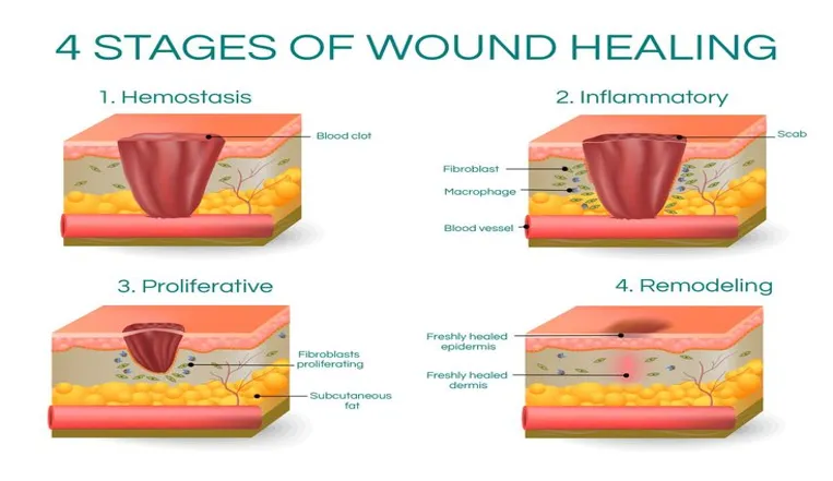


Fig 1: Four stages of wound healing

 
  1. Homeostasis: blood clotting and wound closure

Hemostasis is the first phase of wound healing and occurs immediately after injury to prevent excessive blood loss. Local vasoconstriction reduces blood flow at the injury site, while platelet activation and aggregation lead to the formation of a temporary platelet plug. Simultaneously, the coagulation cascade is activated, resulting in the conversion of fibrinogen to fibrin by thrombin. Cross-linking of fibrin strands by factor XIII stabilizes the clot, which seals the wound, limits blood loss, and provides a protective barrier against microbial invasion.

Preventing wound infection is essential for normal healing, as microbial contamination can prolong inflammation and lead to complications such as cellulitis, osteomyelitis, sepsis, and necrotizing fasciitis. Therefore, maintaining an aseptic wound environment is a key aspect of wound management. The most common wound pathogens include Staphylococcus aureus, Staphylococcus epidermidis, Streptococcus pyogenes, Enterococcus faecalis, Pseudomonas aeruginosa, Escherichia coli, Klebsiella pneumoniae, and Proteus species, making them important targets for antimicrobial wound therapies.


  1. Inflammation

The inflammatory phase is a critical stage of wound healing that enables the immune system to eliminate pathogens, remove damaged tissue, and initiate repair. Following injury, immune and resident cells, including macrophages, neutrophils, fibroblasts, and epithelial cells, release inflammatory mediators such as cytokines, chemokines, prostaglandins, and leukotrienes. Pro-inflammatory cytokines, including interleukin (IL)-1β, IL-2, IL-6, interferon-γ (IFN-γ), and tumor necrosis factor-α (TNF-α), coordinate the inflammatory response, while anti-inflammatory cytokines such as IL-10 help regulate inflammation and prevent excessive tissue damage. Chemokines, including macrophage inflammatory protein-2 (MIP-2) and monocyte chemoattractant protein-1 (MCP-1), recruit leukocytes to the wound site. Additionally, activation of the arachidonic acid pathway through cyclooxygenase (COX) and lipoxygenase (LOX) enzymes generates prostaglandins and leukotrienes, further modulating the inflammatory process. Together, these coordinated events prepare the wound for the subsequent stages of tissue repair and provide important therapeutic targets for enhancing wound healing.

  1. Proliferative Phase

The proliferative phase is characterized by rapid tissue regeneration and restoration of the wound structure. During this stage, chemokines, cytokines, and growth factors stimulate fibroblast migration and proliferation through signaling pathways such as MAPK, YAP, and TGF-β. Fibroblasts synthesize collagen and other extracellular matrix (ECM) components, forming a scaffold for new tissue development. Some fibroblasts differentiate into myofibroblasts, which promote wound contraction and facilitate closure of the injured area. These coordinated processes are essential for effective tissue repair and regeneration.

  1.  Remodeling Phase

The remodeling phase is the final stage of wound healing, during which the newly formed tissue undergoes maturation and reorganization to improve its strength and functionality. This phase may continue for several months or even years. Type III collagen is gradually replaced by the stronger type I collagen, while the extracellular matrix is reorganized to enhance tissue integrity. Continued myofibroblast-mediated wound contraction and collagen remodeling result in the formation of mature scar tissue with increased tensile strength.

VARIOUS PLANTS USED FOR WOUND HEALING

  1. Marsilea minuta Linn.

Marsilea minuta Linn., commonly known as water clover, is an aquatic fern of the family Marsileaceae with traditional uses in treating fever, skin disorders, and inflammation. Its pharmacological activities are attributed to various bioactive phytoconstituents present in the plant. Previous studies have demonstrated its antioxidant and anti-inflammatory properties, suggesting potential wound healing activity. However, its wound healing efficacy remains underexplored, particularly through in vivo studies(5).

The major compounds identified were benzoic acid-4-ethoxy-, ethyl ester (43.39%) and farnesol acetate (18.42%), a sesquiterpene derivative. These compounds were selected for molecular docking studies against TEM-72 and topoisomerase IV to investigate their potential antibacterial mechanisms. Other identified compounds included phenol, 2,4-bis(1,1-dimethylethyl) (8.37%), oxacycloheptadec-8-en-2-one (5.68%), and trans-farnesol (5.11%). The presence of phenolic and other bioactive compounds may contribute to the antioxidant and antibacterial activities of the plant, as phenolic compounds are reported to possess antioxidant, antimicrobial, hepatoprotective, and antidiabetic properties(6).

  1. Cassia fistula

Cassia fistula, commonly known as the golden shower tree, belongs to the family Leguminosae. It is traditionally used by various tribal communities for the treatment of rhinosinusitis and other ailments, including rheumatism, hemoptysis, itching, leukoderma, and gastrointestinal disorders. The plant contains several bioactive secondary metabolites, such as tannins, terpenes, flavonoids, and carbohydrates, which have been reported to exhibit antimicrobial properties(7).

The leaves of Cassia fistula have demonstrated anti-inflammatory and wound healing potential. They exhibit notable antiseptic properties, supporting their traditional use as broad-spectrum antibacterial agents for preventing infections. Additionally, the plant has been reported to possess anti-inflammatory and glycemic-regulating activities(8).

  1. Ficus religiosa

Ficus religiosa, a prominent member of the family Moraceae, is commonly known as peepal. Various parts of the plant, including the bark, fruits, leaves, and stems, have been widely used in traditional medicine. Its therapeutic properties have been reported for several activities, including antibacterial, antioxidant, anti-inflammatory, wound healing, cardioprotective, antithrombotic, and antimutagenic effects. Additionally, the plant exhibits astringent, antiparasitic, antiulcer, antiviral, and antifungal activities, supporting its traditional use in managing various conditions, including tumors, dermatitis, and other skin disorders(9).

The wound healing activity of Ficus religiosa Linn. is attributed to its bioactive phytoconstituents, including flavonoids (quercetin, kaempferol, and myricetin), tannins, phenolic compounds, terpenoids, alkaloids, saponins, and phytosterols such as β-sitosterol and β-sitosteryl-D-glucoside, which promote wound repair through antioxidant, anti-inflammatory, antimicrobial, and tissue-regenerative effects(10).

  1. Mimosa pudica Linn

Mimosa pudica Linn. Which belongs to the family Fabaceae is mainly known as a sensitive plant, humble plant sleeps sleeping, honteuse, dormidera, morivivi, touch-me-not, marie-honte, mayhont, grass, timawi and having many other sleep names.

The plant contains several bioactive constituents, including mimosine, mimosinamine, tyrosine, 3,4-dihydroxypyridine, D-glucuronic acid 4-O-(3,5-dihydroxybenzoic acid)-β-D-glucuronide, mimosine acid, tubulin, C-glycosylflavones, and phenolic ketones. These phytochemicals contribute to its diverse pharmacological properties, including antimicrobial, antidiabetic, antivirulent, antifertility, antioxidant, hepatoprotective, hypoglycemic, lipid-lowering, antidote, antidepressant, and wound healing activities(11).

Ethanolic extracts of the plant have been reported to significantly reduce blood glucose levels in streptozotocin-induced diabetic rats, demonstrating its potential antidiabetic activity. Additionally, topical application of chloroform and methanolic extracts of the root and shoot showed notable wound healing effects, as evidenced by increased hydroxyproline content, tensile strength, and dry weight of granulation tissue in incision wound models. Due to these therapeutic properties and the presence of bioactive phenolic compounds, the plant was selected for evaluating its wound healing potential in diabetic animal models(11).

The precise mechanism by which Mimosa pudica (lajwanti) extract enhances wound repair and regeneration remains unclear; however, several pathways may contribute to its activity. The extract promotes collagen, hexosamine, and DNA synthesis, which are essential for tissue regeneration. Its wound healing effect may also be associated with the reduction of oxidative stress, as evidenced by increased glutathione (GSH) levels, enhanced catalase and superoxide dismutase (SOD) activities, and decreased lipid peroxidation. The extract may regulate inflammatory responses by modulating NF-κB activation and stimulating the production of growth factors and cytokines, including PDGF, TGF-β, and VEGF, which support cell proliferation and tissue repair. Increased antioxidant activity suggests possible activation of the Nrf2 pathway, which helps control excessive inflammation during wound healing. These effects may be attributed to the presence of bioactive constituents such as flavonoids (quercetin, luteolin, and rutin) and mimosine present in the extract(12).

  1. Murraya koenigii

Murraya koenigii, commonly known as curry leaf, belongs to the family Rutaceae and is valued for its characteristic aroma and medicinal properties. The plant contains various bioactive compounds, including alkaloids, phenolic compounds, quercetin, saponins, polypeptides, and steroids, which contribute to its therapeutic potential. Different parts of the plant have been traditionally used in the management of several ailments and are reported to possess antispasmodic, antidiarrheal, gastroprotective, purgative, and blood-purifying activities(13).

The plant contains abundant carbazole alkaloids, flavonoids, and phenolic compounds, which are responsible for its wide range of pharmacological effects, including antioxidant, anti-inflammatory, antimicrobial, and wound healing activities. Traditionally, its leaves have been utilized for the treatment of wounds, skin infections, and inflammatory disorders(14).

The wound healing potential of Murraya koenigii fruits may be associated with their antioxidant activity, which helps reduce free radical generation at the wound site, thereby decreasing inflammation and promoting angiogenesis and collagen formation. Additionally, triterpenoids and tannins contribute to wound repair through their astringent and antimicrobial properties, facilitating wound contraction and enhancing the process of epithelialization(15).

  1. Musa paradisiaca

Musa paradisiaca, commonly known as banana, belongs to the family Musaceae. Various parts of the plant contain diverse bioactive constituents, including quercetin, tannins, polyphenols, alkaloids, and carbohydrates. Extracts of Musa paradisiaca have demonstrated several pharmacological activities, including antidiarrheal, antioxidant, hypoglycemic, cardiovascular protective, wound healing, anti-allergic, antiseptic, antimalarial, and antivenom properties(16).

The decoction prepared from Musa paradisiaca leaves contain phytoconstituents like quercetin, catechin, gallic acid, chlorogenic acid, rutin, dopamine, β-carotene, ascorbic acid, tannins, and saponins(17) which is traditionally used for the treatment of wounds, scratches, and insect bites. In Nigeria, traditional practitioners have employed banana leaf extracts for managing infectious diseases, gastroenteritis, malaria, abdominal pain, and ulcers. The leaves are also rich in dietary fiber, which may help reduce cholesterol levels, relieve constipation, and support colorectal health by improving intestinal function(16).

Treatment with methanolic and hexane extracts of Musa paradisiaca peel demonstrated enhanced wound healing, characterized by complete epithelialization, increased collagen fiber deposition, and fibroblast infiltration. Furthermore, the methanolic peel extract promoted greater proliferation of blood capillaries, indicating improved tissue regeneration and vascularization during the healing process(18).

  1. Centella asiatica

Centella asiatica, commonly known as Gotu Kola, is a perennial herb belonging to the family Apiaceae (Umbelliferae). The aerial parts of this plant have been extensively used in traditional Asian medicine, particularly for dermatological conditions, due to their therapeutic properties(19).

The major bioactive constituents of C. asiatica are triterpenoids, including glycosides such as madecassoside and asiaticoside, and aglycones such as asiatic acid and madecassic acid(20). These compounds are primarily responsible for its pharmacological effects, especially its wound healing and skin-regenerative activities(21). Due to these properties, C. asiatica extracts and their triterpenoids are widely utilized in pharmaceutical and cosmeceutical applications for promoting tissue repair and improving skin health(22).

  1. Xanthosoma sagittifolium (L.) Schott

Xanthosoma sagittifolium (L.) Schott is a pantropical plant species belonging to the family Araceae, originating from Central and South America and now widely distributed across Africa, Asia, and Oceania(23). Beyond its agricultural and cultural importance, the plant has been traditionally used in ethnomedicine for managing various health conditions, including diabetes, gastrointestinal disorders, abscesses, and wounds. In several African and South American communities, crushed leaves or inflorescences are applied topically to wounds and skin injuries to promote hemostasis and enhance tissue repair, reflecting its long-standing role in traditional wound management practices(24).

The wound-healing activity of Xanthosoma sagittifolium is attributed to the presence of chlorogenic acid, caffeic acid, quercetin, kaempferol, catechin, rutin, tannins, saponins, alkaloids, and ascorbic acid, which possess antioxidant, anti-inflammatory, antimicrobial, and collagen-promoting properties that facilitate wound repair(25),(26).

  1. Boerhavia diffusa L.

Boerhavia diffusa L., commonly known as Punarnava, is a perennial creeping herb belonging to the family Nyctaginaceae and is widely distributed throughout India. It is an important medicinal plant in the traditional Indian system of medicine and has been reported to possess diverse pharmacological activities, including immunomodulatory, anticancer, antidiabetic, antifibrinolytic, anti-inflammatory, diuretic, hepatoprotective, antimicrobial, antifungal, anticonvulsant, and antioxidant properties, many of which have been scientifically validated(27).

The wound-healing activity of Boerhavia diffusa is attributed to the presence of boeravinones (A–F), punarnavine, quercetin, kaempferol, eupalitin, ursolic acid, β-sitosterol, lignans, rotenoids, and flavonoids, which exhibit antioxidant, anti-inflammatory, antimicrobial, and collagen-promoting activities that accelerate wound healing(28).

  1. Andrographis paniculata

Andrographis paniculata, commonly known as the “King of Bitters,” is a medicinal plant belonging to the family Acanthaceae and is native to South and Southeast Asia. It has been traditionally used in AYUSH and Traditional Chinese Medicine systems and is recognized for its diverse pharmacological activities, including anti-inflammatory, antiviral, antibacterial, hepatoprotective, antipyretic, and anticancer effects(29). These properties are mainly attributed to its bioactive secondary metabolites, particularly diterpenoid lactones such as andrographolide, 14-deoxy-11,12-didehydroandrographolide, and neoandrographolide, along with flavonoids and polyphenols. Among these constituents, andrographolide is considered the major active compound due to its potent antioxidant, anti-inflammatory, and immunomodulatory activities(30). These phytochemicals may support wound healing by regulating inflammatory mediators, enhancing fibroblast proliferation, promoting collagen synthesis, and reducing oxidative stress, thereby facilitating tissue regeneration and repair(31).

A study reported that topical treatment with a 10% aqueous leaf extract of Andrographis paniculata significantly accelerated wound closure in rats. The treated animals showed reduced inflammation and scar formation, along with enhanced angiogenesis and increased collagen fiber deposition during wound repair. Andrographolide, a bicyclic diterpenoid isolated from A. paniculata leaves, has also been clinically evaluated and demonstrated beneficial effects in various autoimmune disorders(32).

  1. Amphimas pterocarpoides

Amphimas pterocarpoides, a member of the family Leguminosae, is widely distributed across tropical regions of Africa and is traditionally known as “yaya” in Ghana(32). Various parts of the plant, particularly the leaves and bark, are used in traditional medicine for managing conditions such as cough, pneumonia, venereal diseases, pox infections, swellings, pain, wounds, malaria, and gouty arthritis(33). The reddish bark resin is traditionally used for treating dysentery, anemia, schistosomiasis, hematuria, dysmenorrhea, mumps, and as an antidote for certain poisons. Additionally, twigs and wood preparations are used traditionally for preventing miscarriage and managing impotence(32).

Isoflavonoids such as amphiisoflavone, methoxyisoformononetin derivatives, and isoformononetin from Amphimas pterocarpoides exhibit antimicrobial and antioxidant activities(32). Its wound healing effects are mainly associated with isoflavonoids, including genistein, daidzein, formononetin, biochanin A, afrormosin, coumestrol, and prenylated derivatives, which promote tissue repair through antioxidant, anti-inflammatory, and regenerative actions(34).

  1. Elaeis guineensis Jacquin

Elaeis guineensis Jacq., commonly known as African oil palm, is an evergreen, single-stemmed multipurpose plant belonging to the family Arecaceae. It has long been used in West African traditional medicine, where various parts of the plant are considered therapeutically valuable. The fruit oil is traditionally used as a hair treatment, for headaches, stomach disorders, and as a poison antidote for livestock. It is also utilized for non-medicinal purposes, including roofing, firewood, and fuel for goldsmithing. Studies have reported that mesocarp oil and palm kernel oil are used traditionally for treating dermatitis, regulating body temperature during childhood convulsions, and managing ailments such as wounds, rheumatism, bronchitis, gonorrhea, and menorrhagia(35).

Recent research by Olasunkamin and colleagues in 2018 demonstrated that the root extract of Elaeis guineensis Jacq. exhibits antimicrobial activity against wound-associated pathogens, suggesting its potential role in wound healing. Medicinal plants with wound-healing properties are increasingly being explored because affordable and natural therapies can enhance healing, reduce mortality, lower healthcare costs, and minimize complications such as amputation and adverse effects associated with synthetic drugs(35).

A previous study by Che Zain et al. in 2020 reported that the extract of Elaeis guineensis Jacq. leaves (EGL) exhibited wound-healing potential due to the presence of bioactive phytoconstituents such as flavonoids, phenolic compounds, tannins, terpenoids, alkaloids, saponins, carotenoids, tocopherols, and tocotrienols by enhancing cell proliferation and migration in 3T3 fibroblast cells. The methanolic EGL extract demonstrated significant migratory activity in a scratch-wound assay, achieving approximately 85.83–93.34% wound closure, indicating its potential role in promoting tissue repair(36).

  1. Blumea balsamifera (L.) DC.,

Blumea balsamifera (L.) DC., commonly known as Sambong, is a medicinal herb belonging to the family Asteraceae and is characterized by a high content of essential oils, which have been widely utilized in Traditional Chinese Medicine. Its leaves have traditionally been applied for the management of various disorders, including eczema, dermatitis, skin wounds, bruises, beriberi, lumbago, menorrhagia, and rheumatism. Recent studies have demonstrated that leaf extracts possess several pharmacological activities, including plasmin inhibition, antifungal effects, free radical scavenging, and anti-obesity properties. Historical records, including the ancient Chinese medical text Seeking Herbal Medicines in Lingnan, document the use of B. balsamifera extracts for treating snakebite injuries, skin wounds, and itching(37).

Blumea balsamifera (L.) DC. contains a variety of bioactive phytoconstituents, including flavonoids such as quercetin and luteolin derivatives, phenolic compounds, tannins, terpenoids, essential oil components (monoterpenes and sesquiterpenes), alkaloids, and saponins, which contribute to its diverse pharmacological activities(38). The essential oil obtained from the leaves of Blumea balsamifera (L.) DC. (BB oil) contains L-borneol as its major bioactive component. However, the pharmacological potential of BB oil has not been extensively investigated. Recent research has therefore focused on evaluating its wound-healing activity and exploring the possible mechanisms underlying its therapeutic effects(37).

  1. Martynia annua L

Martynia annua L., belonging to the family Martyniaceae, is a glandular, hairy annual herb commonly known as Bichchhu. It has been traditionally used for various medicinal purposes, including the treatment of epilepsy, sore throat, inflammation, and wounds. The leaf paste has been applied to wounds of domestic animals, while root extracts have demonstrated antifungal activity against Alternaria alternata and Aspergillus niger(39). Phytochemical investigations of M. annua have revealed the presence of glycosides, tannins, carbohydrates, phenolic compounds, flavonoids, and anthocyanins. Specific bioactive compounds identified include cyanidin-3-galactoside in flowers, p-hydroxybenzoic acid, sinapic acid, and gentisic acid in leaves and fruits, chlorogenic acid in leaves, and fatty acids in seeds(40). Additionally, ethanol extracts of M. annua leaves have shown anti-inflammatory and wound-healing activities in animal models, demonstrating their potential in promoting tissue repair(41).

  1. Aloe vera (L.) Burm. f.

Aloe vera (L.) Burm. f. belonging to the family Liliaceae, is one of the most extensively studied medicinal plants due to its therapeutic potential, particularly in skin disorders(42). Traditionally known as the “plant of immortality,” A. vera has been used in various cultures to treat burns, wounds, psoriasis, dermatitis, and infections(43). Numerous in vitro, in vivo, and clinical studies have confirmed its wound-healing properties through different preparations, including gel, latex, juice, and extracts. The healing activity is attributed to the synergistic effects of bioactive constituents such as polysaccharides, acemannan, aloin A and B, and emodin, which promote wound repair through antioxidant, anti-inflammatory, antimicrobial, and tissue-regenerative mechanisms(44),(45). Although the gel and leaf extracts are well studied, A. vera flowers, containing compounds such as apigenin glycoside derivatives with antioxidant and anti-inflammatory properties, remain comparatively less explored for cutaneous wound healing(43).

Aloe vera (L.) Burm. f. is a medicinal plant with diverse pharmacological properties, including anti-inflammatory, antibacterial, antioxidant, hypoglycemic, and wound-healing activities(46),(47). Studies in animal models have demonstrated that A. vera gel accelerates wound repair by reducing inflammation, promoting fibroblast proliferation, collagen synthesis, wound contraction, re-epithelialization, angiogenesis, and enhancing growth factors such as TGF-β1 and VEGF(48). Bioactive compounds such as β-sitosterol and aloesin have been reported to stimulate endothelial cell proliferation and migration, thereby supporting angiogenesis and tissue regeneration. The fresh leaves contain anthraquinone-rich yellow latex and a clear mucilaginous gel composed mainly of water, polysaccharides (including glucomannan and acemannan), proteins, vitamins, minerals, and other bioactive constituents responsible for its therapeutic effects(48).

The polysaccharides and glycoproteins present in the leaf pulp of Aloe vera (L.) Burm. f. contributes significantly to its wound-healing and anti-inflammatory activities. The healing effect is primarily attributed to glucomannan, which enhances fibroblast proliferation and activity, thereby promoting collagen synthesis and secretion(49). Additionally, A. vera extracts exhibit antimicrobial properties due to bioactive compounds such as lupeol, salicylic acid, urea nitrogen, cinnamic acid, phenols, and sulfur, which show inhibitory effects against bacteria, fungi, and viruses. Enzymes present in A. vera, including amylase, lipase, and carbopeptidase, also contribute to its therapeutic effects by aiding digestion and reducing inflammation through bradykinin inactivation(49).

  1. Copaifera L. 

Copaifera L. belongs to the family Fabaceae, tree native to Central and South America, and Africa. Various studies have investigated the pharmacological properties of Copaifera extracts, particularly hydroalcoholic leaf extracts and copaiba oleoresin, demonstrating their potential therapeutic applications(49).

The oleoresin of Copaifera L. contains abundant bioactive terpenoids, particularly sesquiterpenes and diterpenes such as β-caryophyllene, α-humulene, β-bisabolene, caryophyllene oxide, copalic acid, kaurenoic acid, and hardwickiic acid. These compounds contribute to wound healing through their anti-inflammatory, antimicrobial, antioxidant, and tissue-regenerative activities by promoting collagen deposition, re-epithelialization, and protection against microbial infection(50),(51).

The pharmacological effects of Copaifera L. oleoresin are mainly attributed to its high sesquiterpene content, which constitutes more than 90% of its composition. However, its therapeutic activity is not related to a single compound but results from the synergistic interactions among multiple bioactive constituents. Studies have demonstrated that copaiba oil exhibits antibacterial, anti-inflammatory, and anti-psoriatic activities. Additionally, Copaifera langsdorffii oleoresin showed no cytotoxic effects on fibroblasts at 100 µg/mL and promoted wound healing in rabbit and rat models, indicating its potential as a wound-healing agent(51).

  1. Curcuma longa Linn.

Curcuma longa L., commonly known as yellow turmeric, belongs to the family Zingiberaceae and is an important medicinal and economically valuable plant(52). Its major bioactive compound, curcumin, possesses potent antioxidant, anti-inflammatory, and anticancer properties(53). The rhizome of C. longa has traditionally been used for treating diarrhea, vomiting, fever, boils, sores, and various skin disorders due to its therapeutic effects(54).

Curcuma longa L. contains several bioactive constituents, including curcumin, demethoxycurcumin, bisdemethoxycurcumin (curcuminoids), and volatile oil components such as arturmerone, α-turmerone, β-turmerone, zingiberene, and atlantone. These compounds exhibit anti-inflammatory, antioxidant, antimicrobial, and collagen-promoting properties, contributing to wound healing by reducing oxidative stress, enhancing fibroblast proliferation, stimulating collagen synthesis, promoting angiogenesis, and accelerating re-epithelialization(55),(56).

The wound-healing activity of Curcuma longa L. extract is mainly attributed to the inhibition of inflammatory mediators, including cyclooxygenase-2 (COX-2) and lipoxygenase (LOX), which reduces inflammation and promotes tissue repair. Curcumin enhances re-epithelialization, cell proliferation, granulation tissue formation, and collagen synthesis during wound healing. This bioactive compound is considered safe and has been widely used in traditional Ayurvedic and Chinese medicine for managing inflammation and various disorders. Its therapeutic effects are mediated through regulation of multiple molecular pathways, including NF-κB, STAT3, transforming growth factor-β (TGF-β), pro-inflammatory cytokines, and oxidative stress-related pathways, supporting fibroblast activity and extracellular matrix formation in wound repair(57).

  1. Ocimum tenuiflorum L. 

Ocimum tenuiflorum L. commonly known as Tulasi, belongs to the family Lamiaceae and is widely distributed in tropical and subtropical regions of India. Various parts of the plant are traditionally used in Ayurveda and Siddha medicine for treating infections, skin disorders, liver-related conditions, and as a remedy for snake and scorpion bites(58). Leaf extracts of O. sanctum exhibit anti-inflammatory, analgesic, and immunomodulatory activities(59). Its flavonoids possess strong free radical scavenging and antioxidant properties, which contribute to wound healing by reducing oxidative stress and protecting tissues from free radical-mediated damage(60).

Ocimum tenuiflorum L. contains several bioactive phytoconstituents, including eugenol, ursolic acid, rosmarinic acid, oleanolic acid, apigenin, luteolin, carvacrol, linalool, β-caryophyllene, flavonoids, and tannins. These compounds contribute to its wound-healing potential through antioxidant, anti-inflammatory, and antimicrobial activities by reducing oxidative stress, controlling inflammation, preventing microbial infection, enhancing fibroblast proliferation and collagen synthesis, and promoting angiogenesis and re-epithelialization(61),(62).

Free radical scavenging activity is considered one of the key mechanisms through which Ocimum tenuiflorum L. protects cells from oxidative damage. Studies in animal models suggest that O. sanctum can modulate immune responses by influencing antibody production, hypersensitivity mediator release, and tissue responses. Additionally, O. sanctum has demonstrated antimicrobial activity against pathogenic microorganisms in vitro. Traditional herbal formulations containing O. sanctum have shown wound-healing effects comparable to standard treatments, such as nitrofurazone and propamidine creams, in infected wound models(63).

  1. Calendula officinalis L.

Calendula officinalis L., a member of the family Asteraceae, is a widely recognized medicinal plant with diverse therapeutic applications. Bioactive compounds present in its flowers exhibit antiviral, antifungal, antibacterial, antigenotoxic, and anti-inflammatory activities. Due to its skin-protective and regenerative properties, C. officinalis flower preparations are commonly applied to damaged and irritated skin. Additionally, marigold promotes collagen synthesis, thereby improving skin strength, maintaining hydration, and accelerating the healing of wounds and surgical scars(64).

The flowers of Calendula officinalis L. contain several bioactive constituents, including triterpenoids, saponins, flavonoids, carotenoids, fatty acids, polysaccharides, and essential oils. Although the levels of these compounds may vary depending on geographical origin, the therapeutic effects of topical C. officinalis preparations remain consistent. In vitro studies have demonstrated the cytotoxic and tumor-inhibitory potential of C. officinalis extracts against various cancer cell lines, including cervical, breast, leukemia, prostate, and fibrosarcoma models. Traditionally, C. officinalis has also been used internally for managing gastrointestinal conditions such as peptic and duodenal ulcers, gastritis, colitis, and mucosal inflammation(65),(66).

In Ayurvedic medicine, Calendula officinalis L. is recognized for its scar-reducing and emollient properties, which are associated with enhanced collagen metabolism and increased angiogenesis at wound sites. Studies have demonstrated that C. officinalis flower extract can improve scar reduction and skin-softening effects(67). Experimental investigations using ethanolic, hexane, and dichloromethane fractions of C. officinalis have shown significant wound-healing and angiogenic activities, with ethanolic extract promoting new blood vessel formation and reducing wound size compared with control treatments(68).

Studies have demonstrated that Calendula officinalis L. extract enhances epithelialization in chronic venous ulcers and promotes wound repair in burn models. Treatment with ethanolic flower extract improved healing by increasing collagen hydroxyproline and hexosamine levels, while reducing tissue damage markers, acute-phase proteins, and lipid peroxidation through its antioxidant activity(69). The ethanolic extract also enhances collagen synthesis and blood circulation at wound sites(70). Due to its antimicrobial, antioxidant, antiseptic, and anti-inflammatory properties, C. officinalis gel and ointment preparations have shown improved wound healing and are suitable for topical application because of their stability, skin penetration, and ease of use(71).

Although the wound-healing mechanisms of Calendula officinalis L. are not completely understood, studies have shown that its extracts promote fibroblast migration and proliferation through a PI3K-dependent pathway. Flower extracts of C. officinalis enhance granulation tissue formation by regulating the expression of connective tissue growth factor (CTGF) and α-smooth muscle actin (α-SMA) in excisional wound models. Additionally, C. officinalis has been reported to stimulate angiogenesis, as demonstrated in chicken chorioallantoic membrane assays and rat skin wound-healing models(71).

  1. Arctium lappa L.

Arctium lappa L., commonly known as burdock, is a perennial herb belonging to the family Asteraceae and is widely cultivated for medicinal use. Traditionally, it has been used in North America, Europe, and Asia for treating sore throat and various skin disorders, including boils, rashes, and acne. Scientific studies have reported that A. lappa exhibits antioxidant, anti-inflammatory, antidiabetic, antimicrobial, antiviral, anticancer, and hepatoprotective activities. Additionally, its root extract has been shown to improve dermal extracellular matrix metabolism by regulating glycosaminoglycan turnover and reducing skin aging-related changes(72).

Arctium lappa L. contains several bioactive phytoconstituents, including lignans such as arctiin and arctigenin, chlorogenic acid, caffeic acid, quercetin, rutin, inulin, polyphenols, flavonoids, tannins, and sesquiterpene lactones. These compounds contribute to its antioxidant, anti-inflammatory, antimicrobial, and wound-healing properties by promoting fibroblast proliferation, enhancing collagen synthesis, modulating extracellular matrix remodeling, and facilitating re-epithelialization during tissue repair(73).

Arctium lappa L. has been reported to influence cell adhesion and gene expression in canine dermal fibroblasts by modulating the Wnt/β-catenin signaling pathway, which plays an important role in wound repair(74). Additionally, a pilot clinical study involving a commercial ointment containing A. lappa demonstrated improved pain management and enhanced healing of first- and second-degree burns compared with control treatment(75).

  1. Astragalus propinquus and Rehmannia glutinosa

Astragalus propinquus, commonly known as Mongolian milkvetch in English is a flowering plant in the family Fabaceae.

Rehmannia glutinosa is a flowering broomrape, belonging to the family Orobanchaceae.

Astragalus propinquus contains several bioactive phytoconstituents, including astragalosides I–IV (particularly astragaloside IV), calycosin, formononetin, ononin, isoflavonoids, astragalus polysaccharides, flavonoids, and saponins. These compounds exhibit antioxidant, anti-inflammatory, immunomodulatory, and pro-angiogenic activities, contributing to wound healing by promoting fibroblast proliferation, collagen synthesis, angiogenesis, and re-epithelialization(76).

Rehmannia glutinosa contains several bioactive phytoconstituents, including catalpol, rehmanniosides, acteoside (verbascoside), aucubin, iridoid glycosides, phenylethanoid glycosides, flavonoids, and polysaccharides. These compounds possess antioxidant, anti-inflammatory, immunomodulatory, and tissue-regenerative properties, contributing to wound healing by enhancing fibroblast proliferation, collagen deposition, angiogenesis, and reducing oxidative stress(77).

The roots of Astragalus propinquus and Rehmannia glutinosa are widely used in Traditional Chinese Medicine for the management of urinary retention, edema, and diabetes-related disorders(78). A herbal formulation containing both roots has shown clinical efficacy in treating diabetic foot ulcers, with subsequent studies in diabetic rats confirming its wound-healing potential(79). The combined extract promotes diabetic wound repair by enhancing angiogenesis, reducing oxidative stress, activating the TGF-β1 signaling pathway, and stimulating extracellular matrix deposition in human skin fibroblasts(80).

  1. Ampelopsis japonica

Ampelopsis japonica is a perennial climbing vine belonging to the family Vitaceae and is native to China, with distribution across East Asia and eastern North America(81). The roots of A. japonica have been traditionally used to treat burns, ulcers, and other inflammatory conditions. Pharmacological studies have demonstrated that the plant possesses neuroprotective, antimicrobial, and anticancer activities, supporting its therapeutic potential(82),(83).

Ampelopsis japonica contains several bioactive phytoconstituents, including resveratrol, ε-viniferin, ampelopsin (dihydromyricetin), catechin, epicatechin, quercetin, kaempferol, gallic acid, and other polyphenols and flavonoids. These compounds exhibit antioxidant, anti-inflammatory, antimicrobial, and tissue-regenerative properties, contributing to wound healing by reducing oxidative stress and inflammation, promoting fibroblast proliferation, enhancing collagen synthesis, stimulating angiogenesis, and accelerating re-epithelialization(84).

Lee et al. demonstrated that ethanolic root extracts of Ampelopsis japonica significantly accelerated the healing of cutaneous scald wounds in rats. Treatment enhanced re-epithelialization, granulation tissue formation, angiogenesis, and collagen deposition compared with Vaseline and silver sulfadiazine. The wound-healing effect was associated with the regulation of inflammatory cytokines, characterized by early increases in TNF-α and TGF-β1 followed by elevated IL-10 during the later stages of healing, facilitating tissue repair and wound closure(85).

  1. Angelica sinensis

Angelica sinensis, commonly known as dong quai or female ginseng, is a herb belonging to the family Apiaceae, indigenous to China.

Angelica sinensis is widely used for treating gynecological disorders, inflammation, headaches, mild anemia, fatigue, and hypertension(86). It exhibits diverse pharmacological activities, including anti-inflammatory, antioxidant, anticancer, and immunomodulatory effects. Studies have shown that A. sinensis extracts promote wound healing by enhancing fibroblast proliferation, collagen secretion, cell migration, glycolysis, and cell viability(87). Some reports also suggest that aqueous extracts stimulate angiogenesis through activation of JNK1/2, p38, and VEGF signaling pathways(88),(89), although the effects on blood vessel formation remain controversial, as the isolated compound n-butylidenephthalide has been reported to inhibit angiogenesis and induce apoptosis(90).

Angelica sinensis contains several bioactive phytoconstituents, including ferulic acid, Z-ligustilide, butylidenephthalide, senkyunolide A, polysaccharides, coniferyl ferulate, flavonoids, and volatile oils. These compounds possess antioxidant, anti-inflammatory, antimicrobial, pro-angiogenic, and tissue-regenerative properties, contributing to wound healing by promoting fibroblast proliferation, collagen synthesis, angiogenesis, and re-epithelialization while reducing oxidative stress and inflammation(91).

  1. Boswellia sacra

Boswellia sacra, also known as Boswellia carteri and others, and commonly called the frankincense tree or the olibanum tree, is a tree in the genus Boswellia, in the Burseraceae family.

Boswellia sacra produces frankincense resin, which has been traditionally used in Africa, India, and the Middle East for managing trauma and inflammatory disorders such as rheumatoid arthritis(92),(93). Bioactive compounds such as boswellic acid derivatives exhibit therapeutic effects, including modulation of inflammation and cellular responses. Frankincense is also a component of the traditional formulation ANBP, containing Agrimonia eupatoriaNelumbo nuciferaBoswellia sacra, and Typha angustifolia pollen. ANBP promotes wound repair by activating TGF-β1/Smad signaling, regulating inflammation, enhancing organized granulation tissue formation, re-epithelialization, and extracellular matrix maturation(94). Studies have further shown that ANBP reduces scar formation and accelerates wound closure in diabetic wound models by promoting neovascularization(95).

Boswellia sacra is rich in bioactive phytoconstituents, including pentacyclic triterpenoids (α- and β-boswellic acid, acetyl-β-boswellic acid, 11-keto-β-boswellic acid [KBA], and 3-O-acetyl-11-keto-β-boswellic acid [AKBA]), monoterpenes (α-pinene, limonene, myrcene, and p-cymene), sesquiterpenes, diterpenes, and essential oils. These phytoconstituents promote wound healing(96).

  1. Caesalpinia sappan L.

Caesalpinia sappan L. is a species of flowering tree in the legume family, Fabaceae, that is native to tropical Asia. Common names in English include sappanwood and Indian redwood. It is used in Traditional Chinese Medicine to improve blood circulation and alleviate edema and pain(97). Its bioactive homoisoflavonoids exhibit antiallergic, anti-inflammatory, and antiviral activities(98),(99). Ethanolic extracts of C. sappan demonstrate broad-spectrum antibacterial effects against various wound-associated pathogens, including Staphylococcus aureus, MRSA, Pseudomonas aeruginosaAcinetobacter baumanniiEscherichia coli, and Klebsiella pneumoniae(100). Additionally, root extracts enhance dermal fibroblast proliferation, migration, and collagen synthesis, thereby promoting cutaneous wound healing(101).

Caesalpinia sappan L. contains several bioactive phytoconstituents, including homoisoflavonoids (brazilin and brazilein), flavonoids (sappanchalcone, protosappanin A and B), phenolic compounds, tannins, saponins, terpenoids, and gallic acid derivatives that promote wound healing(102),(103).

  1. Camellia sinensis

Camellia sinensis is a species of evergreen shrub or small tree in the flowering plant family Theaceae. It is rich in bioactive phytoconstituents, including catechins [epigallocatechin-3-gallate (EGCG), epigallocatechin (EGC), epicatechin gallate (ECG), and epicatechin (EC)], flavonoids, quercetin, kaempferol, phenolic acids, tannins, caffeine, and L-theanine. These phytoconstituents promote wound healing through their antioxidant, anti-inflammatory, antimicrobial, collagen-stimulating, angiogenic, and re-epithelialization activities(104).

Camellia sinensis, the source of green tea, is widely consumed in Asia due to its health-promoting properties(105). Its pharmacological effects, including antioxidant, anti-inflammatory, antimicrobial, anticancer, antiaging, antiobesity, cardioprotective, and neuroprotective activities, are mainly attributed to polyphenolic catechins, particularly epigallocatechin-3-gallate (EGCG)(105). EGCG promotes keratinocyte proliferation and differentiation, modulates TGF-β signaling, reduces matrix metalloproteinase expression, and regulates collagen synthesis, contributing to scar prevention(106). Additionally, C. sinensis extracts enhance fibroblast proliferation, collagen production(107), angiogenesis, and wound closure, demonstrating potential in improving normal and diabetic wound healing(108).

  1. Carthamus tinctorius

Carthamus tinctorius also false saffron, is a highly branched, herbaceous, thistle-like annual plant in the family Asteraceae. It contains several bioactive phytoconstituents involved in wound healing, including quinochalcone glycosides such as hydroxysafflor yellow A (HSYA), safflor yellow A and B, and carthamin; flavonoids such as quercetin, luteolin, kaempferol, and acacetin; phenolic acids including chlorogenic acid and ferulic acid; polysaccharides, serotonin derivatives, and seed oil constituents such as linoleic acid, oleic acid, tocopherols, and phytosterols. These phytochemicals collectively contribute to wound healing through antioxidant, anti-inflammatory, antimicrobial, angiogenic, collagen-promoting, and re-epithelialization activities(109).

Carthamus tinctorius seeds are widely used as a source of edible oil and have a long history in Traditional Chinese Medicine for managing blood-related disorders. Studies have demonstrated that C. tinctorius exhibits diverse pharmacological activities, including antioxidant, anti-inflammatory, immunomodulatory, vasodilatory, anticoagulant, anticancer, and analgesic effects(110). Hydroxysafflor yellow A (HSYA), a major water-soluble pigment component of safflower, possesses antioxidant, anti-inflammatory, pro-angiogenic, and anti-apoptotic properties. Topical application of low-dose HSYA has been shown to enhance diabetic wound healing by promoting angiogenesis, re-epithelialization, and granulation tissue formation, although higher concentrations may inhibit wound repair(111).

  1. Celosia argentea

Celosia argentea L., commonly known as silver cockscomb, belongs to the family Amaranthaceae and is traditionally used for treating skin sores, ulcers, eruptions, and other dermatological disorders(112). Leaf extracts of C. argentea exhibit antioxidant, hepatoprotective, antidiabetic, and antimicrobial activities(113). Studies have shown that alcoholic extracts of C. argentea accelerate burn wound healing in rats by enhancing collagen and hexosamine levels in granulation tissue and promoting dermal fibroblast proliferation and migration(112).

Celosia argentea contains several phytoconstituents that are relevant to wound healing, including flavonoids (quercetin, kaempferol), phenolic compounds, saponins, alkaloids, tannins, terpenoids, glycosides, steroids, polysaccharides, and betalain pigments (betacyanins and betaxanthins). These bioactive compounds contribute to wound repair by exerting antioxidant, anti-inflammatory, antimicrobial, collagen-stimulating, angiogenic, and re-epithelialization effects, thereby accelerating tissue regeneration and wound closure(112).

  1. Cinnamomum cassia

Cinnamomum cassia (L.), a member of the family Lauraceae, is widely used as a spice and traditional medicine. Its bark has been traditionally used to improve blood circulation and relieve pain. C. cassia is also included in several traditional formulations, such as Shexiang Baoxin Pill, used for cardiovascular disorders(114). Cinnamaldehyde, the major bioactive compound of C. cassia, exhibits antimicrobial, anti-inflammatory, antidiabetic, antioxidant, and neuroprotective activities(115). It promotes angiogenesis by activating PI3K/AKT and MAPK signaling pathways, increasing VEGF expression, and has been shown to enhance wound healing in experimental models(116).

Cinnamomum cassia contains several major phytoconstituents associated with wound healing, including cinnamaldehyde (the principal bioactive compound), eugenol, cinnamic acid, cinnamyl acetate, coumarin, procyanidins, catechins, epicatechin, quercetin, kaempferol, and other polyphenols. These compounds promote wound healing through antioxidant, anti-inflammatory, antimicrobial, collagen-stimulating, angiogenic, and re-epithelialization activities, thereby enhancing tissue repair and reducing the risk of wound infection(117).

  1. Commiphora myrrha

Commiphora myrrha, called myrrh, Somali myrrh, herabol myrrh, common myrrh is a tree in the family Burseraceae. It contains several bioactive phytoconstituents that contribute to wound healing, including sesquiterpenes (furanoeudesma-1,3-diene, curzerene, lindestrene), terpenoids, furanosesquiterpenes, triterpenes, steroids, volatile oils, resins, guggulsterones, flavonoids, phenolic compounds, and tannins. These phytochemicals promote wound healing through antimicrobial, anti-inflammatory, antioxidant, analgesic, collagen-stimulating, and re-epithelialization activities, thereby accelerating tissue regeneration and reducing wound infection(118).

Commiphora myrrha produces myrrh resin, which possesses well-established antioxidant, anti-inflammatory, antibacterial, and analgesic properties(119). Traditionally, myrrh has been used for managing gastrointestinal disorders, fractures, arthritis, parasitic infections, and for topical wound care to reduce swelling and pain(120). Studies have shown that myrrh-based formulations, including combinations with other medicinal plants such as Adiantum capillus-venerisAloe vera, and Lawsonia inermis, enhance wound healing in diabetic animal models(121). Additionally, myrrh may promote tissue repair by regulating wound-healing mediators, including TGF-β1 and VEGF, in dermal fibroblasts(122).

  1. Daphne genkwa

Daphne genkwa a member of the family Thymelaeaceae and one of the fundamental herbs in Traditional Chinese Medicine, is widely distributed in regions of China along the Yellow and Yangtze Rivers. It has traditionally been used for its anticonvulsant, analgesic, diuretic, antitussive, expectorant, and sedative properties(123). The major bioactive constituents of D. genkwa, including biflavonoids, coumarins, diterpenes, and triterpenes, contribute to its anti-inflammatory, antitumor, immunomodulatory, and antimelanogenic activities(123). Flavonoid extracts from D. genkwa flowers enhance wound healing by activating the ERK/MEK signaling pathway, promoting fibroblast proliferation and increasing collagen-related gene expression, including COL1A1 and COL3A1(124).

It contains several bioactive phytoconstituents associated with wound-healing activity, including daphnane-type diterpenes (genkwadaphnin, yuanhuacine, yuanhuadine), flavonoids (apigenin, luteolin, quercetin derivatives), coumarins, lignans, phenolic compounds, triterpenoids, and steroids. These constituents exhibit anti-inflammatory, antioxidant, antimicrobial, and tissue-regenerative properties that may support wound healing by reducing oxidative stress, modulating inflammation, and promoting repair processes(125).

  1. Entada phaseoloides

Entada phaseoloides commonly known as St. Thomas bean, is a climbing vine belonging to the family Fabaceae and is distributed across tropical forests of Africa, Australia, Asia, and the Western Pacific. The bark and seeds are rich in saponins and tannins and have traditionally been used as analgesic, antimicrobial, hemostatic, anticancer, and topical agents for skin injuries(126),(127). Studies have shown that tannin-enriched extracts of E. phaseoloides accelerate the healing of infected wounds in rats through antibacterial effects and by promoting cell proliferation and migration. However, further clinical studies are required to confirm its wound-healing efficacy in humans(128).

It contains several phytoconstituents that may contribute to wound healing, including saponins, flavonoids (quercetin and kaempferol derivatives), tannins, phenolic compounds, alkaloids, terpenoids, steroids, triterpenes, and fatty acids. These constituents exhibit antioxidant, anti-inflammatory, antimicrobial, and collagen-promoting activities, which support wound contraction, fibroblast proliferation, collagen deposition, and tissue regeneration(129).

  1. Hibiscus rosa-sinensis

Hibiscus rosa-sinensis L., commonly known as shoeblackplant, is an evergreen shrub belonging to the family Malvaceae and native to tropical Southeast Asia(130). Traditionally, its leaves and flowers have been used for promoting hair growth and preventing premature graying(131). Studies have reported that H. rosa-sinensis exhibits antibacterial and wound-healing properties. Its extracts promote tissue repair by reducing inflammation, enhancing fibroblast proliferation and collagen deposition, and increasing the expression of wound-healing mediators such as VEGF and TGF-β1 in excisional wound models(132).

It contains several phytoconstituents associated with wound-healing activity, including flavonoids (quercetin, kaempferol, anthocyanins), phenolic compounds, tannins, saponins, alkaloids, terpenoids, steroids, mucilage, and vitamin C. These constituents contribute to wound healing through antioxidant, anti-inflammatory, antimicrobial, collagen synthesis, fibroblast proliferation, and re-epithelialization activities, promoting faster wound contraction and tissue regeneration(130).

  1. Ganoderma lucidum

Ganoderma lucidum commonly known as lingzhi or the “mushroom of immortality,” is a medicinal fungus belonging to the family Ganodermataceae and is traditionally used in Chinese, Korean, and Japanese medicine to enhance immunity. It exhibits diverse pharmacological activities, including immunomodulatory, anti-inflammatory, antioxidant, anti-infective, cardioprotective, and lipid-regulating effects(133),(134). Polysaccharide extracts from the fruiting body of G. lucidum have demonstrated wound-healing potential in diabetic rat models by promoting fibroblast proliferation and migration(135), enhancing angiogenesis, and reducing oxidative stress(136). These effects may also be associated with its ability to modulate immune responses and improve tissue repair mechanisms.

It contains several bioactive phytoconstituents (mycochemicals) associated with wound healing, including triterpenoids (ganoderic acids, ganodermanontriol), polysaccharides (β-glucans), proteins, peptides, sterols, nucleosides, phenolic compounds, and flavonoids. These constituents contribute to wound repair through immunomodulatory, antioxidant, anti-inflammatory, antimicrobial, angiogenic, and collagen-promoting activities, enhancing fibroblast proliferation, extracellular matrix formation, and tissue regeneration(137).

  1. Ligusticum striatum 

Ligusticum striatum belongs to the family Apiaceae (Umbelliferae). It contains several bioactive phytoconstituents associated with wound healing, including phthalides (ligustilide, butylidenephthalide, senkyunolide A), phenolic acids (ferulic acid, caffeic acid), volatile oils, flavonoids, coumarins, terpenoids, and polysaccharides. These compounds contribute to wound repair through anti-inflammatory, antioxidant, antimicrobial, angiogenic, and blood circulation-enhancing effects, which may support granulation tissue formation, collagen deposition, and tissue regeneration(138).

It has a long history of use in traditional medicine for promoting cardiovascular and cerebrovascular health and is commonly used for managing ischemic conditions, menstrual disorders, and headaches. More than 170 chemical constituents have been identified from L. striatum, with phthalide lactones and alkaloids representing the major pharmacologically active groups(139). Essential oils derived from L. striatum have demonstrated potential anti-scarring effects by reducing dermal scar formation in rabbit ear wound models(140).

  1. Lonicera japonica

Lonicera japonica commonly known as honeysuckle, belongs to the family Caprifoliaceae and has been extensively used in traditional medicine in Japan, Korea, and China for treating infectious diseases(141). Pharmacological studies have revealed that L. japonica exhibits antimicrobial, anti-inflammatory, antipyretic, antioxidant, anticancer, hepatoprotective, and lipid-regulating activities(142),(143). Ethanol extracts from its flowering aerial parts have been reported to enhance cutaneous wound healing by promoting re-epithelialization, angiogenesis, granulation tissue formation, and wound contraction(144). Although traditionally consumed as a health-promoting herb, excessive doses may cause adverse neurological effects(142).

It contains several phytoconstituents associated with wound healing, including chlorogenic acid, caffeic acid, flavonoids (luteolin, quercetin, rutin), iridoid glycosides (loganin, secologanin), triterpenoid saponins, phenolic compounds, essential oils, and anthocyanins. These constituents contribute to wound repair through antioxidant, anti-inflammatory, antimicrobial, and tissue-regenerative activities by reducing oxidative stress, inhibiting inflammatory mediators, preventing microbial infection, and promoting collagen formation and re-epithelialization(142).

  1. Paeonia suffruticosa

Paeonia suffruticosa, commonly known as moutan peony, belongs to the family Paeoniaceae and has been cultivated for centuries. The root bark is the primary source of bioactive compounds used Pharmacological studies have demonstrated that P. suffruticosa possesses antioxidant, neuroprotective, antitumor, anti-inflammatory, and antidiabetic activities(145). Traditionally, its dried root is applied to cracked skin to promote healing and relieve pain(146). In vitro studies have shown that low concentrations of P. suffruticosa extracts enhance the viability and proliferation of human dermal fibroblasts and HaCaT keratinocytes, indicating its potential role in wound repair(147).

It contains several bioactive phytoconstituents associated with wound healing, including monoterpene glycosides (paeoniflorin, oxypaeoniflorin), phenolic compounds (paeonol, gallic acid), flavonoids, tannins, phenolic acids, terpenoids, and polysaccharides. These constituents contribute to wound healing through anti-inflammatory, antioxidant, antimicrobial, and collagen-promoting activities, supporting fibroblast proliferation, extracellular matrix formation, reduction of oxidative damage, and tissue regeneration(148).

  1. Panax ginseng

Panax ginseng belonging to the family Araliaceae, is a widely used medicinal plant in China, Japan, Korea, and Eastern Siberia for improving cognitive function, memory, immunity, physical endurance, and reducing fatigue. It has traditionally been used for managing conditions such as depression, anxiety, and chronic fatigue syndrome. Pharmacological studies have demonstrated that P. ginseng exhibits vasodilatory, lipid-regulating, anti-inflammatory, antioxidant, anticancer, antibacterial, antiallergic, antiaging, and immunomodulatory activities. Its major bioactive constituents are ginsenosides (also known as panaxosides), a group of saponins considered responsible for many of its therapeutic effects(149).

It contains several bioactive phytoconstituents associated with wound healing, including ginsenosides (Rb1, Rg1, Rg3, Re, Rd), polysaccharides, flavonoids, phenolic compounds, peptides, polyacetylenes, and saponins. These constituents promote wound healing through anti-inflammatory, antioxidant, antimicrobial, angiogenic, immunomodulatory, and collagen-stimulating activities by enhancing fibroblast proliferation, keratinocyte migration, angiogenesis, and extracellular matrix formation(150).

Root extracts of Panax ginseng have demonstrated protective effects against UVB-induced skin damage and have been shown to accelerate healing in laser-induced burns and excisional wound models(151). Studies indicate that P. ginseng extracts enhance keratinocyte migration, promote cell proliferation, and stimulate collagen synthesis in human dermal fibroblasts(152). Additionally, ginsenoside Rb2, a bioactive compound isolated from P. ginseng, promotes epidermal formation by increasing the expression of key wound-healing mediators, including epidermal growth factor, fibronectin, keratin, and collagen-related factors(153).

CURRENT CHALLENGES AND FUTURE PROSPECTS

The integration of herbal therapeutics with polymer-based delivery systems offers promising opportunities for developing advanced wound care therapies(154),(72). However, the development and commercialization of herbo-polymeric wound care products face several challenges, including formulation stability, improved bioavailability, regulatory requirements, and ethical concerns related to sustainability and traditional herbal knowledge(155),(156). Advances in material science, biotechnology, regulatory frameworks, and collaborative approaches in integrative medicine provide new possibilities to overcome these limitations. These developments may support the creation of safe, effective, accessible, and ethically sustainable wound healing therapies(157),(158).

CONCLUSION

The medicinal plants reviewed in this article demonstrate significant wound-healing potential owing to their diverse phytoconstituents and multiple pharmacological activities. Bioactive compounds such as flavonoids, terpenoids, alkaloids, tannins, phenolic compounds, saponins, and glycosides contribute to wound repair by exerting antioxidant, anti-inflammatory, antimicrobial, angiogenic, and collagen-promoting effects. These phytoconstituents collectively enhance fibroblast proliferation, collagen synthesis, re-epithelialization, and tissue remodeling, thereby accelerating the wound-healing process. Although the findings from experimental studies are promising, further standardization and well-designed clinical studies are required to validate their therapeutic efficacy and ensure their successful translation into clinical wound management.


REFERENCES

  1. Cedillo-Cortezano M, Martinez-Cuevas LR, López JAM, Barrera López IL, Escutia-Perez S, Petricevich VL. Use of Medicinal Plants in the Process of Wound Healing: A Literature Review. Pharmaceuticals [Internet]. 2024 Feb 27;17(3):303. Available from: https://www.mdpi.com/1424-8247/17/3/303
  2. Las Heras K, Igartua M, Santos-Vizcaino E, Hernandez RM. Chronic wounds: Current status, available strategies and emerging therapeutic solutions. J Control Release [Internet]. 2020 Dec;328:532–50. Available from: https://linkinghub.elsevier.com/retrieve/pii/S0168365920305538
  3. Dorai AA. Wound care with traditional, complementary and alternative medicine. Indian J Plast Surg [Internet]. 2012 May;45(2):418–24. Available from: http://www.ncbi.nlm.nih.gov/pubmed/23162243
  4. Cock IE, Van Vuuren SF. A review of southern African medicinal plants used traditionally to treat wounds, and studies into their wound healing properties. South African J Bot [Internet]. 2026 Apr;191:112–38. Available from: https://linkinghub.elsevier.com/retrieve/pii/S0254629926000670
  5. Sachan K, Singh A, Sharma S, Mehta S, Rao K, Bhidonia PR, et al. Evaluation of Marsilea minuta Linn. for wound healing activity through the excision wound model. Adv Biomark Sci Technol [Internet]. 2026;8:363–78. Available from: https://linkinghub.elsevier.com/retrieve/pii/S2543106426000189
  6. Arokiyaraj S, Bharanidharan R, Agastian P, Shin H. Chemical composition, antioxidant activity and antibacterial mechanism of action from Marsilea minuta leaf hexane: methanol extract. Chem Cent J [Internet]. 2018 Dec 20;12(1):105. Available from: https://bmcchem.biomedcentral.com/articles/10.1186/s13065-018-0476-4
  7. Satpute SM, Bhamburdekar SB, Kutwal DN, Waghmare SR, And, D.K.Gaikwad. EVALUATION OF ANTIBACTERIAL POTENTIAL OF CASSIA FISTULA L. WORLD J Pharm Pharm Sci. 2015;4(02):635–40.
  8. Panda S, Padhi L, Mohanty G. Antibacterial activities and phytochemical analysis of Cassia fistula (Linn.) leaf. J Adv Pharm Technol Res [Internet]. 2011;2(1):62. Available from: https://journals.lww.com/10.4103/2231-4040.79814
  9. Chandrasekar SB, Bhanumathy M, Pawar AT, Somasundaram T. Phytopharmacology of Ficus religiosa. Pharmacogn Rev [Internet]. 2010 Jul;4(8):195–9. Available from: http://www.ncbi.nlm.nih.gov/pubmed/22228961
  10. Kumar D, Gupta J, Gupta A, Kumar S, Arya R. A review on chemical and biological properties of Cayratia trifolia Linn. (Vitaceae). Pharmacogn Rev [Internet]. 2011;5(10):184. Available from: https://www.phcogrev.com/PharmacognRev-5-10-184
  11. Chandra AA, Darmayanti NPA. EVALUATION OF THE WOUND HEALING PROPERTIES OF MIMOSA PUDICA LINN. IN STREPTOZOTOCIN-INDUCED DIABETES MELLITUS IN RATS. Int J Pharm Sci Res. 2019;27(2):58–66.
  12. Chandra Jagetia G. Mimosa pudica (Lajwanti) Accelerates Repair and Regeneration of Deep Dermal Excision Wound in Swiss Albino Mice. Int J Complement Altern Med [Internet]. 2017 Nov 1;9(2). Available from: https://medcraveonline.com/IJCAM/mimosa-pudica-lajwantiaccelerates-repair-and-regeneration-of-deep-dermal-excision-wound-in-swiss-albino-mice.html
  13. Abdullah R, Younas Q, Kaleem A, Iqtedar M, Aftab M, Saleem F. Phytochemical and antimicrobial properties of different plants and in silico investigation of their bioactive compounds in wound healing and rheumatism. Saudi J Biol Sci [Internet]. 2024 Mar;31(3):103942. Available from: https://linkinghub.elsevier.com/retrieve/pii/S1319562X24000202
  14. Singh AK, Yadav A, Rajput DS GN. Evaluation of wound healing and antioxidant activity of Murraya koenigii leaves with biochemical and histopathological validation. J Chem Heal Risks. 2023;13(5):1043–52.
  15. Pitchaiah G, Samiya S, Lakshmi KNSV SP. Anti-Inflammatory and wound healing activity of hydroalcoholic extract of Murraya koenigii fruits in rats. Int J Herb Med. 2016;4(3):40–4.
  16. Padilla-Camberos E, Flores-Fernández JM, Canales-Aguirre AA, Barragán-Álvarez CP, Gutiérrez-Mercado Y, Lugo-Cervantes E. Wound healing and antioxidant capacity of Musa paradisiaca Linn. peel extracts. J Pharm Pharmacogn Res [Internet]. 2016 Jan 1;4(1):165–73. Available from: https://jppres.com/jppres/wound-healing-and-antioxidant-capacity-of-musa-paradisiaca-peel/
  17. Pereira A, Maraschin M. Banana (Musa spp) from peel to pulp: Ethnopharmacology, source of bioactive compounds and its relevance for human health. J Ethnopharmacol [Internet]. 2015 Feb;160:149–63. Available from: https://linkinghub.elsevier.com/retrieve/pii/S037887411400782X
  18. Amutha K, Selvakumari U. Wound healing activity of methanolic stem extract of Musa paradisiaca Linn. (Banana) in Wistar albino rats. Int Wound J [Internet]. 2016 Oct 16;13(5):763–7. Available from: https://onlinelibrary.wiley.com/doi/10.1111/iwj.12371
  19. Thong-on W, Sakchaisri K, Prathanturarug S. The effects of glycoside-rich green extract from Centella asiatica (L.) Urban on wound healing and anti-aging activity. Phytomedicine Plus [Internet]. 2024 Nov;4(4):100628. Available from: https://linkinghub.elsevier.com/retrieve/pii/S2667031324001015
  20. Azerad R. Chemical structures, production and enzymatic transformations of sapogenins and saponins from Centella asiatica (L.) Urban. Fitoterapia [Internet]. 2016 Oct;114:168–87. Available from: https://linkinghub.elsevier.com/retrieve/pii/S0367326X1630168X
  21. Gray NE, Alcazar Magana A, Lak P, Wright KM, Quinn J, Stevens JF, et al. Centella asiatica: phytochemistry and mechanisms of neuroprotection and cognitive enhancement. Phytochem Rev [Internet]. 2018 Feb 20;17(1):161–94. Available from: http://link.springer.com/10.1007/s11101-017-9528-y
  22. Azis HA, Taher M, Ahmed AS, Sulaiman WMAW, Susanti D, Chowdhury SR, et al. In vitro and In vivo wound healing studies of methanolic fraction of Centella asiatica extract. South African J Bot [Internet]. 2017 Jan;108:163–74. Available from: https://linkinghub.elsevier.com/retrieve/pii/S0254629916302617
  23. Boakye AA, Wireko‐Manu FD, Oduro I, Ellis WO, Gudjónsdóttir M, Chronakis IS. Utilizing cocoyam ( Xanthosoma sagittifolium ) for food and nutrition security: A review. Food Sci Nutr [Internet]. 2018 Jun 13;6(4):703–13. Available from: https://onlinelibrary.wiley.com/doi/10.1002/fsn3.602
  24. Soares Píccolo Cupertino M, Sarandy MM, Gonçalves RV, Gusmão LJ, Junior DTS, Purgato GA, et al. Ethnopharmacological relevance of Xanthosoma sagittifolium (L.) Schott: experimental validation of its wound healing potential. J Ethnopharmacol [Internet]. 2026 Aug;367:121744. Available from: https://linkinghub.elsevier.com/retrieve/pii/S0378874126005969
  25. Chen W-C, Liou S-S, Tzeng T-F, Lee S-L, Liu I-M. Effect of Topical Application of Chlorogenic Acid on Excision Wound Healing in Rats. Planta Med [Internet]. 2013 Apr 8;79(08):616–21. Available from: http://www.thieme-connect.de/DOI/DOI?10.1055/s-0032-1328364
  26. Molefe PF, Ghasemishahrestani Z, Mbele M, Khanyile S, Farrant J, Khumalo NP, et al. African Medicinal Plants in Cutaneous Wound Repair: A Comprehensive Analysis of the Role of Phytochemicals. Int Wound J [Internet]. 2025 Aug 17;22(8). Available from: https://onlinelibrary.wiley.com/doi/10.1111/iwj.70742
  27. Juneja K, Mishra R, Chauhan S, Gupta S, Roy P, Sircar D. Metabolite profiling and wound-healing activity of Boerhavia diffusa leaf extracts using in vitro and in vivo models. J Tradit Complement Med [Internet]. 2020 Jan;10(1):52–9. Available from: https://linkinghub.elsevier.com/retrieve/pii/S2225411018303420
  28. Mishra S, Aeri V, Gaur PK, Jachak SM. Phytochemical, Therapeutic, and Ethnopharmacological Overview for a Traditionally Important Herb: Boerhavia diffusa Linn. Biomed Res Int [Internet]. 2014;2014:1–19. Available from: http://www.hindawi.com/journals/bmri/2014/808302/
  29. Veldman LBM, Belt-Van Zoen E, Baars EW. Mechanistic Evidence of Andrographis paniculata (Burm. f.) Wall. ex Nees, Pelargonium sidoides DC., Echinacea Species and a Combination of Hedera helix L., Primula veris L./Primula elatior L. and Thymus vulgaris L./Thymus zygis L. in the Treatment of Acut. Pharmaceuticals [Internet]. 2023 Aug 24;16(9):1206. Available from: https://www.mdpi.com/1424-8247/16/9/1206
  30. Tundis R, Patra JK, Bonesi M, Das S, Nath R, Das Talukdar A, et al. Anti-Cancer Agent: The Labdane Diterpenoid-Andrographolide. Plants [Internet]. 2023 May 12;12(10):1969. Available from: https://www.mdpi.com/2223-7747/12/10/1969
  31. Raghavan R, Cheriyamundath S, Madassery J. 14-Deoxy-11,12-didehydroandrographolide inhibits proliferation and induces GSH-dependent cell death of human promonocytic leukemic cells. J Nat Med [Internet]. 2014 Apr 24;68(2):387–94. Available from: http://link.springer.com/10.1007/s11418-014-0815-2
  32. Asante-Kwatia E, Adjei S, Jibira Y, Gyimah, Lord, Adjei-Hinneh G, Amponsah IK, et al. Amphimas pterocarpoides harms.: An Evaluation of flavonoid and phenolic contents, wound healing, anthelmintic and antioxidant activities of the leaves and stem bark. Heliyon [Internet]. 2021 Nov;7(11):e08261. Available from: https://linkinghub.elsevier.com/retrieve/pii/S2405844021023641
  33. Wickens GE, Burkill HM. The Useful Plants of West Tropical Africa. Kew Bull [Internet]. 1986;41(2):471. Available from: https://www.jstor.org/stable/4102963?origin=crossref
  34. Asante-Kwatia E, Adjei S, Jibira Y, Gyimah, Lord, Adjei-Hinneh G, Amponsah IK, et al. Amphimas pterocarpoides harms.: An Evaluation of flavonoid and phenolic contents, wound healing, anthelmintic and antioxidant activities of the leaves and stem bark. Heliyon [Internet]. 2021 Nov;7(11):e08261. Available from: http://www.ncbi.nlm.nih.gov/pubmed/34765780
  35. Awolola GV, Emmanuel SS, Adesibikan AA. Evaluation of phytoconstituent and wound-healing potential of methanolic waste shell extract of Elaeis guineensis Jacquin in female rats. Phytomedicine Plus [Internet]. 2021 Nov;1(4):100126. Available from: https://linkinghub.elsevier.com/retrieve/pii/S2667031321001081
  36. Sulistiarini R, Helmi H, Narsa AC. Elaeis guineensis Jacq. leaves are a potential biomass for herbal medicine resources: A mini review. J Appl Pharm Sci [Internet]. 2022; Available from: https://japsonline.com/abstract.php?article_id=3754&sts=2
  37. Pang Y, Wang D, Hu X, Wang H, Fu W, Fan Z, et al. Effect of volatile oil from Blumea Balsamifera (L.) DC. leaves on wound healing in mice. J Tradit Chinese Med [Internet]. 2014 Dec;34(6):716–24. Available from: https://linkinghub.elsevier.com/retrieve/pii/S025462721530087X
  38. Kim HH, Oh MH, Park KJ, Heo JH, Lee MW. Anti-inflammatory activity of sulfate-containing phenolic compounds isolated from the leaves of Myrica rubra. Fitoterapia [Internet]. 2014 Jan;92:188–93. Available from: https://linkinghub.elsevier.com/retrieve/pii/S0367326X13002724
  39. Lodhi S, Singhai AK. Wound healing effect of flavonoid rich fraction and luteolin isolated from Martynia annua Linn. on streptozotocin induced diabetic rats. Asian Pac J Trop Med [Internet]. 2013 Apr;6(4):253–9. Available from: http://linkinghub.elsevier.com/retrieve/pii/S199576451360053X
  40. Mali P., Ansari A., Chaturvedi M. Antifertility effect of chronically administered Martynia annua root extract on male rats. J Ethnopharmacol [Internet]. 2002 Oct;82(2–3):61–7. Available from: https://linkinghub.elsevier.com/retrieve/pii/S0378874102000843
  41. Santram L, Singhai AK. Preliminary pharmacological evaluation of Martynia annua Linn leaves for wound healing. Asian Pac J Trop Biomed [Internet]. 2011 Dec;1(6):421–7. Available from: http://www.ncbi.nlm.nih.gov/pubmed/23569806
  42. Radha MH, Laxmipriya NP. Evaluation of biological properties and clinical effectiveness of Aloe vera: A systematic review. J Tradit Complement Med [Internet]. 2015 Jan;5(1):21–6. Available from: https://linkinghub.elsevier.com/retrieve/pii/S2225411014000078
  43. Razia S, Park H, Shin E, Shim K-S, Cho E, Kang MC, et al. Synergistic effect of Aloe vera flower and Aloe gel on cutaneous wound healing targeting MFAP4 and its associated signaling pathway: In-vitro study. J Ethnopharmacol [Internet]. 2022 May;290:115096. Available from: https://linkinghub.elsevier.com/retrieve/pii/S0378874122001349
  44. Burusapat C, Supawan M, Pruksapong C, Pitiseree A, Suwantemee C. Topical Aloe Vera Gel for Accelerated Wound Healing of Split-Thickness Skin Graft Donor Sites: A Double-Blind, Randomized, Controlled Trial and Systematic Review. Plast Reconstr Surg [Internet]. 2018 Jul;142(1):217–26. Available from: https://journals.lww.com/00006534-201807000-00037
  45. Leiva-Cala C, Lorenzo-Pouso AI, Centenera-Centenera B, López-Palafox J, Gándara-Vila P, García-García A, et al. Clinical efficacy of an Aloe Vera gel versus a 0.12% chlorhexidine gel in preventing traumatic ulcers in patients with fixed orthodontic appliances: a double-blind randomized clinical trial. Odontology [Internet]. 2020 Jul 29;108(3):470–8. Available from: http://link.springer.com/10.1007/s10266-019-00468-w
  46. Spoorthy N. B, Ayesha N. Bioactive constituents of the genus Aloe and their potential therapeutic and pharmacological applications: A review. J Appl Pharm Sci [Internet]. 2020 Nov 5; Available from: https://www.japsonline.com/abstract.php?article_id=3248&sts=2
  47. Sánchez M, González-Burgos E, Iglesias I, Gómez-Serranillos MP. Pharmacological Update Properties of Aloe Vera and its Major Active Constituents. Molecules [Internet]. 2020 Mar 13;25(6):1324. Available from: https://www.mdpi.com/1420-3049/25/6/1324
  48. Kaewsrisung S, Sukpat S, Issarasena N, Patumraj S, Somboonwong J. The effects of oral Aloe vera on the efficacy of transplanted human endothelial cells and the expression of matrix metalloproteinases in diabetic wound healing. Heliyon [Internet]. 2021 Dec;7(12):e08533. Available from: https://linkinghub.elsevier.com/retrieve/pii/S2405844021026360
  49. Genesi BP, de Melo Barbosa R, Severino P, Rodas ACD, Yoshida CMP, Mathor MB, et al. Aloe vera and copaiba oleoresin-loaded chitosan films for wound dressings: microbial permeation, cytotoxicity, and in vivo proof of concept. Int J Pharm [Internet]. 2023 Mar;634:122648. Available from: https://linkinghub.elsevier.com/retrieve/pii/S0378517323000686
  50. Leandro LM, De Sousa Vargas F, Barbosa PCS, Neves JKO, Da Silva JA, Da Veiga-Junior VF. Chemistry and Biological Activities of Terpenoids from Copaiba (Copaifera spp.) Oleoresins. Molecules [Internet]. 2012 Mar 30;17(4):3866–89. Available from: https://www.mdpi.com/1420-3049/17/4/3866
  51. Da Trindade R, Da Silva JK, Setzer WN. Copaifera of the Neotropics: A Review of the Phytochemistry and Pharmacology. Int J Mol Sci [Internet]. 2018 May 18;19(5):1511. Available from: https://www.mdpi.com/1422-0067/19/5/1511
  52. Verma S, Singh S, Sharma S, Tewari SK, Roy RK, Goel AK, et al. Assessment of genetic diversity in indigenous turmeric (Curcuma longa) germplasm from India using molecular markers. Physiol Mol Biol Plants [Internet]. 2015 Apr 20;21(2):233–42. Available from: http://link.springer.com/10.1007/s12298-015-0286-2
  53. Kocaadam B, Şanlier N. Curcumin, an active component of turmeric ( Curcuma longa ), and its effects on health. Crit Rev Food Sci Nutr [Internet]. 2017 Sep 2;57(13):2889–95. Available from: https://www.tandfonline.com/doi/full/10.1080/10408398.2015.1077195
  54. Mutia WON, Usman AN, Jaqin N, Prihantono, Rahman L, Ahmad M. Potency of complemeter therapy to the healing process of perineal wound; turmeric (Curcuma longa Linn) Infusa. Gac Sanit [Internet]. 2021;35:S322–6. Available from: https://linkinghub.elsevier.com/retrieve/pii/S0213911121002375
  55. Li S. Chemical Composition and Product Quality Control of Turmeric (Curcuma longa L.). Pharm Crop [Internet]. 2011 Jul 6;5(1):28–54. Available from: http://benthamopen.com/ABSTRACT/TOPHARMCJ-2-28
  56. Akbik D, Ghadiri M, Chrzanowski W, Rohanizadeh R. Curcumin as a wound healing agent. Life Sci [Internet]. 2014 Oct;116(1):1–7. Available from: https://linkinghub.elsevier.com/retrieve/pii/S0024320514007036
  57. Hewlings S, Kalman D. Curcumin: A Review of Its Effects on Human Health. Foods [Internet]. 2017 Oct 22;6(10):92. Available from: https://www.mdpi.com/2304-8158/6/10/92
  58. Godhwani S, Godhwani JL, Was DS. Ocimum sanctum— A preliminary study evaluating its immunoregulatory profile in albino rats. J Ethnopharmacol [Internet]. 1988 Dec;24(2–3):193–8. Available from: https://linkinghub.elsevier.com/retrieve/pii/0378874188901511
  59. Godhwani S, Godhwani JL, Vyas DS. Ocimum sanctum: An experimental study evaluating its anti-inflammatory, analgesic and antipyretic activity in animals. J Ethnopharmacol [Internet]. 1987 Nov;21(2):153–63. Available from: https://linkinghub.elsevier.com/retrieve/pii/0378874187901255
  60. Shetty S, Udupa S, Udupa L. Evaluation of Antioxidant and Wound Healing Effects of Alcoholic and Aqueous Extract of Ocimum sanctum Linn in Rats. Evidence-Based Complement Altern Med [Internet]. 2008 Jan 11;5(1):95–101. Available from: https://onlinelibrary.wiley.com/doi/10.1093/ecam/nem004
  61. Cohen M. Tulsi - Ocimum sanctum: A herb for all reasons. J Ayurveda Integr Med [Internet]. 2014;5(4):251. Available from: http://www.jaim.in/text.asp?2014/5/4/251/146554
  62. Pattanayak P, Behera P, Das D, Panda S. Ocimum sanctum Linn. A reservoir plant for therapeutic applications: An overview. Pharmacogn Rev [Internet]. 2010;4(7):95. Available from: https://www.phcogrev.com/PharmacognRev-4-7-95
  63. Jamshidi N, Cohen MM. The Clinical Efficacy and Safety of Tulsi in Humans: A Systematic Review of the Literature. Rigano D, editor. Evidence-Based Complement Altern Med [Internet]. 2017 Jan 16;2017(1). Available from: https://onlinelibrary.wiley.com/doi/10.1155/2017/9217567
  64. Sridhanya MS, Sundarraj S, Chandrasekaran NS, Girigoswami K. PEG entrapped ZnO synthesized using Calendula officinalis flower extract- physicochemical characterization, in vitro and in vivo biocompatibility assessment, and antibiofilm activity. Next Nanotechnol [Internet]. 2026 Jun;9:100528. Available from: https://linkinghub.elsevier.com/retrieve/pii/S2949829526001671
  65. Muley B, Khadabadi S, Banarase N. Phytochemical Constituents and Pharmacological Activities of Calendula officinalis Linn (Asteraceae): A Review. Trop J Pharm Res [Internet]. 2009 Nov 24;8(5). Available from: http://www.ajol.info/index.php/tjpr/article/view/48090
  66. Jiménez-Medina E, Garcia-Lora A, Paco L, Algarra I, Collado A, Garrido F. A new extract of the plant calendula officinalis produces a dual in vitroeffect: cytotoxic anti-tumor activity and lymphocyte activation. BMC Cancer [Internet]. 2006 Dec 5;6(1):119. Available from: https://bmccancer.biomedcentral.com/articles/10.1186/1471-2407-6-119
  67. Fronza M, Heinzmann B, Hamburger M, Laufer S, Merfort I. Determination of the wound healing effect of Calendula extracts using the scratch assay with 3T3 fibroblasts. J Ethnopharmacol [Internet]. 2009 Dec;126(3):463–7. Available from: https://linkinghub.elsevier.com/retrieve/pii/S0378874109005741
  68. Parente LML, Andrade MA, Brito LAB, Moura VMBD de, Miguel MP, Lino-Júnior R de S, et al. Angiogenic activity of Calendula officinalis flowers L. in rats. Acta Cir Bras [Internet]. 2011 Feb;26(1):19–24. Available from: http://www.scielo.br/scielo.php?script=sci_arttext&pid=S0102-86502011000100005&lng=en&tlng=en
  69. K. Chandran P, Kuttan R. Effect of Calendula officinalis Flower Extract on Acute Phase Proteins, Antioxidant Defense Mechanism and Granuloma Formation During Thermal Burns. J Clin Biochem Nutr [Internet]. 2008;43(2):58–64. Available from: http://www.jstage.jst.go.jp/article/jcbn/43/2/43_2008043/_article
  70. Fonseca YM, Catini CD, Vicentini FTMC, Nomizo A, Gerlach RF, Fonseca MJV. Protective effect of Calendula officinalis extract against UVB-induced oxidative stress in skin: Evaluation of reduced glutathione levels and matrix metalloproteinase secretion. J Ethnopharmacol [Internet]. 2010 Feb;127(3):596–601. Available from: https://linkinghub.elsevier.com/retrieve/pii/S0378874109007740
  71. Bhargab Deka, Bedanta Bhattacharjee, Anshul Shakya, Abu Md Ashif Ikbal, Chayanika Goswami, Santa Sarma. Mechanism of Action of Wound Healing Activity of Calendula officinalis: A Comprehensive Review. Pharm Biosci J [Internet]. 2021 Jan 3;28–44. Available from: http://pharmabiosciencejournal.com/index.php/pbj/article/view/1700
  72. Shedoeva A, Leavesley D, Upton Z, Fan C. Wound Healing and the Use of Medicinal Plants. Evidence-Based Complement Altern Med [Internet]. 2019 Sep 22;2019:1–30. Available from: https://www.hindawi.com/journals/ecam/2019/2684108/
  73. Luquis F, Silva AAO, Wadt NSY, Hi EMB, Martins AMCC, Bach EE. Arctium lappa L.: Chemical composition, antioxidants, phytochemical compounds and use for healing activity. South Florida J Dev [Internet]. 2021 May 17;2(2):2063–71. Available from: https://southfloridapublishing.com/ojs/index.php/jdev/article/view/318
  74. Pomari E, Stefanon B, Colitti M. Effect of Arctium lappa (burdock) extract on canine dermal fibroblasts. Vet Immunol Immunopathol [Internet]. 2013 Dec;156(3–4):159–66. Available from: https://linkinghub.elsevier.com/retrieve/pii/S0165242713002778
  75. Kolacz NM, Jaroch MT, Bear ML, Hess RF. The Effect of Burns & Wounds (B&W)/Burdock Leaf Therapy on Burn-Injured Amish Patients. J Holist Nurs [Internet]. 2014 Dec 25;32(4):327–40. Available from: https://journals.sagepub.com/doi/10.1177/0898010114525683
  76. Fu J, Wang Z, Huang L, Zheng S, Wang D, Chen S, et al. Review of the Botanical Characteristics, Phytochemistry, and Pharmacology of <scp> Astragalus membranaceus </scp> (Huangqi). Phyther Res [Internet]. 2014 Sep 2;28(9):1275–83. Available from: https://onlinelibrary.wiley.com/doi/10.1002/ptr.5188
  77. Zhang R-X, Li M-X, Jia Z-P. Rehmannia glutinosa: Review of botany, chemistry and pharmacology. J Ethnopharmacol [Internet]. 2008 May;117(2):199–214. Available from: https://linkinghub.elsevier.com/retrieve/pii/S037887410800069X
  78. LIU P-P, SHAN G-S, ZHANG F, CHEN J-N, JIA T-Z. Metabolomics analysis and rapid identification of changes in chemical ingredients in crude and processed Astragali Radix by UPLC-QTOF-MS combined with novel informatics UNIFI platform. Chin J Nat Med [Internet]. 2018 Sep;16(9):714–20. Available from: https://linkinghub.elsevier.com/retrieve/pii/S1875536418301110
  79. Wong MW, Leung PC, Wong WC. Limb salvage in extensive diabetic foot ulceration-a preliminary clinical study using simple debridement and herbal drinks. Hong Kong Med J = Xianggang yi xue za zhi [Internet]. 2001 Dec;7(4):403–7. Available from: http://www.ncbi.nlm.nih.gov/pubmed/11773675
  80. Zhang Q, Fong CC, Yu WK, Chen Y, Wei F, Koon CM, et al. Herbal formula Astragali Radix and Rehmanniae Radix exerted wound healing effect on human skin fibroblast cell line Hs27 via the activation of transformation growth factor (TGF-β) pathway and promoting extracellular matrix (ECM) deposition. Phytomedicine [Internet]. 2012 Dec;20(1):9–16. Available from: https://linkinghub.elsevier.com/retrieve/pii/S0944711312003169
  81. Mi J, Wu C, Li C, Xi F, Wu Z, Chen W. Two new triterpenoids from Ampelopsis japonica (Thunb.) Makino. Nat Prod Res [Internet]. 2014 Jan 2;28(1):52–6. Available from: http://www.tandfonline.com/doi/abs/10.1080/14786419.2013.838237
  82. Park H, Shim JS, Kim HG, Lee H, Oh MS. Ampelopsis Radix Protects Dopaminergic Neurons against 1-Methyl-4-phenylpyridinium/1-methyl-4-phenyl-1,2,3,6-tetrahydropyridine-Induced Toxicity in Parkinson’s Disease Models In Vitro and In Vivo. Evidence-Based Complement Altern Med [Internet]. 2013;2013:1–9. Available from: http://www.hindawi.com/journals/ecam/2013/346438/
  83. NHO KJ, CHUN JM, KIM D-S, KIM HK. Ampelopsis japonica ethanol extract suppresses migration and invasion in human MDA-MB-231 breast cancer cells. Mol Med Rep [Internet]. 2015 May;11(5):3722–8. Available from: https://www.spandidos-publications.com/10.3892/mmr.2015.3179
  84. Liang J-H, Lin H-R, Yang C-S, Liaw C-C, Wang I-C, Chen J-J. Bioactive Components from Ampelopsis japonica with Antioxidant, Anti-α-Glucosidase, and Antiacetylcholinesterase Activities. Antioxidants (Basel, Switzerland) [Internet]. 2022 Jun 23;11(7). Available from: http://www.ncbi.nlm.nih.gov/pubmed/35883719
  85. Lee K, Lee B, Lee M-H, Kim B, Chinannai KS, Ham I, et al. Effect of Ampelopsis Radix on wound healing in scalded rats. BMC Complement Altern Med [Internet]. 2015 Dec 8;15(1):213. Available from: https://bmccomplementalternmed.biomedcentral.com/articles/10.1186/s12906-015-0751-z
  86. Jin M, Zhao K, Huang Q, Xu C, Shang P. Isolation, structure and bioactivities of the polysaccharides from Angelica sinensis (Oliv.) Diels: A review. Carbohydr Polym [Internet]. 2012 Jul;89(3):713–22. Available from: https://linkinghub.elsevier.com/retrieve/pii/S014486171200389X
  87. Hsiao C-Y, Hung C-Y, Tsai T-H, Chak K-F. A Study of the Wound Healing Mechanism of a Traditional Chinese Medicine, Angelica sinensis , Using a Proteomic Approach. Evidence-Based Complement Altern Med [Internet]. 2012;2012:1–14. Available from: http://www.hindawi.com/journals/ecam/2012/467531/
  88. Wang J, Yuan Z, Zhao H, Ju D, Chen Y, Chen X, et al. Ferulic acid promotes endothelial cells proliferation through up-regulating cyclin D1 and VEGF. J Ethnopharmacol [Internet]. 2011 Sep;137(2):992–7. Available from: https://linkinghub.elsevier.com/retrieve/pii/S0378874111004958
  89. Lam H, Lin H, Lao S, Gao J, Hong S, Leong C, et al. The angiogenic effects of Angelica sinensis extract on HUVEC in vitro and zebrafish in vivo. J Cell Biochem [Internet]. 2008 Jan 11;103(1):195–211. Available from: https://onlinelibrary.wiley.com/doi/10.1002/jcb.21403
  90. Yeh J-C, Cindrova-Davies T, Belleri M, Morbidelli L, Miller N, Cho C-WC, et al. The natural compound n-butylidenephthalide derived from the volatile oil of Radix Angelica sinensis inhibits angiogenesis in vitro and in vivo. Angiogenesis [Internet]. 2011 May 15;14(2):187–97. Available from: http://link.springer.com/10.1007/s10456-011-9202-8
  91. Wei W-L, Zeng R, Gu C-M, Qu Y, Huang L-F. Angelica sinensis in China-A review of botanical profile, ethnopharmacology, phytochemistry and chemical analysis. J Ethnopharmacol [Internet]. 2016 Aug;190:116–41. Available from: https://linkinghub.elsevier.com/retrieve/pii/S0378874116302951
  92. Banno N, Akihisa T, Yasukawa K, Tokuda H, Tabata K, Nakamura Y, et al. Anti-inflammatory activities of the triterpene acids from the resin of Boswellia carteri. J Ethnopharmacol [Internet]. 2006 Sep;107(2):249–53. Available from: https://linkinghub.elsevier.com/retrieve/pii/S0378874106001310
  93. Jahandideh M, Hajimehdipoor H, Mortazavi SA, Dehpour A, Hassanzadeh G. A Wound Healing Formulation Based on Iranian Traditional Medicine and Its HPTLC Fingerprint. Iran J Pharm Res  IJPR [Internet]. 2016;15(Suppl):149–57. Available from: http://www.ncbi.nlm.nih.gov/pubmed/28228812
  94. Hou Q, He W-J, Hao H-J, Han Q-W, Chen L, Dong L, et al. The Four-Herb Chinese Medicine ANBP Enhances Wound Healing and Inhibits Scar Formation via Bidirectional Regulation of Transformation Growth Factor Pathway. Langsley G, editor. PLoS One [Internet]. 2014 Dec 9;9(12):e112274. Available from: https://dx.plos.org/10.1371/journal.pone.0112274
  95. Hou Q, He W-J, Chen L, Hao H-J, Liu J-J, Dong L, et al. Effects of the Four-Herb Compound ANBP on Wound Healing Promotion in Diabetic Mice. Int J Low Extrem Wounds [Internet]. 2015 Dec 20;14(4):335–42. Available from: https://journals.sagepub.com/doi/10.1177/1534734615575244
  96. Miran M, Amirshahrokhi K, Ajanii Y, Zadali R, Rutter MW, Enayati A, et al. Taxonomical Investigation, Chemical Composition, Traditional Use in Medicine, and Pharmacological Activities of Boswellia sacra Flueck. Bourhia M, editor. Evidence-Based Complement Altern Med [Internet]. 2022 Feb 18;2022:1–14. Available from: https://www.hindawi.com/journals/ecam/2022/8779676/
  97. Zhao H, Wang X, Li W, Koike K, Bai H. A new minor homoisoflavonoid from Caesalpinia sappan. Nat Prod Res [Internet]. 2014 Jan 17;28(2):102–5. Available from: https://www.tandfonline.com/doi/full/10.1080/14786419.2013.847439
  98. Min BS, Cuong TD, Hung TM, Min BK, Shin BS, Woo MH. Compounds from the heartwood of Caesalpinia sappan and their anti-inflammatory activity. Bioorg Med Chem Lett [Internet]. 2012 Dec;22(24):7436–9. Available from: https://linkinghub.elsevier.com/retrieve/pii/S0960894X12013455
  99. Jeong HJ, Kim YM, Kim JH, Kim JY, Park J-Y, Park S-J, et al. Homoisoflavonoids from Caesalpinia sappan Displaying Viral Neuraminidases Inhibition. Biol Pharm Bull [Internet]. 2012;35(5):786–90. Available from: https://www.jstage.jst.go.jp/article/bpb/35/5/35_5_786/_article
  100. Temrangsee P, Kondo S, Itharat A. Antibacterial activity of extracts from five medicinal plants and their formula against bacteria that cause chronic wound infection. J Med Assoc Thai [Internet]. 2011 Dec;94 Suppl 7:S166-71. Available from: http://www.ncbi.nlm.nih.gov/pubmed/22619924
  101. Tewtrakul S, Tungcharoen P, Sudsai T, Karalai C, Ponglimanont C, Yodsaoue O. Antiinflammatory and Wound Healing Effects of Caesalpinia sappan L. Phyther Res [Internet]. 2015 Jun 11;29(6):850–6. Available from: https://onlinelibrary.wiley.com/doi/10.1002/ptr.5321
  102. Nirmal NP, Rajput MS, Prasad RGSV, Ahmad M. Brazilin from Caesalpinia sappan heartwood and its pharmacological activities: A review. Asian Pac J Trop Med [Internet]. 2015 Jun;8(6):421–30. Available from: http://linkinghub.elsevier.com/retrieve/pii/S1995764515000541
  103. kongkham  supranee, Kaenu A. Antioxidant activity of Caesalpinia sappan heartwood extracts against hydrogen peroxide-induced oxidative damage in MRC-5 cell lines. Chulalongkorn Med J [Internet]. 2025 Jun 15;69(3). Available from: https://digital.car.chula.ac.th/clmjournal/vol69/iss3/6
  104. Musial C, Kuban-Jankowska A, Gorska-Ponikowska M. Beneficial Properties of Green Tea Catechins. Int J Mol Sci [Internet]. 2020 Mar 4;21(5):1744. Available from: https://www.mdpi.com/1422-0067/21/5/1744
  105. Yang CS, Chen G, Wu Q. Recent Scientific Studies of a Traditional Chinese Medicine, Tea, on Prevention of Chronic Diseases. J Tradit Complement Med [Internet]. 2014 Jan;4(1):17–23. Available from: https://linkinghub.elsevier.com/retrieve/pii/S222541101630205X
  106. Klass BR, Branford OA, Grobbelaar AO, Rolfe KJ. The effect of epigallocatechin-3-gallate, a constituent of green tea, on transforming growth factor-Î21â&#128;&#147;stimulated wound contraction. Wound Repair Regen [Internet]. 2010 Jan;18(1):80–8. Available from: https://onlinelibrary.wiley.com/doi/10.1111/j.1524-475X.2009.00552.x
  107. Er S, Dikmen M. Camellia sinensis increased apoptosis on U2OS osteosarcoma cells and wound healing potential on NIH3T3 fibroblast cells. Cytotechnology [Internet]. 2017 Dec 16;69(6):901–14. Available from: http://link.springer.com/10.1007/s10616-017-0105-4
  108. Kim H, Kawazoe T, Han D, Matsumara K, Suzuki S, Tsutsumi S, et al. Enhanced wound healing by an epigallocatechin gallate‐incorporated collagen sponge in diabetic mice. Wound Repair Regen [Internet]. 2008 Sep 3;16(5):714–20. Available from: https://onlinelibrary.wiley.com/doi/10.1111/j.1524-475X.2008.00422.x
  109. Zhou X, Tang L, Xu Y, Zhou G, Wang Z. Towards a better understanding of medicinal uses of Carthamus tinctorius L. in traditional Chinese medicine: A phytochemical and pharmacological review. J Ethnopharmacol [Internet]. 2014 Jan;151(1):27–43. Available from: https://linkinghub.elsevier.com/retrieve/pii/S0378874113007757
  110. Yao D, Wang Z, Miao L, Wang L. Effects of extracts and isolated compounds from safflower on some index of promoting blood circulation and regulating menstruation. J Ethnopharmacol [Internet]. 2016 Sep;191:264–72. Available from: http://linkinghub.elsevier.com/retrieve/pii/S0378874116303695
  111. Gao S-Q, Chang C, Niu X-Q, Li L-J, Zhang Y, Gao J-Q. Topical application of Hydroxysafflor Yellow A accelerates the wound healing in streptozotocin induced T1DM rats. Eur J Pharmacol [Internet]. 2018 Mar;823:72–8. Available from: https://linkinghub.elsevier.com/retrieve/pii/S0014299918300244
  112. Priya KS, Arumugam G, Rathinam B, Wells A, Babu M. Celosia argentea Linn. leaf extract improves wound healing in a rat burn wound model. Wound Repair Regen [Internet]. 2004 Nov 12;12(6):618–25. Available from: https://onlinelibrary.wiley.com/doi/10.1111/j.1067-1927.2004.12603.x
  113. Tang Y, Xin H, Guo M. Review on research of the phytochemistry and pharmacological activities of Celosia argentea. Rev Bras Farmacogn [Internet]. 2016 Nov;26(6):787–96. Available from: https://linkinghub.elsevier.com/retrieve/pii/S0102695X1630182X
  114. Zhang K-J, Zhu J-Z, Bao X-Y, Zheng Q, Zheng G, Wang Y. Shexiang Baoxin Pills for Coronary Heart Disease in Animal Models: Preclinical Evidence and Promoting Angiogenesis Mechanism. Front Pharmacol [Internet]. 2017 Jun 28;8. Available from: http://journal.frontiersin.org/article/10.3389/fphar.2017.00404/full
  115. Zhang C, Fan L, Fan S, Wang J, Luo T, Tang Y, et al. Cinnamomum cassia Presl: A Review of Its Traditional Uses, Phytochemistry, Pharmacology and Toxicology. Molecules [Internet]. 2019 Sep 25;24(19):3473. Available from: https://www.mdpi.com/1420-3049/24/19/3473
  116. Yuan X, Han L, Fu P, Zeng H, Lv C, Chang W, et al. Cinnamaldehyde accelerates wound healing by promoting angiogenesis via up-regulation of PI3K and MAPK signaling pathways. Lab Investig [Internet]. 2018 Jun;98(6):783–98. Available from: https://linkinghub.elsevier.com/retrieve/pii/S0023683722014350
  117. Rao PV, Gan SH. Cinnamon: A Multifaceted Medicinal Plant. Kamal MA, editor. Evidence-Based Complement Altern Med [Internet]. 2014 Jan 10;2014(1). Available from: https://onlinelibrary.wiley.com/doi/10.1155/2014/642942
  118. Hanuš LO, Řezanka T, Dembitsky VM, Moussaieff A. Myrrh - Commiphora chemistry. Biomed Pap [Internet]. 2005 Jul 1;149(1):3–28. Available from: http://biomed.papers.upol.cz/doi/10.5507/bp.2005.001.html
  119. El Ashry ESH, Rashed N, Salama OM, Saleh A. Components, therapeutic value and uses of myrrh. Pharmazie [Internet]. 2003 Mar;58(3):163–8. Available from: http://www.ncbi.nlm.nih.gov/pubmed/12685809
  120. Shen T, Li G-H, Wang X-N, Lou H-X. The genus Commiphora: A review of its traditional uses, phytochemistry and pharmacology. J Ethnopharmacol [Internet]. 2012 Jul;142(2):319–30. Available from: https://linkinghub.elsevier.com/retrieve/pii/S0378874112003352
  121. Galehdari H, Negahdari S, Kesmati M, Rezaie A, Shariati G. Effect of the herbal mixture composed of Aloe Vera, Henna, Adiantum capillus-veneris, and Myrrha on wound healing in streptozotocin-induced diabetic rats. BMC Complement Altern Med [Internet]. 2016 Dec 6;16(1):386. Available from: http://bmccomplementalternmed.biomedcentral.com/articles/10.1186/s12906-016-1359-7
  122. Negahdari S, Galehdari H, Kesmati M, Rezaie A, Shariati G. Wound Healing Activity of Extracts and Formulations of Aloe vera, Henna, Adiantum capillus-veneris, and Myrrh on Mouse Dermal Fibroblast Cells. Int J Prev Med [Internet]. 2017 Jan;8(1). Available from: https://journals.lww.com/01439446-201708000-00018
  123. Hu Y, Pan R, Wang Y, Ma M, Peng Y, Fan W, et al. Daphne genkwa: Ethnopharmacology, phytochemistry and pharmacology of an important traditional Chinese medicine. Fitoterapia [Internet]. 2024 Sep;177:106089. Available from: https://linkinghub.elsevier.com/retrieve/pii/S0367326X24002727
  124. Yang D, Xu J, Shi R. Root extractive from Daphne genkwa benefits in wound healing of anal fistula through up-regulation of collagen genes in human skin fibroblasts. Biosci Rep [Internet]. 2017 Apr 30;37(2). Available from: https://portlandpress.com/bioscirep/article/37/2/BSR20170182/83429/Root-extractive-from-Daphne-genkwa-benefits-in
  125. de Queiroz Dias D, Sales DL, Andrade JC, da Silva ARP, Tintino SR, Oliveira-Tintino CD de M, et al. Antibacterial and antibiotic modifying activity evaluation of ruminants’ body fat used as zootherapeutics in ethnoveterinary practices in Northeast Brazil. J Ethnopharmacol [Internet]. 2019 Apr;233:87–93. Available from: https://linkinghub.elsevier.com/retrieve/pii/S0378874118341904
  126. Widsten P, Cruz CD, Fletcher GC, Pajak MA, McGhie TK. Tannins and Extracts of Fruit Byproducts: Antibacterial Activity against Foodborne Bacteria and Antioxidant Capacity. J Agric Food Chem [Internet]. 2014 Nov 19;62(46):11146–56. Available from: https://pubs.acs.org/doi/10.1021/jf503819t
  127. Figueroa-Espinoza MC, Zafimahova A, Alvarado PGM, Dubreucq E, Poncet-Legrand C. Grape seed and apple tannins: Emulsifying and antioxidant properties. Food Chem [Internet]. 2015 Jul;178:38–44. Available from: https://linkinghub.elsevier.com/retrieve/pii/S0308814615000588
  128. Su X, Liu X, Wang S, Li B, Pan T, Liu D, et al. Wound-healing promoting effect of total tannins from Entada phaseoloides (L.) Merr. in rats. Burns [Internet]. 2017 Jun;43(4):830–8. Available from: https://linkinghub.elsevier.com/retrieve/pii/S030541791630434X
  129. Vitale S, Colanero S, Placidi M, Di Emidio G, Tatone C, Amicarelli F, et al. Phytochemistry and Biological Activity of Medicinal Plants in Wound Healing: An Overview of Current Research. Molecules [Internet]. 2022 Jun 1;27(11):3566. Available from: https://www.mdpi.com/1420-3049/27/11/3566
  130. Bhaskar A, Nithya V. Evaluation of the wound-healing activity of Hibiscus rosa sinensis L (Malvaceae) in Wistar albino rats. Indian J Pharmacol [Internet]. 2012;44(6):694. Available from: https://journals.lww.com/10.4103/0253-7613.103252
  131. Adhirajan N, Ravi Kumar T, Shanmugasundaram N, Babu M. In vivo and in vitro evaluation of hair growth potential of Hibiscus rosa-sinensis Linn. J Ethnopharmacol [Internet]. 2003 Oct;88(2–3):235–9. Available from: https://linkinghub.elsevier.com/retrieve/pii/S0378874103002319
  132. Shen H-M, Chen C, Jiang J-Y, Zheng Y-L, Cai W-F, Wang B, et al. The N-butyl alcohol extract from Hibiscus rosa-sinensis L. flowers enhances healing potential on rat excisional wounds. J Ethnopharmacol [Internet]. 2017 Feb;198:291–301. Available from: https://linkinghub.elsevier.com/retrieve/pii/S0378874117301332
  133. Sanodiya B, Thakur G, Baghel R, Prasad G, Bisen P. Ganoderma lucidum: A Potent Pharmacological Macrofungus. Curr Pharm Biotechnol [Internet]. 2009 Dec 1;10(8):717–42. Available from: http://www.eurekaselect.com/openurl/content.php?genre=article&issn=1389-2010&volume=10&issue=8&spage=717
  134. Swallah MS, Bondzie-Quaye P, Wu Y, Acheampong A, Sossah FL, Elsherbiny SM, et al. Therapeutic potential and nutritional significance of Ganoderma lucidum - a comprehensive review from 2010 to 2022. Food Funct [Internet]. 2023 Feb 21;14(4):1812–38. Available from: http://www.ncbi.nlm.nih.gov/pubmed/36734035
  135. Cheng P-G, Phan C-W, Sabaratnam V, Abdullah N, Abdulla MA, Kuppusamy UR. Polysaccharides-Rich Extract of Ganoderma lucidum (M.A. Curtis:Fr.) P. Karst Accelerates Wound Healing in Streptozotocin-Induced Diabetic Rats. Evidence-Based Complement Altern Med [Internet]. 2013;2013:1–9. Available from: http://www.hindawi.com/journals/ecam/2013/671252/
  136. Tie L, Yang H-Q, An Y, Liu S-Q, Han J, Xu Y, et al. Ganoderma Lucidum Polysaccharide Accelerates Refractory Wound Healing by Inhibition of Mitochondrial Oxidative Stress in Type 1 Diabetes. Cell Physiol Biochem [Internet]. 2012;29(3–4):583–94. Available from: https://karger.com/article/doi/10.1159/000338512
  137. Boh B, Berovic M, Zhang J, Zhi-Bin L. Ganoderma lucidum and its pharmaceutically active compounds. In 2007. p. 265–301. Available from: https://linkinghub.elsevier.com/retrieve/pii/S1387265607130106
  138. Sunil C, Irudayaraj SS, Duraipandiyan V, Alrashood ST, Alharbi SA, Ignacimuthu S. Friedelin exhibits antidiabetic effect in diabetic rats via modulation of glucose metabolism in liver and muscle. J Ethnopharmacol [Internet]. 2021 Mar;268:113659. Available from: https://linkinghub.elsevier.com/retrieve/pii/S0378874120335479
  139. Chen Z, Zhang C, Gao F, Fu Q, Fu C, He Y, et al. A systematic review on the rhizome of Ligusticum chuanxiong Hort. (Chuanxiong). Food Chem Toxicol [Internet]. 2018 Sep;119:309–25. Available from: https://linkinghub.elsevier.com/retrieve/pii/S0278691518301248
  140. Wu J-G, Wei Y-J, Ran X, Zhang H, Nian H, Qin L-P. Inhibitory effects of essential oil from rhizomes of Ligusticum chuanxiong on hypertrophic scarring in the rabbit ear model. Pharm Biol [Internet]. 2011 Jul 3;49(7):764–9. Available from: http://www.tandfonline.com/doi/full/10.3109/13880209.2010.542761
  141. Fu J, Yang L, Khan MA, Mei Z. Genetic characterization and authentication of Lonicera japonica Thunb. by using improved RAPD analysis. Mol Biol Rep [Internet]. 2013 Oct 22;40(10):5993–9. Available from: http://link.springer.com/10.1007/s11033-013-2703-3
  142. Shang X, Pan H, Li M, Miao X, Ding H. Lonicera japonica Thunb.: Ethnopharmacology, phytochemistry and pharmacology of an important traditional Chinese medicine. J Ethnopharmacol [Internet]. 2011 Oct;138(1):1–21. Available from: https://linkinghub.elsevier.com/retrieve/pii/S0378874111005897
  143. Li Y, Cai W, Weng X, Li Q, Wang Y, Chen Y, et al. Lonicerae Japonicae Flos and Lonicerae Flos: A Systematic Pharmacology Review. Evidence-Based Complement Altern Med [Internet]. 2015;2015:1–16. Available from: http://www.hindawi.com/journals/ecam/2015/905063/
  144. Chen W-C, Liou S-S, Tzeng T-F, Lee S-L, Liu I-M. Wound repair and anti-inflammatory potential of Lonicera japonica in excision wound-induced rats. BMC Complement Altern Med [Internet]. 2012 Dec 23;12(1):226. Available from: https://bmccomplementalternmed.biomedcentral.com/articles/10.1186/1472-6882-12-226
  145. Diovu EO, Ayoka TO, Onah CM, Okorie NH, Nnadi CO. Biochemical and histological insights of 1,4-polyisoprene isolated from Sphenocentrum jollyanum pierre (menispermaceae) stem in wound healing activity in streptozotocin-induced diabetic rats. J Ethnopharmacol [Internet]. 2023 May;307:116248. Available from: https://linkinghub.elsevier.com/retrieve/pii/S0378874123001162
  146. He D-Y, Dai S-M. Anti-Inflammatory and Immunomodulatory Effects of Paeonia Lactiflora Pall., a Traditional Chinese Herbal Medicine. Front Pharmacol [Internet]. 2011;2. Available from: http://journal.frontiersin.org/article/10.3389/fphar.2011.00010/abstract
  147. Wang R, Lechtenberg M, Sendker J, Petereit F, Deters A, Hensel A. Wound-healing plants from TCM: in vitro investigations on selected TCM plants and their influence on human dermal fibroblasts and keratinocytes. Fitoterapia [Internet]. 2013 Jan;84:308–17. Available from: https://linkinghub.elsevier.com/retrieve/pii/S0367326X12003474
  148. Nandy S, Mukherjee A, Pandey DK, Ray P, Dey A. Indian Sarsaparilla (Hemidesmus indicus): Recent progress in research on ethnobotany, phytochemistry and pharmacology. J Ethnopharmacol [Internet]. 2020 May;254:112609. Available from: https://linkinghub.elsevier.com/retrieve/pii/S0378874119309900
  149. Ratan ZA, Haidere MF, Hong YH, Park SH, Lee J-O, Lee J, et al. Pharmacological potential of ginseng and its major component ginsenosides. J Ginseng Res [Internet]. 2021 Mar;45(2):199–210. Available from: https://linkinghub.elsevier.com/retrieve/pii/S1226845320300592
  150. Attele AS, Wu JA, Yuan C-S. Ginseng pharmacology. Biochem Pharmacol [Internet]. 1999 Dec;58(11):1685–93. Available from: https://linkinghub.elsevier.com/retrieve/pii/S0006295299002129
  151. Kim W-K, Song S-Y, Oh WK, Kaewsuwan S, Tran TL, Kim W-S, et al. Wound-healing effect of ginsenoside Rd from leaves of Panax ginseng via cyclic AMP-dependent protein kinase pathway. Eur J Pharmacol [Internet]. 2013 Feb;702(1–3):285–93. Available from: https://linkinghub.elsevier.com/retrieve/pii/S001429991300068X
  152. Chen X, Wang M, Xu X, Liu J, Mei B, Fu P, et al. Panax ginseng total protein promotes proliferation and secretion of collagen in NIH/3T3 cells by activating extracellular signal-related kinase pathway. J Ginseng Res [Internet]. 2017 Jul;41(3):411–8. Available from: https://linkinghub.elsevier.com/retrieve/pii/S1226845316302603
  153. Choi S. Epidermis proliferative effect of thePanax ginseng Ginsenoside Rb2. Arch Pharm Res [Internet]. 2002 Feb;25(1):71–6. Available from: http://link.springer.com/10.1007/BF02975265
  154. Boateng JS, Matthews KH, Stevens HNE, Eccleston GM. Wound Healing Dressings and Drug Delivery Systems: A Review. J Pharm Sci [Internet]. 2008 Aug;97(8):2892–923. Available from: https://linkinghub.elsevier.com/retrieve/pii/S0022354916326521
  155. Quader S, Cabral H, Mochida Y, Ishii T, Liu X, Toh K, et al. Selective intracellular delivery of proteasome inhibitors through pH-sensitive polymeric micelles directed to efficient antitumor therapy. J Control Release [Internet]. 2014 Aug;188:67–77. Available from: https://linkinghub.elsevier.com/retrieve/pii/S0168365914003514
  156. Prado RF do, de Oliveira FS, Nascimento RD, de Vasconcellos LMR, Carvalho YR, Cairo CAA. Osteoblast response to porous titanium and biomimetic surface: In vitro analysis. Mater Sci Eng C [Internet]. 2015 Jul;52:194–203. Available from: https://linkinghub.elsevier.com/retrieve/pii/S0928493115002106
  157. Sood A, Granick MS, Tomaselli NL. Wound Dressings and Comparative Effectiveness Data. Adv Wound Care [Internet]. 2014 Aug;3(8):511–29. Available from: https://journals.sagepub.com/doi/full/10.1089/wound.2012.0401
  158. Boateng J, Catanzano O. Advanced Therapeutic Dressings for Effective Wound Healing—A Review. J Pharm Sci [Internet]. 2015 Nov;104(11):3653–80. Available from: https://linkinghub.elsevier.com/retrieve/pii/S0022354916301538

Reference

  1. Cedillo-Cortezano M, Martinez-Cuevas LR, López JAM, Barrera López IL, Escutia-Perez S, Petricevich VL. Use of Medicinal Plants in the Process of Wound Healing: A Literature Review. Pharmaceuticals [Internet]. 2024 Feb 27;17(3):303. Available from: https://www.mdpi.com/1424-8247/17/3/303
  2. Las Heras K, Igartua M, Santos-Vizcaino E, Hernandez RM. Chronic wounds: Current status, available strategies and emerging therapeutic solutions. J Control Release [Internet]. 2020 Dec;328:532–50. Available from: https://linkinghub.elsevier.com/retrieve/pii/S0168365920305538
  3. Dorai AA. Wound care with traditional, complementary and alternative medicine. Indian J Plast Surg [Internet]. 2012 May;45(2):418–24. Available from: http://www.ncbi.nlm.nih.gov/pubmed/23162243
  4. Cock IE, Van Vuuren SF. A review of southern African medicinal plants used traditionally to treat wounds, and studies into their wound healing properties. South African J Bot [Internet]. 2026 Apr;191:112–38. Available from: https://linkinghub.elsevier.com/retrieve/pii/S0254629926000670
  5. Sachan K, Singh A, Sharma S, Mehta S, Rao K, Bhidonia PR, et al. Evaluation of Marsilea minuta Linn. for wound healing activity through the excision wound model. Adv Biomark Sci Technol [Internet]. 2026;8:363–78. Available from: https://linkinghub.elsevier.com/retrieve/pii/S2543106426000189
  6. Arokiyaraj S, Bharanidharan R, Agastian P, Shin H. Chemical composition, antioxidant activity and antibacterial mechanism of action from Marsilea minuta leaf hexane: methanol extract. Chem Cent J [Internet]. 2018 Dec 20;12(1):105. Available from: https://bmcchem.biomedcentral.com/articles/10.1186/s13065-018-0476-4
  7. Satpute SM, Bhamburdekar SB, Kutwal DN, Waghmare SR, And, D.K.Gaikwad. EVALUATION OF ANTIBACTERIAL POTENTIAL OF CASSIA FISTULA L. WORLD J Pharm Pharm Sci. 2015;4(02):635–40.
  8. Panda S, Padhi L, Mohanty G. Antibacterial activities and phytochemical analysis of Cassia fistula (Linn.) leaf. J Adv Pharm Technol Res [Internet]. 2011;2(1):62. Available from: https://journals.lww.com/10.4103/2231-4040.79814
  9. Chandrasekar SB, Bhanumathy M, Pawar AT, Somasundaram T. Phytopharmacology of Ficus religiosa. Pharmacogn Rev [Internet]. 2010 Jul;4(8):195–9. Available from: http://www.ncbi.nlm.nih.gov/pubmed/22228961
  10. Kumar D, Gupta J, Gupta A, Kumar S, Arya R. A review on chemical and biological properties of Cayratia trifolia Linn. (Vitaceae). Pharmacogn Rev [Internet]. 2011;5(10):184. Available from: https://www.phcogrev.com/PharmacognRev-5-10-184
  11. Chandra AA, Darmayanti NPA. EVALUATION OF THE WOUND HEALING PROPERTIES OF MIMOSA PUDICA LINN. IN STREPTOZOTOCIN-INDUCED DIABETES MELLITUS IN RATS. Int J Pharm Sci Res. 2019;27(2):58–66.
  12. Chandra Jagetia G. Mimosa pudica (Lajwanti) Accelerates Repair and Regeneration of Deep Dermal Excision Wound in Swiss Albino Mice. Int J Complement Altern Med [Internet]. 2017 Nov 1;9(2). Available from: https://medcraveonline.com/IJCAM/mimosa-pudica-lajwantiaccelerates-repair-and-regeneration-of-deep-dermal-excision-wound-in-swiss-albino-mice.html
  13. Abdullah R, Younas Q, Kaleem A, Iqtedar M, Aftab M, Saleem F. Phytochemical and antimicrobial properties of different plants and in silico investigation of their bioactive compounds in wound healing and rheumatism. Saudi J Biol Sci [Internet]. 2024 Mar;31(3):103942. Available from: https://linkinghub.elsevier.com/retrieve/pii/S1319562X24000202
  14. Singh AK, Yadav A, Rajput DS GN. Evaluation of wound healing and antioxidant activity of Murraya koenigii leaves with biochemical and histopathological validation. J Chem Heal Risks. 2023;13(5):1043–52.
  15. Pitchaiah G, Samiya S, Lakshmi KNSV SP. Anti-Inflammatory and wound healing activity of hydroalcoholic extract of Murraya koenigii fruits in rats. Int J Herb Med. 2016;4(3):40–4.
  16. Padilla-Camberos E, Flores-Fernández JM, Canales-Aguirre AA, Barragán-Álvarez CP, Gutiérrez-Mercado Y, Lugo-Cervantes E. Wound healing and antioxidant capacity of Musa paradisiaca Linn. peel extracts. J Pharm Pharmacogn Res [Internet]. 2016 Jan 1;4(1):165–73. Available from: https://jppres.com/jppres/wound-healing-and-antioxidant-capacity-of-musa-paradisiaca-peel/
  17. Pereira A, Maraschin M. Banana (Musa spp) from peel to pulp: Ethnopharmacology, source of bioactive compounds and its relevance for human health. J Ethnopharmacol [Internet]. 2015 Feb;160:149–63. Available from: https://linkinghub.elsevier.com/retrieve/pii/S037887411400782X
  18. Amutha K, Selvakumari U. Wound healing activity of methanolic stem extract of Musa paradisiaca Linn. (Banana) in Wistar albino rats. Int Wound J [Internet]. 2016 Oct 16;13(5):763–7. Available from: https://onlinelibrary.wiley.com/doi/10.1111/iwj.12371
  19. Thong-on W, Sakchaisri K, Prathanturarug S. The effects of glycoside-rich green extract from Centella asiatica (L.) Urban on wound healing and anti-aging activity. Phytomedicine Plus [Internet]. 2024 Nov;4(4):100628. Available from: https://linkinghub.elsevier.com/retrieve/pii/S2667031324001015
  20. Azerad R. Chemical structures, production and enzymatic transformations of sapogenins and saponins from Centella asiatica (L.) Urban. Fitoterapia [Internet]. 2016 Oct;114:168–87. Available from: https://linkinghub.elsevier.com/retrieve/pii/S0367326X1630168X
  21. Gray NE, Alcazar Magana A, Lak P, Wright KM, Quinn J, Stevens JF, et al. Centella asiatica: phytochemistry and mechanisms of neuroprotection and cognitive enhancement. Phytochem Rev [Internet]. 2018 Feb 20;17(1):161–94. Available from: http://link.springer.com/10.1007/s11101-017-9528-y
  22. Azis HA, Taher M, Ahmed AS, Sulaiman WMAW, Susanti D, Chowdhury SR, et al. In vitro and In vivo wound healing studies of methanolic fraction of Centella asiatica extract. South African J Bot [Internet]. 2017 Jan;108:163–74. Available from: https://linkinghub.elsevier.com/retrieve/pii/S0254629916302617
  23. Boakye AA, Wireko?Manu FD, Oduro I, Ellis WO, Gudjónsdóttir M, Chronakis IS. Utilizing cocoyam ( Xanthosoma sagittifolium ) for food and nutrition security: A review. Food Sci Nutr [Internet]. 2018 Jun 13;6(4):703–13. Available from: https://onlinelibrary.wiley.com/doi/10.1002/fsn3.602
  24. Soares Píccolo Cupertino M, Sarandy MM, Gonçalves RV, Gusmão LJ, Junior DTS, Purgato GA, et al. Ethnopharmacological relevance of Xanthosoma sagittifolium (L.) Schott: experimental validation of its wound healing potential. J Ethnopharmacol [Internet]. 2026 Aug;367:121744. Available from: https://linkinghub.elsevier.com/retrieve/pii/S0378874126005969
  25. Chen W-C, Liou S-S, Tzeng T-F, Lee S-L, Liu I-M. Effect of Topical Application of Chlorogenic Acid on Excision Wound Healing in Rats. Planta Med [Internet]. 2013 Apr 8;79(08):616–21. Available from: http://www.thieme-connect.de/DOI/DOI?10.1055/s-0032-1328364
  26. Molefe PF, Ghasemishahrestani Z, Mbele M, Khanyile S, Farrant J, Khumalo NP, et al. African Medicinal Plants in Cutaneous Wound Repair: A Comprehensive Analysis of the Role of Phytochemicals. Int Wound J [Internet]. 2025 Aug 17;22(8). Available from: https://onlinelibrary.wiley.com/doi/10.1111/iwj.70742
  27. Juneja K, Mishra R, Chauhan S, Gupta S, Roy P, Sircar D. Metabolite profiling and wound-healing activity of Boerhavia diffusa leaf extracts using in vitro and in vivo models. J Tradit Complement Med [Internet]. 2020 Jan;10(1):52–9. Available from: https://linkinghub.elsevier.com/retrieve/pii/S2225411018303420
  28. Mishra S, Aeri V, Gaur PK, Jachak SM. Phytochemical, Therapeutic, and Ethnopharmacological Overview for a Traditionally Important Herb: Boerhavia diffusa Linn. Biomed Res Int [Internet]. 2014;2014:1–19. Available from: http://www.hindawi.com/journals/bmri/2014/808302/
  29. Veldman LBM, Belt-Van Zoen E, Baars EW. Mechanistic Evidence of Andrographis paniculata (Burm. f.) Wall. ex Nees, Pelargonium sidoides DC., Echinacea Species and a Combination of Hedera helix L., Primula veris L./Primula elatior L. and Thymus vulgaris L./Thymus zygis L. in the Treatment of Acut. Pharmaceuticals [Internet]. 2023 Aug 24;16(9):1206. Available from: https://www.mdpi.com/1424-8247/16/9/1206
  30. Tundis R, Patra JK, Bonesi M, Das S, Nath R, Das Talukdar A, et al. Anti-Cancer Agent: The Labdane Diterpenoid-Andrographolide. Plants [Internet]. 2023 May 12;12(10):1969. Available from: https://www.mdpi.com/2223-7747/12/10/1969
  31. Raghavan R, Cheriyamundath S, Madassery J. 14-Deoxy-11,12-didehydroandrographolide inhibits proliferation and induces GSH-dependent cell death of human promonocytic leukemic cells. J Nat Med [Internet]. 2014 Apr 24;68(2):387–94. Available from: http://link.springer.com/10.1007/s11418-014-0815-2
  32. Asante-Kwatia E, Adjei S, Jibira Y, Gyimah, Lord, Adjei-Hinneh G, Amponsah IK, et al. Amphimas pterocarpoides harms.: An Evaluation of flavonoid and phenolic contents, wound healing, anthelmintic and antioxidant activities of the leaves and stem bark. Heliyon [Internet]. 2021 Nov;7(11):e08261. Available from: https://linkinghub.elsevier.com/retrieve/pii/S2405844021023641
  33. Wickens GE, Burkill HM. The Useful Plants of West Tropical Africa. Kew Bull [Internet]. 1986;41(2):471. Available from: https://www.jstor.org/stable/4102963?origin=crossref
  34. Asante-Kwatia E, Adjei S, Jibira Y, Gyimah, Lord, Adjei-Hinneh G, Amponsah IK, et al. Amphimas pterocarpoides harms.: An Evaluation of flavonoid and phenolic contents, wound healing, anthelmintic and antioxidant activities of the leaves and stem bark. Heliyon [Internet]. 2021 Nov;7(11):e08261. Available from: http://www.ncbi.nlm.nih.gov/pubmed/34765780
  35. Awolola GV, Emmanuel SS, Adesibikan AA. Evaluation of phytoconstituent and wound-healing potential of methanolic waste shell extract of Elaeis guineensis Jacquin in female rats. Phytomedicine Plus [Internet]. 2021 Nov;1(4):100126. Available from: https://linkinghub.elsevier.com/retrieve/pii/S2667031321001081
  36. Sulistiarini R, Helmi H, Narsa AC. Elaeis guineensis Jacq. leaves are a potential biomass for herbal medicine resources: A mini review. J Appl Pharm Sci [Internet]. 2022; Available from: https://japsonline.com/abstract.php?article_id=3754&sts=2
  37. Pang Y, Wang D, Hu X, Wang H, Fu W, Fan Z, et al. Effect of volatile oil from Blumea Balsamifera (L.) DC. leaves on wound healing in mice. J Tradit Chinese Med [Internet]. 2014 Dec;34(6):716–24. Available from: https://linkinghub.elsevier.com/retrieve/pii/S025462721530087X
  38. Kim HH, Oh MH, Park KJ, Heo JH, Lee MW. Anti-inflammatory activity of sulfate-containing phenolic compounds isolated from the leaves of Myrica rubra. Fitoterapia [Internet]. 2014 Jan;92:188–93. Available from: https://linkinghub.elsevier.com/retrieve/pii/S0367326X13002724
  39. Lodhi S, Singhai AK. Wound healing effect of flavonoid rich fraction and luteolin isolated from Martynia annua Linn. on streptozotocin induced diabetic rats. Asian Pac J Trop Med [Internet]. 2013 Apr;6(4):253–9. Available from: http://linkinghub.elsevier.com/retrieve/pii/S199576451360053X
  40. Mali P., Ansari A., Chaturvedi M. Antifertility effect of chronically administered Martynia annua root extract on male rats. J Ethnopharmacol [Internet]. 2002 Oct;82(2–3):61–7. Available from: https://linkinghub.elsevier.com/retrieve/pii/S0378874102000843
  41. Santram L, Singhai AK. Preliminary pharmacological evaluation of Martynia annua Linn leaves for wound healing. Asian Pac J Trop Biomed [Internet]. 2011 Dec;1(6):421–7. Available from: http://www.ncbi.nlm.nih.gov/pubmed/23569806
  42. Radha MH, Laxmipriya NP. Evaluation of biological properties and clinical effectiveness of Aloe vera: A systematic review. J Tradit Complement Med [Internet]. 2015 Jan;5(1):21–6. Available from: https://linkinghub.elsevier.com/retrieve/pii/S2225411014000078
  43. Razia S, Park H, Shin E, Shim K-S, Cho E, Kang MC, et al. Synergistic effect of Aloe vera flower and Aloe gel on cutaneous wound healing targeting MFAP4 and its associated signaling pathway: In-vitro study. J Ethnopharmacol [Internet]. 2022 May;290:115096. Available from: https://linkinghub.elsevier.com/retrieve/pii/S0378874122001349
  44. Burusapat C, Supawan M, Pruksapong C, Pitiseree A, Suwantemee C. Topical Aloe Vera Gel for Accelerated Wound Healing of Split-Thickness Skin Graft Donor Sites: A Double-Blind, Randomized, Controlled Trial and Systematic Review. Plast Reconstr Surg [Internet]. 2018 Jul;142(1):217–26. Available from: https://journals.lww.com/00006534-201807000-00037
  45. Leiva-Cala C, Lorenzo-Pouso AI, Centenera-Centenera B, López-Palafox J, Gándara-Vila P, García-García A, et al. Clinical efficacy of an Aloe Vera gel versus a 0.12% chlorhexidine gel in preventing traumatic ulcers in patients with fixed orthodontic appliances: a double-blind randomized clinical trial. Odontology [Internet]. 2020 Jul 29;108(3):470–8. Available from: http://link.springer.com/10.1007/s10266-019-00468-w
  46. Spoorthy N. B, Ayesha N. Bioactive constituents of the genus Aloe and their potential therapeutic and pharmacological applications: A review. J Appl Pharm Sci [Internet]. 2020 Nov 5; Available from: https://www.japsonline.com/abstract.php?article_id=3248&sts=2
  47. Sánchez M, González-Burgos E, Iglesias I, Gómez-Serranillos MP. Pharmacological Update Properties of Aloe Vera and its Major Active Constituents. Molecules [Internet]. 2020 Mar 13;25(6):1324. Available from: https://www.mdpi.com/1420-3049/25/6/1324
  48. Kaewsrisung S, Sukpat S, Issarasena N, Patumraj S, Somboonwong J. The effects of oral Aloe vera on the efficacy of transplanted human endothelial cells and the expression of matrix metalloproteinases in diabetic wound healing. Heliyon [Internet]. 2021 Dec;7(12):e08533. Available from: https://linkinghub.elsevier.com/retrieve/pii/S2405844021026360
  49. Genesi BP, de Melo Barbosa R, Severino P, Rodas ACD, Yoshida CMP, Mathor MB, et al. Aloe vera and copaiba oleoresin-loaded chitosan films for wound dressings: microbial permeation, cytotoxicity, and in vivo proof of concept. Int J Pharm [Internet]. 2023 Mar;634:122648. Available from: https://linkinghub.elsevier.com/retrieve/pii/S0378517323000686
  50. Leandro LM, De Sousa Vargas F, Barbosa PCS, Neves JKO, Da Silva JA, Da Veiga-Junior VF. Chemistry and Biological Activities of Terpenoids from Copaiba (Copaifera spp.) Oleoresins. Molecules [Internet]. 2012 Mar 30;17(4):3866–89. Available from: https://www.mdpi.com/1420-3049/17/4/3866
  51. Da Trindade R, Da Silva JK, Setzer WN. Copaifera of the Neotropics: A Review of the Phytochemistry and Pharmacology. Int J Mol Sci [Internet]. 2018 May 18;19(5):1511. Available from: https://www.mdpi.com/1422-0067/19/5/1511
  52. Verma S, Singh S, Sharma S, Tewari SK, Roy RK, Goel AK, et al. Assessment of genetic diversity in indigenous turmeric (Curcuma longa) germplasm from India using molecular markers. Physiol Mol Biol Plants [Internet]. 2015 Apr 20;21(2):233–42. Available from: http://link.springer.com/10.1007/s12298-015-0286-2
  53. Kocaadam B, ?anlier N. Curcumin, an active component of turmeric ( Curcuma longa ), and its effects on health. Crit Rev Food Sci Nutr [Internet]. 2017 Sep 2;57(13):2889–95. Available from: https://www.tandfonline.com/doi/full/10.1080/10408398.2015.1077195
  54. Mutia WON, Usman AN, Jaqin N, Prihantono, Rahman L, Ahmad M. Potency of complemeter therapy to the healing process of perineal wound; turmeric (Curcuma longa Linn) Infusa. Gac Sanit [Internet]. 2021;35:S322–6. Available from: https://linkinghub.elsevier.com/retrieve/pii/S0213911121002375
  55. Li S. Chemical Composition and Product Quality Control of Turmeric (Curcuma longa L.). Pharm Crop [Internet]. 2011 Jul 6;5(1):28–54. Available from: http://benthamopen.com/ABSTRACT/TOPHARMCJ-2-28
  56. Akbik D, Ghadiri M, Chrzanowski W, Rohanizadeh R. Curcumin as a wound healing agent. Life Sci [Internet]. 2014 Oct;116(1):1–7. Available from: https://linkinghub.elsevier.com/retrieve/pii/S0024320514007036
  57. Hewlings S, Kalman D. Curcumin: A Review of Its Effects on Human Health. Foods [Internet]. 2017 Oct 22;6(10):92. Available from: https://www.mdpi.com/2304-8158/6/10/92
  58. Godhwani S, Godhwani JL, Was DS. Ocimum sanctum— A preliminary study evaluating its immunoregulatory profile in albino rats. J Ethnopharmacol [Internet]. 1988 Dec;24(2–3):193–8. Available from: https://linkinghub.elsevier.com/retrieve/pii/0378874188901511
  59. Godhwani S, Godhwani JL, Vyas DS. Ocimum sanctum: An experimental study evaluating its anti-inflammatory, analgesic and antipyretic activity in animals. J Ethnopharmacol [Internet]. 1987 Nov;21(2):153–63. Available from: https://linkinghub.elsevier.com/retrieve/pii/0378874187901255
  60. Shetty S, Udupa S, Udupa L. Evaluation of Antioxidant and Wound Healing Effects of Alcoholic and Aqueous Extract of Ocimum sanctum Linn in Rats. Evidence-Based Complement Altern Med [Internet]. 2008 Jan 11;5(1):95–101. Available from: https://onlinelibrary.wiley.com/doi/10.1093/ecam/nem004
  61. Cohen M. Tulsi - Ocimum sanctum: A herb for all reasons. J Ayurveda Integr Med [Internet]. 2014;5(4):251. Available from: http://www.jaim.in/text.asp?2014/5/4/251/146554
  62. Pattanayak P, Behera P, Das D, Panda S. Ocimum sanctum Linn. A reservoir plant for therapeutic applications: An overview. Pharmacogn Rev [Internet]. 2010;4(7):95. Available from: https://www.phcogrev.com/PharmacognRev-4-7-95
  63. Jamshidi N, Cohen MM. The Clinical Efficacy and Safety of Tulsi in Humans: A Systematic Review of the Literature. Rigano D, editor. Evidence-Based Complement Altern Med [Internet]. 2017 Jan 16;2017(1). Available from: https://onlinelibrary.wiley.com/doi/10.1155/2017/9217567
  64. Sridhanya MS, Sundarraj S, Chandrasekaran NS, Girigoswami K. PEG entrapped ZnO synthesized using Calendula officinalis flower extract- physicochemical characterization, in vitro and in vivo biocompatibility assessment, and antibiofilm activity. Next Nanotechnol [Internet]. 2026 Jun;9:100528. Available from: https://linkinghub.elsevier.com/retrieve/pii/S2949829526001671
  65. Muley B, Khadabadi S, Banarase N. Phytochemical Constituents and Pharmacological Activities of Calendula officinalis Linn (Asteraceae): A Review. Trop J Pharm Res [Internet]. 2009 Nov 24;8(5). Available from: http://www.ajol.info/index.php/tjpr/article/view/48090
  66. Jiménez-Medina E, Garcia-Lora A, Paco L, Algarra I, Collado A, Garrido F. A new extract of the plant calendula officinalis produces a dual in vitroeffect: cytotoxic anti-tumor activity and lymphocyte activation. BMC Cancer [Internet]. 2006 Dec 5;6(1):119. Available from: https://bmccancer.biomedcentral.com/articles/10.1186/1471-2407-6-119
  67. Fronza M, Heinzmann B, Hamburger M, Laufer S, Merfort I. Determination of the wound healing effect of Calendula extracts using the scratch assay with 3T3 fibroblasts. J Ethnopharmacol [Internet]. 2009 Dec;126(3):463–7. Available from: https://linkinghub.elsevier.com/retrieve/pii/S0378874109005741
  68. Parente LML, Andrade MA, Brito LAB, Moura VMBD de, Miguel MP, Lino-Júnior R de S, et al. Angiogenic activity of Calendula officinalis flowers L. in rats. Acta Cir Bras [Internet]. 2011 Feb;26(1):19–24. Available from: http://www.scielo.br/scielo.php?script=sci_arttext&pid=S0102-86502011000100005&lng=en&tlng=en
  69. K. Chandran P, Kuttan R. Effect of Calendula officinalis Flower Extract on Acute Phase Proteins, Antioxidant Defense Mechanism and Granuloma Formation During Thermal Burns. J Clin Biochem Nutr [Internet]. 2008;43(2):58–64. Available from: http://www.jstage.jst.go.jp/article/jcbn/43/2/43_2008043/_article
  70. Fonseca YM, Catini CD, Vicentini FTMC, Nomizo A, Gerlach RF, Fonseca MJV. Protective effect of Calendula officinalis extract against UVB-induced oxidative stress in skin: Evaluation of reduced glutathione levels and matrix metalloproteinase secretion. J Ethnopharmacol [Internet]. 2010 Feb;127(3):596–601. Available from: https://linkinghub.elsevier.com/retrieve/pii/S0378874109007740
  71. Bhargab Deka, Bedanta Bhattacharjee, Anshul Shakya, Abu Md Ashif Ikbal, Chayanika Goswami, Santa Sarma. Mechanism of Action of Wound Healing Activity of Calendula officinalis: A Comprehensive Review. Pharm Biosci J [Internet]. 2021 Jan 3;28–44. Available from: http://pharmabiosciencejournal.com/index.php/pbj/article/view/1700
  72. Shedoeva A, Leavesley D, Upton Z, Fan C. Wound Healing and the Use of Medicinal Plants. Evidence-Based Complement Altern Med [Internet]. 2019 Sep 22;2019:1–30. Available from: https://www.hindawi.com/journals/ecam/2019/2684108/
  73. Luquis F, Silva AAO, Wadt NSY, Hi EMB, Martins AMCC, Bach EE. Arctium lappa L.: Chemical composition, antioxidants, phytochemical compounds and use for healing activity. South Florida J Dev [Internet]. 2021 May 17;2(2):2063–71. Available from: https://southfloridapublishing.com/ojs/index.php/jdev/article/view/318
  74. Pomari E, Stefanon B, Colitti M. Effect of Arctium lappa (burdock) extract on canine dermal fibroblasts. Vet Immunol Immunopathol [Internet]. 2013 Dec;156(3–4):159–66. Available from: https://linkinghub.elsevier.com/retrieve/pii/S0165242713002778
  75. Kolacz NM, Jaroch MT, Bear ML, Hess RF. The Effect of Burns &amp; Wounds (B&amp;W)/Burdock Leaf Therapy on Burn-Injured Amish Patients. J Holist Nurs [Internet]. 2014 Dec 25;32(4):327–40. Available from: https://journals.sagepub.com/doi/10.1177/0898010114525683
  76. Fu J, Wang Z, Huang L, Zheng S, Wang D, Chen S, et al. Review of the Botanical Characteristics, Phytochemistry, and Pharmacology of <scp> Astragalus membranaceus </scp> (Huangqi). Phyther Res [Internet]. 2014 Sep 2;28(9):1275–83. Available from: https://onlinelibrary.wiley.com/doi/10.1002/ptr.5188
  77. Zhang R-X, Li M-X, Jia Z-P. Rehmannia glutinosa: Review of botany, chemistry and pharmacology. J Ethnopharmacol [Internet]. 2008 May;117(2):199–214. Available from: https://linkinghub.elsevier.com/retrieve/pii/S037887410800069X
  78. LIU P-P, SHAN G-S, ZHANG F, CHEN J-N, JIA T-Z. Metabolomics analysis and rapid identification of changes in chemical ingredients in crude and processed Astragali Radix by UPLC-QTOF-MS combined with novel informatics UNIFI platform. Chin J Nat Med [Internet]. 2018 Sep;16(9):714–20. Available from: https://linkinghub.elsevier.com/retrieve/pii/S1875536418301110
  79. Wong MW, Leung PC, Wong WC. Limb salvage in extensive diabetic foot ulceration-a preliminary clinical study using simple debridement and herbal drinks. Hong Kong Med J = Xianggang yi xue za zhi [Internet]. 2001 Dec;7(4):403–7. Available from: http://www.ncbi.nlm.nih.gov/pubmed/11773675
  80. Zhang Q, Fong CC, Yu WK, Chen Y, Wei F, Koon CM, et al. Herbal formula Astragali Radix and Rehmanniae Radix exerted wound healing effect on human skin fibroblast cell line Hs27 via the activation of transformation growth factor (TGF-β) pathway and promoting extracellular matrix (ECM) deposition. Phytomedicine [Internet]. 2012 Dec;20(1):9–16. Available from: https://linkinghub.elsevier.com/retrieve/pii/S0944711312003169
  81. Mi J, Wu C, Li C, Xi F, Wu Z, Chen W. Two new triterpenoids from Ampelopsis japonica (Thunb.) Makino. Nat Prod Res [Internet]. 2014 Jan 2;28(1):52–6. Available from: http://www.tandfonline.com/doi/abs/10.1080/14786419.2013.838237
  82. Park H, Shim JS, Kim HG, Lee H, Oh MS. Ampelopsis Radix Protects Dopaminergic Neurons against 1-Methyl-4-phenylpyridinium/1-methyl-4-phenyl-1,2,3,6-tetrahydropyridine-Induced Toxicity in Parkinson’s Disease Models In Vitro and In Vivo. Evidence-Based Complement Altern Med [Internet]. 2013;2013:1–9. Available from: http://www.hindawi.com/journals/ecam/2013/346438/
  83. NHO KJ, CHUN JM, KIM D-S, KIM HK. Ampelopsis japonica ethanol extract suppresses migration and invasion in human MDA-MB-231 breast cancer cells. Mol Med Rep [Internet]. 2015 May;11(5):3722–8. Available from: https://www.spandidos-publications.com/10.3892/mmr.2015.3179
  84. Liang J-H, Lin H-R, Yang C-S, Liaw C-C, Wang I-C, Chen J-J. Bioactive Components from Ampelopsis japonica with Antioxidant, Anti-α-Glucosidase, and Antiacetylcholinesterase Activities. Antioxidants (Basel, Switzerland) [Internet]. 2022 Jun 23;11(7). Available from: http://www.ncbi.nlm.nih.gov/pubmed/35883719
  85. Lee K, Lee B, Lee M-H, Kim B, Chinannai KS, Ham I, et al. Effect of Ampelopsis Radix on wound healing in scalded rats. BMC Complement Altern Med [Internet]. 2015 Dec 8;15(1):213. Available from: https://bmccomplementalternmed.biomedcentral.com/articles/10.1186/s12906-015-0751-z
  86. Jin M, Zhao K, Huang Q, Xu C, Shang P. Isolation, structure and bioactivities of the polysaccharides from Angelica sinensis (Oliv.) Diels: A review. Carbohydr Polym [Internet]. 2012 Jul;89(3):713–22. Available from: https://linkinghub.elsevier.com/retrieve/pii/S014486171200389X
  87. Hsiao C-Y, Hung C-Y, Tsai T-H, Chak K-F. A Study of the Wound Healing Mechanism of a Traditional Chinese Medicine, Angelica sinensis , Using a Proteomic Approach. Evidence-Based Complement Altern Med [Internet]. 2012;2012:1–14. Available from: http://www.hindawi.com/journals/ecam/2012/467531/
  88. Wang J, Yuan Z, Zhao H, Ju D, Chen Y, Chen X, et al. Ferulic acid promotes endothelial cells proliferation through up-regulating cyclin D1 and VEGF. J Ethnopharmacol [Internet]. 2011 Sep;137(2):992–7. Available from: https://linkinghub.elsevier.com/retrieve/pii/S0378874111004958
  89. Lam H, Lin H, Lao S, Gao J, Hong S, Leong C, et al. The angiogenic effects of Angelica sinensis extract on HUVEC in vitro and zebrafish in vivo. J Cell Biochem [Internet]. 2008 Jan 11;103(1):195–211. Available from: https://onlinelibrary.wiley.com/doi/10.1002/jcb.21403
  90. Yeh J-C, Cindrova-Davies T, Belleri M, Morbidelli L, Miller N, Cho C-WC, et al. The natural compound n-butylidenephthalide derived from the volatile oil of Radix Angelica sinensis inhibits angiogenesis in vitro and in vivo. Angiogenesis [Internet]. 2011 May 15;14(2):187–97. Available from: http://link.springer.com/10.1007/s10456-011-9202-8
  91. Wei W-L, Zeng R, Gu C-M, Qu Y, Huang L-F. Angelica sinensis in China-A review of botanical profile, ethnopharmacology, phytochemistry and chemical analysis. J Ethnopharmacol [Internet]. 2016 Aug;190:116–41. Available from: https://linkinghub.elsevier.com/retrieve/pii/S0378874116302951
  92. Banno N, Akihisa T, Yasukawa K, Tokuda H, Tabata K, Nakamura Y, et al. Anti-inflammatory activities of the triterpene acids from the resin of Boswellia carteri. J Ethnopharmacol [Internet]. 2006 Sep;107(2):249–53. Available from: https://linkinghub.elsevier.com/retrieve/pii/S0378874106001310
  93. Jahandideh M, Hajimehdipoor H, Mortazavi SA, Dehpour A, Hassanzadeh G. A Wound Healing Formulation Based on Iranian Traditional Medicine and Its HPTLC Fingerprint. Iran J Pharm Res  IJPR [Internet]. 2016;15(Suppl):149–57. Available from: http://www.ncbi.nlm.nih.gov/pubmed/28228812
  94. Hou Q, He W-J, Hao H-J, Han Q-W, Chen L, Dong L, et al. The Four-Herb Chinese Medicine ANBP Enhances Wound Healing and Inhibits Scar Formation via Bidirectional Regulation of Transformation Growth Factor Pathway. Langsley G, editor. PLoS One [Internet]. 2014 Dec 9;9(12):e112274. Available from: https://dx.plos.org/10.1371/journal.pone.0112274
  95. Hou Q, He W-J, Chen L, Hao H-J, Liu J-J, Dong L, et al. Effects of the Four-Herb Compound ANBP on Wound Healing Promotion in Diabetic Mice. Int J Low Extrem Wounds [Internet]. 2015 Dec 20;14(4):335–42. Available from: https://journals.sagepub.com/doi/10.1177/1534734615575244
  96. Miran M, Amirshahrokhi K, Ajanii Y, Zadali R, Rutter MW, Enayati A, et al. Taxonomical Investigation, Chemical Composition, Traditional Use in Medicine, and Pharmacological Activities of Boswellia sacra Flueck. Bourhia M, editor. Evidence-Based Complement Altern Med [Internet]. 2022 Feb 18;2022:1–14. Available from: https://www.hindawi.com/journals/ecam/2022/8779676/
  97. Zhao H, Wang X, Li W, Koike K, Bai H. A new minor homoisoflavonoid from Caesalpinia sappan. Nat Prod Res [Internet]. 2014 Jan 17;28(2):102–5. Available from: https://www.tandfonline.com/doi/full/10.1080/14786419.2013.847439
  98. Min BS, Cuong TD, Hung TM, Min BK, Shin BS, Woo MH. Compounds from the heartwood of Caesalpinia sappan and their anti-inflammatory activity. Bioorg Med Chem Lett [Internet]. 2012 Dec;22(24):7436–9. Available from: https://linkinghub.elsevier.com/retrieve/pii/S0960894X12013455
  99. Jeong HJ, Kim YM, Kim JH, Kim JY, Park J-Y, Park S-J, et al. Homoisoflavonoids from Caesalpinia sappan Displaying Viral Neuraminidases Inhibition. Biol Pharm Bull [Internet]. 2012;35(5):786–90. Available from: https://www.jstage.jst.go.jp/article/bpb/35/5/35_5_786/_article
  100. Temrangsee P, Kondo S, Itharat A. Antibacterial activity of extracts from five medicinal plants and their formula against bacteria that cause chronic wound infection. J Med Assoc Thai [Internet]. 2011 Dec;94 Suppl 7:S166-71. Available from: http://www.ncbi.nlm.nih.gov/pubmed/22619924
  101. Tewtrakul S, Tungcharoen P, Sudsai T, Karalai C, Ponglimanont C, Yodsaoue O. Antiinflammatory and Wound Healing Effects of Caesalpinia sappan L. Phyther Res [Internet]. 2015 Jun 11;29(6):850–6. Available from: https://onlinelibrary.wiley.com/doi/10.1002/ptr.5321
  102. Nirmal NP, Rajput MS, Prasad RGSV, Ahmad M. Brazilin from Caesalpinia sappan heartwood and its pharmacological activities: A review. Asian Pac J Trop Med [Internet]. 2015 Jun;8(6):421–30. Available from: http://linkinghub.elsevier.com/retrieve/pii/S1995764515000541
  103. kongkham  supranee, Kaenu A. Antioxidant activity of Caesalpinia sappan heartwood extracts against hydrogen peroxide-induced oxidative damage in MRC-5 cell lines. Chulalongkorn Med J [Internet]. 2025 Jun 15;69(3). Available from: https://digital.car.chula.ac.th/clmjournal/vol69/iss3/6
  104. Musial C, Kuban-Jankowska A, Gorska-Ponikowska M. Beneficial Properties of Green Tea Catechins. Int J Mol Sci [Internet]. 2020 Mar 4;21(5):1744. Available from: https://www.mdpi.com/1422-0067/21/5/1744
  105. Yang CS, Chen G, Wu Q. Recent Scientific Studies of a Traditional Chinese Medicine, Tea, on Prevention of Chronic Diseases. J Tradit Complement Med [Internet]. 2014 Jan;4(1):17–23. Available from: https://linkinghub.elsevier.com/retrieve/pii/S222541101630205X
  106. Klass BR, Branford OA, Grobbelaar AO, Rolfe KJ. The effect of epigallocatechin-3-gallate, a constituent of green tea, on transforming growth factor-Î21â&#128;&#147;stimulated wound contraction. Wound Repair Regen [Internet]. 2010 Jan;18(1):80–8. Available from: https://onlinelibrary.wiley.com/doi/10.1111/j.1524-475X.2009.00552.x
  107. Er S, Dikmen M. Camellia sinensis increased apoptosis on U2OS osteosarcoma cells and wound healing potential on NIH3T3 fibroblast cells. Cytotechnology [Internet]. 2017 Dec 16;69(6):901–14. Available from: http://link.springer.com/10.1007/s10616-017-0105-4
  108. Kim H, Kawazoe T, Han D, Matsumara K, Suzuki S, Tsutsumi S, et al. Enhanced wound healing by an epigallocatechin gallate?incorporated collagen sponge in diabetic mice. Wound Repair Regen [Internet]. 2008 Sep 3;16(5):714–20. Available from: https://onlinelibrary.wiley.com/doi/10.1111/j.1524-475X.2008.00422.x
  109. Zhou X, Tang L, Xu Y, Zhou G, Wang Z. Towards a better understanding of medicinal uses of Carthamus tinctorius L. in traditional Chinese medicine: A phytochemical and pharmacological review. J Ethnopharmacol [Internet]. 2014 Jan;151(1):27–43. Available from: https://linkinghub.elsevier.com/retrieve/pii/S0378874113007757
  110. Yao D, Wang Z, Miao L, Wang L. Effects of extracts and isolated compounds from safflower on some index of promoting blood circulation and regulating menstruation. J Ethnopharmacol [Internet]. 2016 Sep;191:264–72. Available from: http://linkinghub.elsevier.com/retrieve/pii/S0378874116303695
  111. Gao S-Q, Chang C, Niu X-Q, Li L-J, Zhang Y, Gao J-Q. Topical application of Hydroxysafflor Yellow A accelerates the wound healing in streptozotocin induced T1DM rats. Eur J Pharmacol [Internet]. 2018 Mar;823:72–8. Available from: https://linkinghub.elsevier.com/retrieve/pii/S0014299918300244
  112. Priya KS, Arumugam G, Rathinam B, Wells A, Babu M. Celosia argentea Linn. leaf extract improves wound healing in a rat burn wound model. Wound Repair Regen [Internet]. 2004 Nov 12;12(6):618–25. Available from: https://onlinelibrary.wiley.com/doi/10.1111/j.1067-1927.2004.12603.x
  113. Tang Y, Xin H, Guo M. Review on research of the phytochemistry and pharmacological activities of Celosia argentea. Rev Bras Farmacogn [Internet]. 2016 Nov;26(6):787–96. Available from: https://linkinghub.elsevier.com/retrieve/pii/S0102695X1630182X
  114. Zhang K-J, Zhu J-Z, Bao X-Y, Zheng Q, Zheng G, Wang Y. Shexiang Baoxin Pills for Coronary Heart Disease in Animal Models: Preclinical Evidence and Promoting Angiogenesis Mechanism. Front Pharmacol [Internet]. 2017 Jun 28;8. Available from: http://journal.frontiersin.org/article/10.3389/fphar.2017.00404/full
  115. Zhang C, Fan L, Fan S, Wang J, Luo T, Tang Y, et al. Cinnamomum cassia Presl: A Review of Its Traditional Uses, Phytochemistry, Pharmacology and Toxicology. Molecules [Internet]. 2019 Sep 25;24(19):3473. Available from: https://www.mdpi.com/1420-3049/24/19/3473
  116. Yuan X, Han L, Fu P, Zeng H, Lv C, Chang W, et al. Cinnamaldehyde accelerates wound healing by promoting angiogenesis via up-regulation of PI3K and MAPK signaling pathways. Lab Investig [Internet]. 2018 Jun;98(6):783–98. Available from: https://linkinghub.elsevier.com/retrieve/pii/S0023683722014350
  117. Rao PV, Gan SH. Cinnamon: A Multifaceted Medicinal Plant. Kamal MA, editor. Evidence-Based Complement Altern Med [Internet]. 2014 Jan 10;2014(1). Available from: https://onlinelibrary.wiley.com/doi/10.1155/2014/642942
  118. Hanuš LO, ?ezanka T, Dembitsky VM, Moussaieff A. Myrrh - Commiphora chemistry. Biomed Pap [Internet]. 2005 Jul 1;149(1):3–28. Available from: http://biomed.papers.upol.cz/doi/10.5507/bp.2005.001.html
  119. El Ashry ESH, Rashed N, Salama OM, Saleh A. Components, therapeutic value and uses of myrrh. Pharmazie [Internet]. 2003 Mar;58(3):163–8. Available from: http://www.ncbi.nlm.nih.gov/pubmed/12685809
  120. Shen T, Li G-H, Wang X-N, Lou H-X. The genus Commiphora: A review of its traditional uses, phytochemistry and pharmacology. J Ethnopharmacol [Internet]. 2012 Jul;142(2):319–30. Available from: https://linkinghub.elsevier.com/retrieve/pii/S0378874112003352
  121. Galehdari H, Negahdari S, Kesmati M, Rezaie A, Shariati G. Effect of the herbal mixture composed of Aloe Vera, Henna, Adiantum capillus-veneris, and Myrrha on wound healing in streptozotocin-induced diabetic rats. BMC Complement Altern Med [Internet]. 2016 Dec 6;16(1):386. Available from: http://bmccomplementalternmed.biomedcentral.com/articles/10.1186/s12906-016-1359-7
  122. Negahdari S, Galehdari H, Kesmati M, Rezaie A, Shariati G. Wound Healing Activity of Extracts and Formulations of Aloe vera, Henna, Adiantum capillus-veneris, and Myrrh on Mouse Dermal Fibroblast Cells. Int J Prev Med [Internet]. 2017 Jan;8(1). Available from: https://journals.lww.com/01439446-201708000-00018
  123. Hu Y, Pan R, Wang Y, Ma M, Peng Y, Fan W, et al. Daphne genkwa: Ethnopharmacology, phytochemistry and pharmacology of an important traditional Chinese medicine. Fitoterapia [Internet]. 2024 Sep;177:106089. Available from: https://linkinghub.elsevier.com/retrieve/pii/S0367326X24002727
  124. Yang D, Xu J, Shi R. Root extractive from Daphne genkwa benefits in wound healing of anal fistula through up-regulation of collagen genes in human skin fibroblasts. Biosci Rep [Internet]. 2017 Apr 30;37(2). Available from: https://portlandpress.com/bioscirep/article/37/2/BSR20170182/83429/Root-extractive-from-Daphne-genkwa-benefits-in
  125. de Queiroz Dias D, Sales DL, Andrade JC, da Silva ARP, Tintino SR, Oliveira-Tintino CD de M, et al. Antibacterial and antibiotic modifying activity evaluation of ruminants’ body fat used as zootherapeutics in ethnoveterinary practices in Northeast Brazil. J Ethnopharmacol [Internet]. 2019 Apr;233:87–93. Available from: https://linkinghub.elsevier.com/retrieve/pii/S0378874118341904
  126. Widsten P, Cruz CD, Fletcher GC, Pajak MA, McGhie TK. Tannins and Extracts of Fruit Byproducts: Antibacterial Activity against Foodborne Bacteria and Antioxidant Capacity. J Agric Food Chem [Internet]. 2014 Nov 19;62(46):11146–56. Available from: https://pubs.acs.org/doi/10.1021/jf503819t
  127. Figueroa-Espinoza MC, Zafimahova A, Alvarado PGM, Dubreucq E, Poncet-Legrand C. Grape seed and apple tannins: Emulsifying and antioxidant properties. Food Chem [Internet]. 2015 Jul;178:38–44. Available from: https://linkinghub.elsevier.com/retrieve/pii/S0308814615000588
  128. Su X, Liu X, Wang S, Li B, Pan T, Liu D, et al. Wound-healing promoting effect of total tannins from Entada phaseoloides (L.) Merr. in rats. Burns [Internet]. 2017 Jun;43(4):830–8. Available from: https://linkinghub.elsevier.com/retrieve/pii/S030541791630434X
  129. Vitale S, Colanero S, Placidi M, Di Emidio G, Tatone C, Amicarelli F, et al. Phytochemistry and Biological Activity of Medicinal Plants in Wound Healing: An Overview of Current Research. Molecules [Internet]. 2022 Jun 1;27(11):3566. Available from: https://www.mdpi.com/1420-3049/27/11/3566
  130. Bhaskar A, Nithya V. Evaluation of the wound-healing activity of Hibiscus rosa sinensis L (Malvaceae) in Wistar albino rats. Indian J Pharmacol [Internet]. 2012;44(6):694. Available from: https://journals.lww.com/10.4103/0253-7613.103252
  131. Adhirajan N, Ravi Kumar T, Shanmugasundaram N, Babu M. In vivo and in vitro evaluation of hair growth potential of Hibiscus rosa-sinensis Linn. J Ethnopharmacol [Internet]. 2003 Oct;88(2–3):235–9. Available from: https://linkinghub.elsevier.com/retrieve/pii/S0378874103002319
  132. Shen H-M, Chen C, Jiang J-Y, Zheng Y-L, Cai W-F, Wang B, et al. The N-butyl alcohol extract from Hibiscus rosa-sinensis L. flowers enhances healing potential on rat excisional wounds. J Ethnopharmacol [Internet]. 2017 Feb;198:291–301. Available from: https://linkinghub.elsevier.com/retrieve/pii/S0378874117301332
  133. Sanodiya B, Thakur G, Baghel R, Prasad G, Bisen P. Ganoderma lucidum: A Potent Pharmacological Macrofungus. Curr Pharm Biotechnol [Internet]. 2009 Dec 1;10(8):717–42. Available from: http://www.eurekaselect.com/openurl/content.php?genre=article&issn=1389-2010&volume=10&issue=8&spage=717
  134. Swallah MS, Bondzie-Quaye P, Wu Y, Acheampong A, Sossah FL, Elsherbiny SM, et al. Therapeutic potential and nutritional significance of Ganoderma lucidum - a comprehensive review from 2010 to 2022. Food Funct [Internet]. 2023 Feb 21;14(4):1812–38. Available from: http://www.ncbi.nlm.nih.gov/pubmed/36734035
  135. Cheng P-G, Phan C-W, Sabaratnam V, Abdullah N, Abdulla MA, Kuppusamy UR. Polysaccharides-Rich Extract of Ganoderma lucidum (M.A. Curtis:Fr.) P. Karst Accelerates Wound Healing in Streptozotocin-Induced Diabetic Rats. Evidence-Based Complement Altern Med [Internet]. 2013;2013:1–9. Available from: http://www.hindawi.com/journals/ecam/2013/671252/
  136. Tie L, Yang H-Q, An Y, Liu S-Q, Han J, Xu Y, et al. Ganoderma Lucidum Polysaccharide Accelerates Refractory Wound Healing by Inhibition of Mitochondrial Oxidative Stress in Type 1 Diabetes. Cell Physiol Biochem [Internet]. 2012;29(3–4):583–94. Available from: https://karger.com/article/doi/10.1159/000338512
  137. Boh B, Berovic M, Zhang J, Zhi-Bin L. Ganoderma lucidum and its pharmaceutically active compounds. In 2007. p. 265–301. Available from: https://linkinghub.elsevier.com/retrieve/pii/S1387265607130106
  138. Sunil C, Irudayaraj SS, Duraipandiyan V, Alrashood ST, Alharbi SA, Ignacimuthu S. Friedelin exhibits antidiabetic effect in diabetic rats via modulation of glucose metabolism in liver and muscle. J Ethnopharmacol [Internet]. 2021 Mar;268:113659. Available from: https://linkinghub.elsevier.com/retrieve/pii/S0378874120335479
  139. Chen Z, Zhang C, Gao F, Fu Q, Fu C, He Y, et al. A systematic review on the rhizome of Ligusticum chuanxiong Hort. (Chuanxiong). Food Chem Toxicol [Internet]. 2018 Sep;119:309–25. Available from: https://linkinghub.elsevier.com/retrieve/pii/S0278691518301248
  140. Wu J-G, Wei Y-J, Ran X, Zhang H, Nian H, Qin L-P. Inhibitory effects of essential oil from rhizomes of Ligusticum chuanxiong on hypertrophic scarring in the rabbit ear model. Pharm Biol [Internet]. 2011 Jul 3;49(7):764–9. Available from: http://www.tandfonline.com/doi/full/10.3109/13880209.2010.542761
  141. Fu J, Yang L, Khan MA, Mei Z. Genetic characterization and authentication of Lonicera japonica Thunb. by using improved RAPD analysis. Mol Biol Rep [Internet]. 2013 Oct 22;40(10):5993–9. Available from: http://link.springer.com/10.1007/s11033-013-2703-3
  142. Shang X, Pan H, Li M, Miao X, Ding H. Lonicera japonica Thunb.: Ethnopharmacology, phytochemistry and pharmacology of an important traditional Chinese medicine. J Ethnopharmacol [Internet]. 2011 Oct;138(1):1–21. Available from: https://linkinghub.elsevier.com/retrieve/pii/S0378874111005897
  143. Li Y, Cai W, Weng X, Li Q, Wang Y, Chen Y, et al. Lonicerae Japonicae Flos and Lonicerae Flos: A Systematic Pharmacology Review. Evidence-Based Complement Altern Med [Internet]. 2015;2015:1–16. Available from: http://www.hindawi.com/journals/ecam/2015/905063/
  144. Chen W-C, Liou S-S, Tzeng T-F, Lee S-L, Liu I-M. Wound repair and anti-inflammatory potential of Lonicera japonica in excision wound-induced rats. BMC Complement Altern Med [Internet]. 2012 Dec 23;12(1):226. Available from: https://bmccomplementalternmed.biomedcentral.com/articles/10.1186/1472-6882-12-226
  145. Diovu EO, Ayoka TO, Onah CM, Okorie NH, Nnadi CO. Biochemical and histological insights of 1,4-polyisoprene isolated from Sphenocentrum jollyanum pierre (menispermaceae) stem in wound healing activity in streptozotocin-induced diabetic rats. J Ethnopharmacol [Internet]. 2023 May;307:116248. Available from: https://linkinghub.elsevier.com/retrieve/pii/S0378874123001162
  146. He D-Y, Dai S-M. Anti-Inflammatory and Immunomodulatory Effects of Paeonia Lactiflora Pall., a Traditional Chinese Herbal Medicine. Front Pharmacol [Internet]. 2011;2. Available from: http://journal.frontiersin.org/article/10.3389/fphar.2011.00010/abstract
  147. Wang R, Lechtenberg M, Sendker J, Petereit F, Deters A, Hensel A. Wound-healing plants from TCM: in vitro investigations on selected TCM plants and their influence on human dermal fibroblasts and keratinocytes. Fitoterapia [Internet]. 2013 Jan;84:308–17. Available from: https://linkinghub.elsevier.com/retrieve/pii/S0367326X12003474
  148. Nandy S, Mukherjee A, Pandey DK, Ray P, Dey A. Indian Sarsaparilla (Hemidesmus indicus): Recent progress in research on ethnobotany, phytochemistry and pharmacology. J Ethnopharmacol [Internet]. 2020 May;254:112609. Available from: https://linkinghub.elsevier.com/retrieve/pii/S0378874119309900
  149. Ratan ZA, Haidere MF, Hong YH, Park SH, Lee J-O, Lee J, et al. Pharmacological potential of ginseng and its major component ginsenosides. J Ginseng Res [Internet]. 2021 Mar;45(2):199–210. Available from: https://linkinghub.elsevier.com/retrieve/pii/S1226845320300592
  150. Attele AS, Wu JA, Yuan C-S. Ginseng pharmacology. Biochem Pharmacol [Internet]. 1999 Dec;58(11):1685–93. Available from: https://linkinghub.elsevier.com/retrieve/pii/S0006295299002129
  151. Kim W-K, Song S-Y, Oh WK, Kaewsuwan S, Tran TL, Kim W-S, et al. Wound-healing effect of ginsenoside Rd from leaves of Panax ginseng via cyclic AMP-dependent protein kinase pathway. Eur J Pharmacol [Internet]. 2013 Feb;702(1–3):285–93. Available from: https://linkinghub.elsevier.com/retrieve/pii/S001429991300068X
  152. Chen X, Wang M, Xu X, Liu J, Mei B, Fu P, et al. Panax ginseng total protein promotes proliferation and secretion of collagen in NIH/3T3 cells by activating extracellular signal-related kinase pathway. J Ginseng Res [Internet]. 2017 Jul;41(3):411–8. Available from: https://linkinghub.elsevier.com/retrieve/pii/S1226845316302603
  153. Choi S. Epidermis proliferative effect of thePanax ginseng Ginsenoside Rb2. Arch Pharm Res [Internet]. 2002 Feb;25(1):71–6. Available from: http://link.springer.com/10.1007/BF02975265
  154. Boateng JS, Matthews KH, Stevens HNE, Eccleston GM. Wound Healing Dressings and Drug Delivery Systems: A Review. J Pharm Sci [Internet]. 2008 Aug;97(8):2892–923. Available from: https://linkinghub.elsevier.com/retrieve/pii/S0022354916326521
  155. Quader S, Cabral H, Mochida Y, Ishii T, Liu X, Toh K, et al. Selective intracellular delivery of proteasome inhibitors through pH-sensitive polymeric micelles directed to efficient antitumor therapy. J Control Release [Internet]. 2014 Aug;188:67–77. Available from: https://linkinghub.elsevier.com/retrieve/pii/S0168365914003514
  156. Prado RF do, de Oliveira FS, Nascimento RD, de Vasconcellos LMR, Carvalho YR, Cairo CAA. Osteoblast response to porous titanium and biomimetic surface: In vitro analysis. Mater Sci Eng C [Internet]. 2015 Jul;52:194–203. Available from: https://linkinghub.elsevier.com/retrieve/pii/S0928493115002106
  157. Sood A, Granick MS, Tomaselli NL. Wound Dressings and Comparative Effectiveness Data. Adv Wound Care [Internet]. 2014 Aug;3(8):511–29. Available from: https://journals.sagepub.com/doi/full/10.1089/wound.2012.0401
  158. Boateng J, Catanzano O. Advanced Therapeutic Dressings for Effective Wound Healing—A Review. J Pharm Sci [Internet]. 2015 Nov;104(11):3653–80. Available from: https://linkinghub.elsevier.com/retrieve/pii/S0022354916301538

Photo
Devika K V
Corresponding author

DEPARTMENT OF PHARMACEUTICS, RAJIV GANDHI INSTITUTE OF PHARMACEUTICAL SCIENCES AND RESEARCH, TRIKARIPUR, KASARAGOD, KERALA, INDIA, 671310.

Photo
Arun Kumar K V
Co-author

DEPARTMENT OF PHARMACEUTICS, RAJIV GANDHI INSTITUTE OF PHARMACEUTICAL SCIENCES AND RESEARCH, TRIKARIPUR, KASARAGOD, KERALA, INDIA, 671310.

Photo
Anjana T V
Co-author

DEPARTMENT OF PHARMACEUTICS, RAJIV GANDHI INSTITUTE OF PHARMACEUTICAL SCIENCES AND RESEARCH, TRIKARIPUR, KASARAGOD, KERALA, INDIA, 671310.

Photo
Arya R
Co-author

DEPARTMENT OF PHARMACEUTICS, RAJIV GANDHI INSTITUTE OF PHARMACEUTICAL SCIENCES AND RESEARCH, TRIKARIPUR, KASARAGOD, KERALA, INDIA, 671310.

Photo
Malavika P R
Co-author

DEPARTMENT OF PHARMACEUTICS, RAJIV GANDHI INSTITUTE OF PHARMACEUTICAL SCIENCES AND RESEARCH, TRIKARIPUR, KASARAGOD, KERALA, INDIA, 671310.

Photo
Prathuish T
Co-author

DEPARTMENT OF PHARMACEUTICS, RAJIV GANDHI INSTITUTE OF PHARMACEUTICAL SCIENCES AND RESEARCH, TRIKARIPUR, KASARAGOD, KERALA, INDIA, 671310.

Devika K V, Arun Kumar K V, Anjana T V, Arya R, Malavika P R, Prathuish T, Therapeutic Potential of Various Medicinal Plants in Wound Management: A Comprehensive Review, Int. J. of Pharm. Sci., 2026, Vol 4, Issue 8, 4094-4139, https://doi.org/10.5281/zenodo.22095252

More related articles
An Extensive Review Of Validation In The Pharmaceu...
Ravi Vaishnov, Mahesh Senghani, Vishal Pathak, Prajapati Shravan...
Formulation and Evaluation of Herbal Anti-Inflamma...
Mayur Partole , Priyanka Sadgir, Somnath Lature, Dipak Thorat, Ha...
Biosimilar Orphan Drugs For Rare Diseases: A Compa...
Neha Meshram, Dr. Milind Umekar, Pranjali Patil, Kunal Kamdi...
Development and Characterization of Anti-Dandruff Shampoo Using Onion Peel Extra...
Shruti Narayankar , Priyanka Bandicchot, K. Kamalapurkar...
Unveiling the Antioxidant and Antibacterial Potential of Moringa oleifera and Cl...
Shashi Ranjan Singh, Dharmendra Kumar, Laliteshwar Pratap Singh, Shankul Kumar, Lovely Kumari...
Next-Gen Glycemic Control: An overview of Soft Jelly Matrices to Deliver SGLT-2 ...
Dinesh Raja A, Dheenadhayalan R, Sankar C, Guruvishnu V...
Related Articles
Knowledge, Attitude, And Practices of Caregivers in Pediatrics Epilepsy Manageme...
Arjun S, Ashad Ullah , Arun, Ardra Sudheesh, J. S. Venkatesh, Dr. Santhosh Uttangi...
Nanoparticle Loaded Hydrogels For Targeted Delivery Of Tofacitinib In Rheumatoid...
Arya Rathnakaran, Prathuish T, Malavika P R, Devika K V, Anjana T V, Santhosh Kumar Chinnayyan...
An Extensive Review Of Validation In The Pharmaceutical Industry: Principles, Ap...
Ravi Vaishnov, Mahesh Senghani, Vishal Pathak, Prajapati Shravan Kumar Y. K....
More related articles
An Extensive Review Of Validation In The Pharmaceutical Industry: Principles, Ap...
Ravi Vaishnov, Mahesh Senghani, Vishal Pathak, Prajapati Shravan Kumar Y. K....
Formulation and Evaluation of Herbal Anti-Inflammatory Cream by using Pomegranat...
Mayur Partole , Priyanka Sadgir, Somnath Lature, Dipak Thorat, Harsh Khare ...
Biosimilar Orphan Drugs For Rare Diseases: A Comparative Review Of Regulatory Fr...
Neha Meshram, Dr. Milind Umekar, Pranjali Patil, Kunal Kamdi...
An Extensive Review Of Validation In The Pharmaceutical Industry: Principles, Ap...
Ravi Vaishnov, Mahesh Senghani, Vishal Pathak, Prajapati Shravan Kumar Y. K....
Formulation and Evaluation of Herbal Anti-Inflammatory Cream by using Pomegranat...
Mayur Partole , Priyanka Sadgir, Somnath Lature, Dipak Thorat, Harsh Khare ...
Biosimilar Orphan Drugs For Rare Diseases: A Comparative Review Of Regulatory Fr...
Neha Meshram, Dr. Milind Umekar, Pranjali Patil, Kunal Kamdi...
